Toggle light / dark theme

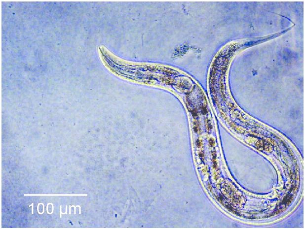
It might not look like much, but this little worm could hold the key to biological regeneration!


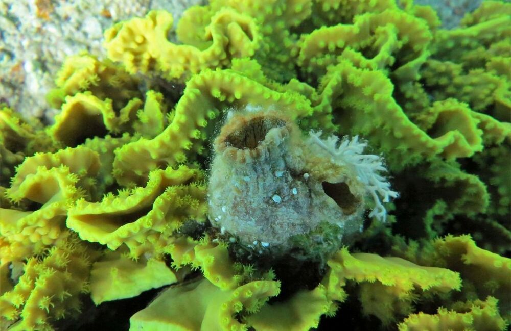
Researchers have made an extraordinary discovery using a creature in common to the Gulf of Eilat. The creature is called Polycarpa mytiligera and is a species of ascidian, which is a marine animal commonly found in the waters of the Gulf that is capable of regenerating its organs. Surprisingly, the researchers discovered the animal can regenerate all of its organs even when dissected into three fragments.

Scientists say that is an astounding discovery because it’s an animal belonging to the Phylum Chordata, which are animals with a dorsal cord, which also includes humans. The ability to regenerate organs itself is not uncommon in the animal kingdom. One example is the gecko able to regrow its tail. However, it’s not common for creatures to be able to regrow entire body systems.

With the Polycarpa mytiligera, scientists have discovered a creature able to regenerate all organs even if separated into three pieces, each piece knowing exactly how to regain the function of all of its missing body systems within a short period. Project researcher Tal Gordon says that by all accounts, the ascidian is a simple organism with two openings in its body, and inside the body is a central organ resembling a pasta strainer.
Antibodies are small protein molecules which are used by the body in order to ‘tag’ foreign pathogens in order for the immune system to identify and destroy them. What is unique about these antibodies is that due to their structure they will only attach themselves to a particular pathogen (on a site known as an antigen). In many cases, the binding of an antibody to a pathogen such as a virus is enough to completely inactive the virus, making it effectively harmless.

Due to the existence of millions upon millions of different types of pathogens, there are quite literally trillions of possibilities for the structures of antibodies, which gives them a near infinite ability to bind with extreme prejudice to just about anything within the body, including our own proteins, such as enzymes and cell receptors.

This ability to bind to biological surfaces allows antibodies to effectively mimic the function of proteins within the body, with the added benefit of often being notably more stable that the protein that they are mimicking. However, there is the slight issue with human antibodies in that they are fairly large, which makes them physically incapable of mimicking many types of proteins. Fortunately, antibodies are not universally the same size within the animal kingdom, and animals such as Llamas have very small antibodies, which are commonly known as nanobodies (very small antibodies).

To answer this question, an internal team of scientists, consisting of researchers affiliated with the Buck Institute for Research on Ageing, and researchers from Nanjing University decided to modify both the Insulin and the rapamycin pathways of a group of C.elegans worms, expecting to see a cumulative result of a 130% increase in lifespan. However, instead of seeing a cumulative effect in lifespan, the worms lived five times longer than they normally would.

“The synergistic extension is really wild. The effect isn’t one plus one equals two, it’s one plus one equals five. Our findings demonstrate that nothing in nature exists in a vacuum; in order to develop the most effective anti-aging treatments we have to look at longevity networks rather than individual pathways.” – Jarad Rollins of Nanjing University.

What could this mean for human regenerative medicine? Humans are not worms, however on a cellular level they do possess very similar biology. Both the insulin pathway and the rapamycin pathway are what is known as ‘conserved’ between humans and C.elegans, meaning that these pathways have been maintained in both organisms. In the distant past, both humans and C.elegans had a common ancestor, in exactly the same way as humans and Chimpanzees have a common ancestor. Evolution has changed our bodies significantly over the millions of years that humans and C.elegans have diverged from one another, but a lot of our fundamental biological functions remain largely unchanged.

The secret to longevity is already in the animals around us.


Some species live unexpectedly long lives. By studying how they do it, researchers hope to pinpoint factors affecting human longevity.

By Bob Holmes.

Life, for most of us, ends far too soon — hence the effort by biomedical researchers to find ways to delay the aging process and extend our stay on Earth. But there’s a paradox at the heart of the science of aging: The vast majority of research focuses on fruit flies, nematode worms and laboratory mice, because they’re easy to work with and lots of genetic tools are available. And yet, a major reason that geneticists chose these species in the first place is because they have short lifespans. In effect, we’ve been learning about longevity from organisms that are the least successful at the game.

Scientists said the findings indicated that the virus likely recently jumped from animals to humans, but stressed that additional studies are necessary.


Scientists have reportedly discovered a new kind of coronavirus that is believed to have originated in dogs – in what may be the eighth unique form of the bug known to cause disease in humans.

Researchers in a study published in the Clinical Infectious Diseases journal said their findings from patients hospitalized with pneumonia in 2017–2018 underscored the public health threat of animal coronaviruses, Reuters reported.

They said they had tested nasal swab samples taken from 301 pneumonia patients at a hospital in the east Malaysian state of Sarawak.

In our ongoing search to continuously improve our health, we occasionally pay lip service to the bacteria that live inside our gut. Normally this concern rarely manifests as anything more than occasionally remembering to buy some of those small bottles of pro-biotic yoghurts while shopping for your…


Recent discoveries have led to the conclusion that the gut plays an important role in cognitive function, with a large amount of research into understanding what is known as the gut-brain axis, which is the collective name given to the biochemical signalling pathways which take place between the gastrointestinal tract and the central nervous system. With an ever-increasing understanding of this pathway, along with an expanded understand of the gut flora (which was found to decline with age), researchers started to ask how the gut flora are involved in the ageing process.

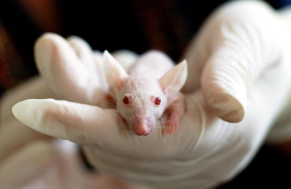
In order to test how exactly ageing gut flora effects the gut-brain axis, researchers at the University of East Anglia conducted a faecal transplant from elderly mice into younger mice. Following this transplant, the young mice were then put through a serious of tests to assess their cognitive abilities. The younger mice showed significant changes in their microbial profiles, as well as significantly impaired capacity for spatial learning, as well as a decreased capacity for memorisation. These mice also showed an altered expression of proteins associated with neurotransmission and neuroplasticity, along with changes in the mice’s hippocampus, which is responsible for allowing the mice to memories new information, as well as recalling previous memories.

This research has successfully proven a link between the changing microbiome of the gut and protein expression within the central nervous system. This discovery is exceptionally good news, as not only is the problem potentially fairly easy to fix (with an aforementioned faecal transplant), but it also provides clues as to how we might compensate for this age related change in the gut microbiome with medication tailors to mimic the role of a young microbiome. Either way, the discovery has opened the door to a number of exciting prospects for regenerative medicine, along with maybe highlighting the fact that we should really start considering our gut bacteria as more than just a collection of microorganisms, and more of a collection of symbiotic organisms that benefit us in ways that we are only just beginning to understand.

To begin with, why do we use mice in medical and biological research? The answer to this question is fairly straight forward. Mice are cheap, they grow quickly, and the public rarely object to experimentations involving mice. However, mice offer something that is far more important than simple pragmatism, as despite being significantly smaller and externally dissimilar to humans, our two species share an awful lot of similarities. Almost every gene found within mice share functions with genes found within humans, with many genes being essentially identical (with the obvious exception of genetic variation found within all species). This means that anatomically mice are remarkably similar to humans.

Now, this is where for the sake of clarity it would be best to break down biomedical research into two categories. Physiological research and pharmaceutical research, as the success of the mouse model should probably be judges separately depending upon the research that is being carried out. Separating the question of the usefulness of the mouse model down into these two categories also solves the function of more accurately focusing the ire of its critics.

The usefulness of the mouse model in the field of physiological research is largely unquestioned at this point. We have quite literally filled entire textbooks with the information we have gained from studying mice, especially in the field of genetics and pathology. The similarities between humans and mice are so prevalent that it is in fact possible to create functioning human/mouse hybrids, known as ‘genetically engineered mouse models’ or ‘GEMMs’. Essentially, GEMMs are mice that have had the mouse version of a particular gene replaced with its human equivalent. This is an exceptionally powerful tool for medical research, and has led to numerous medical breakthroughs, including most notably our current treatment of acute promyelocytic leukaemia (APL), which was created using GEMMs.

Circa 2017


What is the best way to preserve music for future generations to enjoy? Store it in DNA, of course.

That is exactly what Twist Biosciences, which pioneers high-quality DNA synthesis, has facilitated in collaboration with the University of Washington and Microsoft. Two iconic performances – from the iconic Montreux Jazz Festival, one Deep Purple’s playing of Smoke on the Water, and Tutu by Miles Davis, have been ‘encoded onto DNA and read back with 100 percent accuracy.

Legendary music composer and producer, Quincy Jones, said it “absolutely makes my soul smile” to know that the “beauty and history of the Montreux Jazz Festival” has been preserved in this way.

There’s some really interesting CRISPR news out today, and it’s likely to be a forerunner of much more news to come. A research team has demonstrated what looks like robust, long-lasting effects in a primate model after one injection of the CRISPR enzymatic machinery. There have been plenty of rodent reports on various forms of CRISPR, and there are some human trials underway, but these is the first primate numbers that I’m aware of.

The gene they chose to inactivate is PCSK9, which has been a hot topic in drug discovery for some years now. It’s a target validated by several converging lines of evidence from the human population (see the “History” section of that first link). People with overactive PCSK9 have high LDL lipoproteins and cholesterol, and people with mutations that make it inactive have extremely low LDL and seem to be protected from a lot of cardiovascular disease. There are several drugs and drug candidates out there targeting the protein, as well there might be.

It’s a good proof-of-concept, then, because we know exactly what the effects of turning down the expression of active PCSK9 should look like. It’s also got the major advantage of being mostly a liver target – as I’ve mentioned several times on the blog already, many therapies aimed at gene editing or RNA manipulation have a pharmacokinetic complication. The formulations used to get such agents intact into the body (and in a form that they can penetrate cells) tend to get combed out pretty thoroughly by the liver – which after all, is (among other things) in the business of policing the bloodstream for weird, unrecognized stuff that is then targeted for demolition by hepatocytes. Your entire bloodstream goes sluicing through the liver constantly; you’re not going to able to dodge it if your therapy is out there in the circulation. It happens to our small-molecule drugs all the time: hepatic “first pass” metabolism is almost always a factor to reckon with.

The harmfulness of pesticides to beneficial organisms is one of the most serious concerns in agriculture. Therefore scientists are eagerly looking for new, more environmentally friendly and species-specific solutions. Researchers at the Estonian University of Life Sciences, Ghent and the University of Maastricht took a long step forward in this regard.

The detrimental impact of pesticides on non-target organisms is one of the most urgent concerns in current agriculture. Double-stranded RNAs (dsRNAs) represent the most species-specific class of pesticides to date, potentially allowing control of a target pest without effecting other species. The unprecedented target-specificity of dsRNA is due to its nucleotide sequence-specific mode of action that results in post-transcriptional gene silencing, or RNA interference (RNAi), in the target species. The development and field use of dsRNAs, via both the insertion of transgenes into the plant genome and the application of dsRNA sprays, is a rapidly growing area of research. Simultaneously, there exists the growing prospect of harnessing RNAi within integrated pest management schemes.

Using the pollen beetle (Brassicogethes aeneus) and its host crop oilseed rape (Brassica napus) as a model crop-pest system, a team of researchers collectively from Estonian University of Life Sciences, Ghent University and Maastricht University examined how RNAi efficacy depends on duration of dietary exposure to dsRNA. To this end, the authors applied dsRNA (specifically designed to induce RNAi in the pollen beetle) to oilseed rape flowers, and analyzed RNAi-induced mortality between insects chronically fed dsRNA and insects fed dsRNA for 3 days. Most notably, their data suggest that, with chronic dietary exposure to dsRNA, reduced dsRNA concentrations can be applied in order to achieve a similar effect compared to short-term (e.g. 3 days) exposure to higher concentrations. This observation has important implications for optimizing dsRNA spray approaches to managing crop pests.