Toggle light / dark theme

Opinions: Give your opinions in the comments section.

3D printed lab meat, and plant based meats will be more widespread in our future. Would you eat stem cell 3D printed lab meat or plant based meat? Why or why not? What are the differences between natural vs unnatural. Growing up in Texas I know most Texans frown on it, as BBQ is a religion. Is 3D printing meat sustainable\.


Whether it comes from a plant or the cells of an animal, it’s becoming increasingly clear that the meat of the future will probably not be coming from the flesh of slaughtered animals. Instead, whether made from plants or cells, it will be formed into ‘meat’ by a 3D printer. In September of 2021, a Japanese team of researchers at the University of Osaka announced that they had 3D printed Wagyu beef. Beef connoisseurs will recognize the name; Wagyu beef is prized (and suitably priced) for its flavor and fat marbling. Legends abound about the cows such beef derives from, how they are allegedly coddled and massaged, fed a special diet that includes beer — but much of those tales are either exaggerated or pure urban legend. As Joe Heitzeberg, the co-founder and CEO of Crowd Cow explains, There are four breeds native to Japan. Of those four breeds, one of the breeds is genetically unique. It has a genetic predisposition to create this crazy marbling of fat on the inside of muscle tissue. No other livestock does that. The researchers at the University of Osaka used two different types of stem cells from Wagyu cows to create cultured meat, growing living animal cells onto some type of matrix where they are then incubated and grown into animal tissue that has never been part of a living animal. There are currently no reports on the taste of the cultured Wagyu beef but we can assume it’s ‘good’ and given a little time, the technology should be able to produce excellent Wagyu cultured meat — at what price, however, is another big question mark. But there’s another simpler solution that could be a better meat replacement than cultured meat, as even meat grown from stem cells still contains cholesterol and some of the negative health concerns associated with animal protein. Plant-based imitation meat is also being created with 3D printers, and the results are surprising even hardcore meat lovers.

In November 2021, the UK’s Guardian newspaper highlighted the 3D steak produced by Israeli startup Redefine Meat after celebrity chef Marco Pierre White invited chefs, investors connoisseurs, and former winners of the MasterChef cooking show to taste it. This vegan 3D steak made with a 3D printer has a secret formula, but according to the Guardian, it includes soy, pea protein, and other vegetables such as beetroot, chickpeas, and coconut fat. But the reason it’s winning over meat lovers is the unique idea of printing it in layers. With a layer of ‘alternative fat’ made from plant-based materials, and then a layer of ‘alternative muscle’ also made from plants, the imitation meat no longer has a single texture — but like real meat — contains different flavors and textures in different areas. According to the experts gathered by celebrity Chef White, this ‘alt-meat’ is by far the closest synthetic approximation ever.

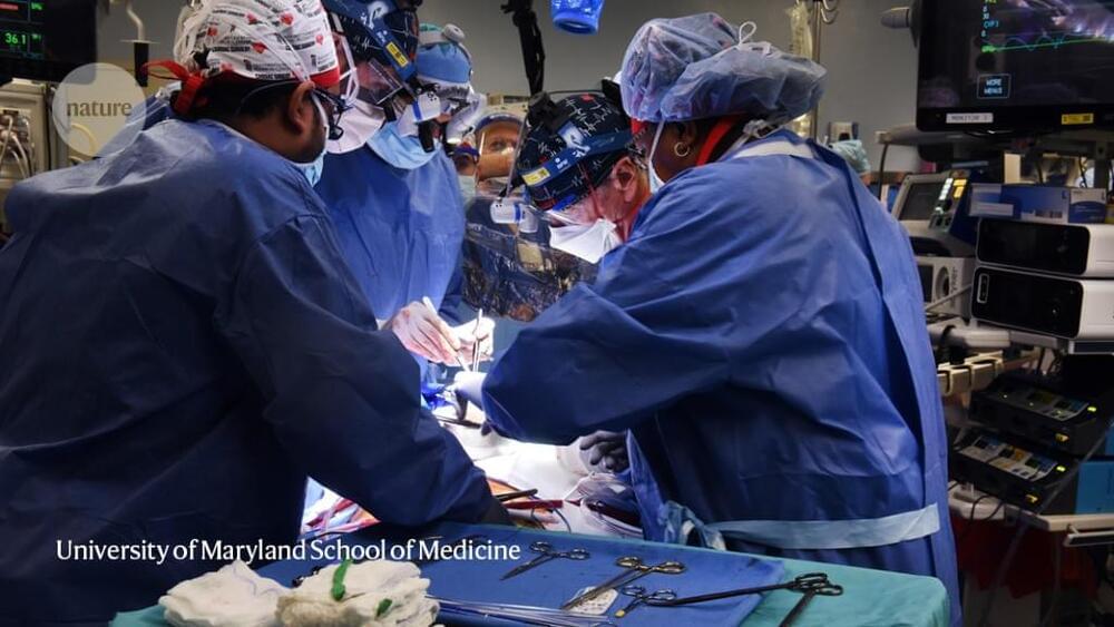
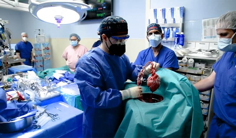
In a first, U.S. surgeons transplant pig heart into human patient.


Unusual opportunity

Last week’s procedure marks the first time that a pig organ has been transplanted into a human who has a chance to survive and recover. In 2021, surgeons at New York University Langone Health transplanted kidneys from the same line of genetically modified pigs into two legally dead people with no discernible brain function. The organs were not rejected, and functioned normally while the deceased recipients were sustained on ventilators.

Aside from that, most research has so far taken place in non-human primates. But researchers hope that the 7 January operation will further kick-start clinical xenotransplantation and help to push it through myriad ethical and regulatory issues.

Gene editing approaches promise to treat a range of diseases, but delivering editing agents to cells in animal models and humans safely and efficiently has proven challenging. Now, researchers led by a team at the Broad Institute of MIT and Harvard have developed a way to get gene editing proteins inside cells in animal models with high enough efficiency to show therapeutic benefit.

In new work published in Cell, the team shows how they have engineered virus-like particles to deliver base editors — proteins that make programmable single-letter changes in DNA — and CRISPR-Cas9 nuclease, a protein that cuts DNA at targeted sites in the genome. In collaboration with ​​research teams led by Krzysztof Palczewski at the University of California, Irvine, and Kiran Musunuru at the Perelman School of Medicine at the University of Pennsylvania, the team used their particles, called engineered virus-like particles (eVLPs), to disable a gene in mice that can be associated with high cholesterol levels, and partially restored visual function to mice harboring a mutation that causes genetic blindness.


Researchers have developed virus-like particles that deliver therapeutic levels of protein to animal models of disease.

A study published by researchers at the University of Illinois Chicago describes a new method for analyzing pyroptosis–the process of cell death that is usually caused by infections and results in excess inflammation in the body–and shows that process, long thought to be irreversible once initiated, can in fact be halted and controlled.

The discovery, which is reported in Nature Communications, means that scientists have a new way to study diseases that are related to malfunctioning cell death processes, like some cancers, and infections that can be complicated by out-of-control inflammation caused by the process. These infections include sepsis, for example, and acute respiratory distress syndrome, which is among the major complications of COVID-19 illness.

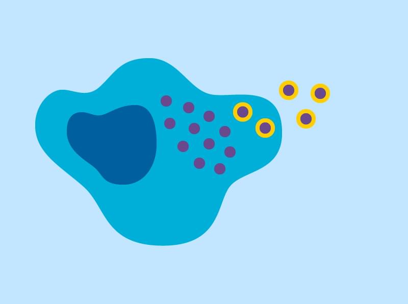
Pyroptosis is a series of biochemical reactions that uses gasdermin, a protein, to open large pores in the cell membrane and destabilize the cell. To understand more about this process, the UIC researchers designed an “optogenetic” gasdermin by genetically engineering the protein to respond to light.

“The cell death process plays an important role in the body, in both healthy states and unhealthy ones, but studying pyroptosis–which is a major type of cell death–has been challenging,” said Gary Mo, UIC assistant professor in the department of pharmacology and regenerative medicine and the department of biomedical engineering at the College of Medicine.

Mo said that methods to examine the pyroptosis mechanisms at play in live cells are difficult to control because they are initiated by unpredictable pathogens, which in turn have disparate effects in different cells and people.

“Our optogenetic gasdermin allowed us to skip over the unpredictable pathogen behavior and the variable cellular response because it mimics at the molecular level what happens in the cell once pyroptosis is initiated,” Mo said.

The researchers applied this tool and used florescent imaging technology to precisely activate gasdermin in cell experiments and observe the pores under various circumstances. They discovered that certain conditions, like specific concentrations of calcium ions, for example, triggered the pores to close within only tens of seconds.

This automatic response to external circumstances provides evidence that pyroptosis dynamically self-regulates.

“This showed us that this form of cell death is not a one-way ticket. The process is actually programmed with a cancel button, an off-switch,” Mo said. “Understanding how to control this process unlocks new avenues for drug discovery, and now we can find drugs that work for both sides–it allows us to think about tuning, either boosting or limiting, this type of cell death in diseases, where we could previously only remove this important process.”

Co-authors of the Nature Communications paper, “Gasdermin D Pores Are Dynamically Regulated by Local Phosphoinositide Circuitry,” are Ana Santa Cruz Garcia, Kevin Schnur and Asrar Malik, all of UIC.

When scientists discovered DNA

DNA, or deoxyribonucleic acid, is a molecule composed of two long strands of nucleotides that coil around each other to form a double helix. It is the hereditary material in humans and almost all other organisms that carries genetic instructions for development, functioning, growth, and reproduction. Nearly every cell in a person’s body has the same DNA. Most DNA is located in the cell nucleus (where it is called nuclear DNA), but a small amount of DNA can also be found in the mitochondria (where it is called mitochondrial DNA or mtDNA).

Houston, TX — Oct 8, 2020 - In a letter published today in the New England Journal of Medicine, a team of physicians from Baylor College of Medicine, Texas Children’s Hospital, and the University of California, San Francisco, describe a remarkable case of a Type 1 diabetes (T1D) patient, who no longer needs insulin to maintain optimal blood sugar levels. The physicians employed a precision/personalized medicine approach to specifically target the underlying genetic mutation, which was the primary driver of this patient’s diabetes.

“To the best of our knowledge, this is the first example of a T1D patient who has experienced a complete reversal of insulin-dependence and we are excited by the prospect that that could be a viable therapeutic strategy for a subset of T1D patients” said corresponding author Dr. Lisa R. Forbes, deputy director for clinical services and community outreach for the Texas Children’s William T. Shearer Center for Human Immunobiology and assistant professor of Pediatrics, Immunology, Allergy and Retrovirology at Baylor.

T1D is a chronic condition in which the pancreas produces little to no insulin, a hormone that maintains sugar levels in the blood. Currently, the treatment options available to T1D patients consist of managing blood sugar levels with insulin, diet and exercise to prevent further complications.

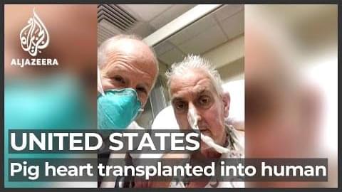
A man with terminal heart disease is responding well three days after being given a genetically modified pig heart in a first-of-its-kind surgery, his doctors reported on Monday.
The surgery, performed by a team at the University of Maryland Medicine in the United States, is among the first to demonstrate the feasibility of a pig-to-human heart transplant, a field made possible by new gene editing tools.
If proven successful, scientists hope pig organs could help alleviate shortages of donor organs.
For David Bennett, a 57-year-old from Maryland, the heart transplant was his last option.

Al Jazeera’s Barbara Angopa reports.

- Follow us on Twitter: https://twitter.com/AJEnglish/
- Find us on Facebook: https://www.facebook.com/aljazeera/
- Check our website: https://www.aljazeera.com/

#HeartTransplant #PigsHeart #DavidBennett

I am happy to say that my recently published computational COVID-19 research has been featured in a major news article by HPCwire! I led this research as CTO of Conduit. My team utilized one of the world’s top supercomputers (Frontera) to study the mechanisms by which the coronavirus’s M proteins and E proteins facilitate budding, an understudied part of the SARS-CoV-2 life cycle. Our results may provide the foundation for new ways of designing antiviral treatments which interfere with budding. Thank you to Ryan Robinson (Conduit’s CEO) and my computational team: Ankush Singhal, Shafat M., David Hill, Jr., Tamer Elkholy, Kayode Ezike, and Ricky Williams.


Conduit, created by MIT graduate (and current CEO) Ryan Robinson, was founded in 2017. But it might not have been until a few years later, when the pandemic started, that Conduit may have found its true calling. While Conduit €™s commercial division is busy developing a Covid-19 test called nanoSPLASH, its nonprofit arm was granted access to one of the most powerful supercomputers in the world €”Frontera, at the Texas Advanced Computing Center (TACC) €”to model the €œbudding € process of SARS-CoV-2.

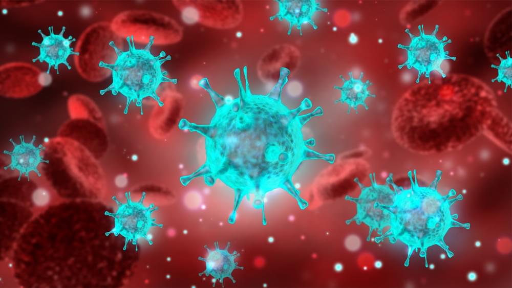
Budding, the researchers explained, is how the virus €™ genetic material is encapsulated in a spherical envelope €”and the process is key to the virus €™ ability to infect. Despite that, they say, it has hitherto been poorly understood:

The Conduit team €”comprised of Logan Thrasher Collins (CTO of Conduit), Tamer Elkholy, Shafat Mubin, David Hill, Ricky Williams, Kayode Ezike and Ankush Singhal €”sought to change that, applying for an allocation from the White House-led Covid-19 High-Performance Computing Consortium to model the budding process on a supercomputer.