Toggle light / dark theme

Bio-Printing Complex Human Tissues & Organs — Dr. Anthony Atala, MD — Director, Wake Forest Institute for Regenerative Medicine, Wake Forest School of Medicine, Wake Forest University.


Dr. Anthony Atala, MD, (https://school.wakehealth.edu/Faculty/A/Anthony-Atala) is the G. Link Professor and Director of the Wake Forest Institute for Regenerative Medicine, and the W. Boyce Professor and Chair of Urology.

A practicing surgeon and a researcher in the area of regenerative medicine, fifteen applications of technologies developed Dr. Atala’s laboratory have been used clinically. He is Editor of 25 books and 3 journals, has published over 800 journal articles, and has received over 250 national and international patents. Dr. Atala was elected to the Institute of Medicine of the National Academies of Sciences, to the National Academy of Inventors as a Charter Fellow, and to the American Institute for Medical and Biological Engineering.

Dr. Atala is a recipient of the US Congress funded Christopher Columbus Foundation Award, bestowed on a living American who is currently working on a discovery that will significantly affect society; the World Technology Award in Health and Medicine, for achieving significant and lasting progress; the Edison Science/Medical Award for innovation, the R&D Innovator of the Year Award, and the Smithsonian Ingenuity Award for Bioprinting Tissue and Organs. Dr. Atala’s work was listed twice as Time Magazine’s Top 10 medical breakthroughs of the year, and once as one of 5 discoveries that will change the future of organ transplants. He was named by Scientific American as one of the world’s most influential people in biotechnology, by U.S. News & World Report as one of 14 Pioneers of Medical Progress in the 21st Century, by Life Sciences Intellectual Property Review as one of the top key influencers in the life sciences intellectual property arena, and by Nature Biotechnology as one of the top 10 translational researchers in the world.

Dr. Atala has led or served several national professional and government committees, including the National Institutes of Health working group on Cells and Developmental Biology, the National Institutes of Health Bioengineering Consortium, and the National Cancer Institute’s Advisory Board. He is a founding member of the Tissue Engineering Society, Regenerative Medicine Foundation, Regenerative Medicine Manufacturing Innovation Consortium, Regenerative Medicine Development Organization, and Regenerative Medicine Manufacturing Society.
A major obstacle to widespread study and clinical use of 3D tissues is their short shelf-life, which may be anywhere from a just few hours to a few days. As in the case of an organ transplant, a bioprinted tissue must be transported rapidly to the location where it is needed, or it will not be viable. In the journal Matter on December 21st, researchers at Brigham and Women’s Hospital and Harvard Medical School describe their work combining 3D bioprinting with cryopreservative techniques to create tissues which can be preserved in a freezer at-196°C and thawed within minutes for immediate use.

“For conventional bioprinting, there is basically no shelf life. It’s really just print, and then use, in most cases,” says lead author Y. Shrike Zhang (@shrikezhang), a biomedical engineer at Brigham and Women’s Hospital. “With cryobioprinting, you can print and store in the frozen state for basically as long as you want.”

The use of 3D bioprinting to create artificial human tissue first appeared twenty years ago. As in conventional 3D printing, an ink is extruded layer by layer through a nozzle into a pre-specified shape. In the case of bioprinting, the ink is typically made up of a gelatin-like scaffolding embedded with living cells. Cryobioprinting works the same way, except the printing is performed directly onto a cold plate held at temperatures down to-20°C. After the tissues are printed, they are immediately moved to cryogenic conditions for long-term storage.

The 24thSpaceX cargo resupply services mission, targeted to launch in late December from NASA’s Kennedy Space Center in Florida, carries scientific research and technology demonstrations to the International Space Station. The experiments aboard include studies of bioprinting, crystallization of monoclonal antibodies, changes in immune function, plant gene expression changes, laundering clothes in space, processing alloys, and student citizen science projects.

A team of researchers from Texas A&M University’s Department of Biomedical Engineering has designed and 3D bioprinted a highly realistic model of a blood vessel.

The model is made of a newly nanoengineered, purpose-built hydrogel bioink and closely mimics the natural vascular function of a real blood vessel, as well as its disease response. The team hopes its work can pave the way for advanced cardiovascular drug development, expediting treatment approval while eliminating the need for animal and human testing altogether.

“A remarkably unique characteristic of this nanoengineered bioink is that regardless of cell density, it demonstrates a high printability and ability to protect encapsulated cells against high shear forces in the bioprinting process,” said Akhilesh Gaharwar, associate professor at the university and co-author of the study. “Remarkably, 3D bioprinted cells maintain a healthy phenotype and remain viable for nearly one month post-fabrication.”

The world of lab-grown meats is fast filling with all kinds of tasty bites, from burgers, to chicken breasts, to a series of increasingly complex cuts of steak. Expanding the scope of cultured beef are scientists from Japan’s Osaka University, who have leveraged cutting-edge bioprinting techniques to produce the first lab-grown “beef” that resembles the marbled texture of the country’s famed Wagyu cows.

From humble beginnings that resembled soggy pork back in 2,009 to the classic steaks and rib-eyes we’ve seen pop up in the last few years, lab-grown meat has come along in leaps and bounds. The most sophisticated examples use bioprinting to “print” living cells, which are nurtured to grow and differentiate into different cell types, ultimately building up into the tissues of the desired animal.

The Osaka University team used two types of stem cells harvested from Wagyu cows as their starting point, bovine satellite cells and adipose-derived stem cells. These cells were incubated and coaxed into becoming the different cell types needed to form individual fibers for muscle, fat and blood vessels. These were then arranged into a 3D stack to resemble the high intramuscular fat content of Wagyu, better known as marbling, or sashi in Japan.

Scientists from the University at Buffalo have developed a rapid new 3D bioprinting method that could represent a significant step towards fully-printed human organs.

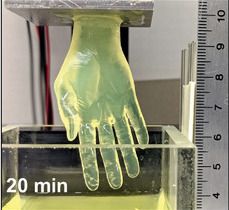
Using a novel vat-SLA-based approach, the team have been able to reduce the time it takes to create cell-laden hydrogel structures, from over 6 hours to just 19 minutes. The expedited biofabrication method also enables the production of embedded blood vessel networks, potentially making it a significant step towards the lifesaving 3D printed organs needed by those on transplant waiting lists.

“Our method allows for the rapid printing of centimeter-sized hydrogel models,” explained the study’s lead co-author, Chi Zhou. “It significantly reduces part deformation and cellular injuries caused by the prolonged exposure to the environmental stresses you commonly see in conventional 3D printing.”

Bioprinting in seconds.


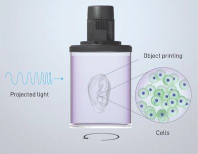
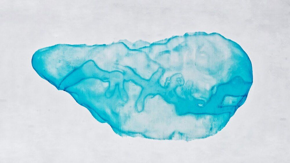
Biofabrication technologies, including stereolithography and extrusion-based printing, are revolutionizing the creation of complex engineered tissues. The current paradigm in bioprinting relies on the additive layer-by-layer deposition and assembly of repetitive building blocks, typically cell-laden hydrogel fibers or voxels, single cells, or cellular aggregates. The scalability of these additive manufacturing technologies is limited by their printing velocity, as lengthy biofabrication processes impair cell functionality. Overcoming such limitations, the volumetric bioprinting of clinically relevant sized, anatomically shaped constructs, in a time frame ranging from seconds to tens of seconds is described. An optical-tomography-inspired printing approach, based on visible light projection, is developed to generate cell-laden tissue constructs with high viability (85%) from gelatin-based photoresponsive hydrogels. Free-form architectures, difficult to reproduce with conventional printing, are obtained, including anatomically correct trabecular bone models with embedded angiogenic sprouts and meniscal grafts. The latter undergoes maturation in vitro as the bioprinted chondroprogenitor cells synthesize neo-fibrocartilage matrix. Moreover, free-floating structures are generated, as demonstrated by printing functional hydrogel-based ball-and-cage fluidic valves. Volumetric bioprinting permits the creation of geometrically complex, centimeter-scale constructs at an unprecedented printing velocity, opening new avenues for upscaling the production of hydrogel-based constructs and for their application in tissue engineering, regenerative medicine, and soft robotics.

Volumetric 3D bioprinter manufacturer and EPFL spin-out Readily3D has taken the first step towards developing a 3D printed living model of the human pancreas for testing diabetes medicines.

Readily3D’s novel technology is being deployed within the EU-funded Enlight project and is reportedly capable of 3D printing a biological tissue containing human stem cells in just 30 seconds.

As the project’s official bioprinter manufacturer, the company has adapted its proprietary contactless tomographic illumination technology to suit the specific needs of pancreatic tissue structures.

Circa 2020


The FRESH technique of 3D bioprinting was invented in Feinberg’s lab to fill an unfilled demand for 3D printed soft polymers, which lack the rigidity to stand unsupported as in a normal print. FRESH 3D printing uses a needle to inject bioink into a bath of soft hydrogel, which supports the object as it prints. Once finished, a simple application of heat causes the hydrogel to melt away, leaving only the 3D bioprinted object.

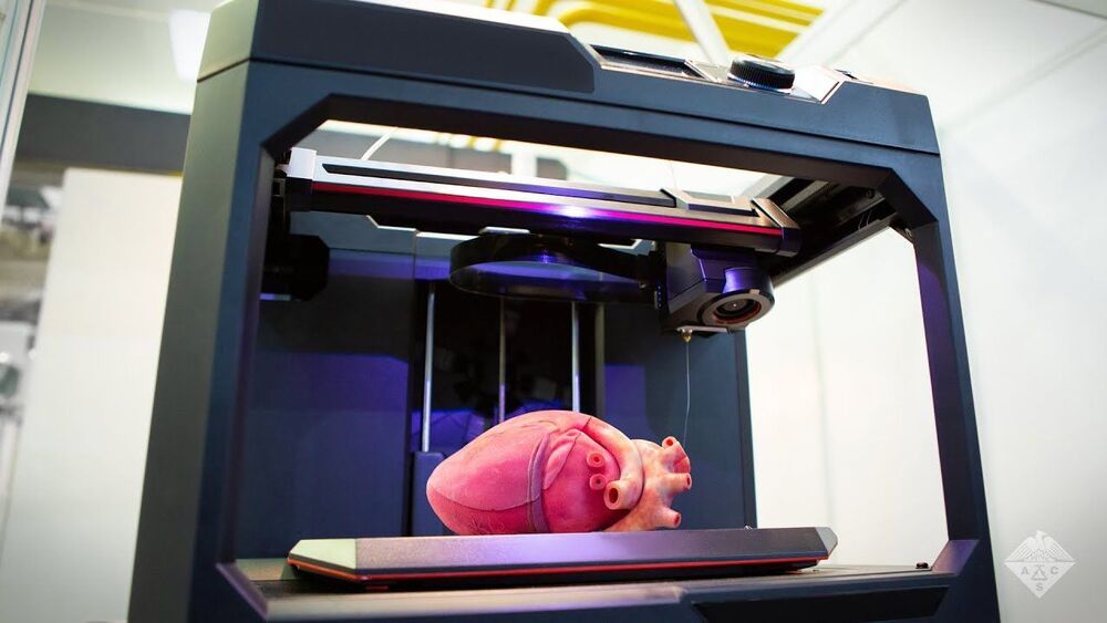
While Feinberg, a professor of biomedical engineering and materials science and engineering, has proven both the versatility and the fidelity of the FRESH technique, the major obstacle to achieving this milestone was printing a human heart at full scale. This necessitated the building of a new 3D printer custom made to hold a gel support bath large enough to print at the desired size, as well as minor software changes to maintain the speed and fidelity of the print.