Toggle light / dark theme

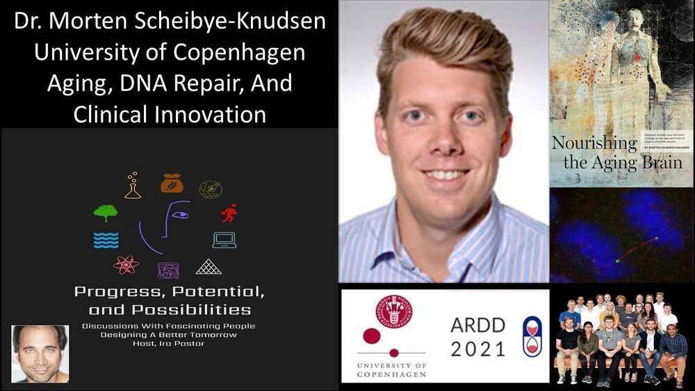
Aging, DNA Repair, And Clinical Innovation — Dr. Morten Scheibye-Knudsen — University of Copenhagen.


Dr. Morten Scheibye-Knudsen is an Associate Professor at the Department of Cellular and Molecular Medicine, and at the Center for Healthy Aging (CEHA), at the University of Copenhagen.

Dr. Scheibye-Knudsen did his MD at the University of Copenhagen and worked briefly as a physician in Denmark and Greenland before turning to science. He did his post-doctoral fellowship at Vilhelm Bohr’s lab at the National Institute on Aging, National Institutes of Health, USA, where he utilized state-of-the art approaches to understand how DNA damage contributes to aging, discovering that neurodegeneration in several premature aging diseases is partly caused by hyperactivation of a DNA damage responsive enzyme called polyADP-ribose polymerase 1 (PARP1). This activation leads to loss of vital metabolites such as Nicotinamide Adenine Dinucleotide (NAD+) and acetyl-CoA. Importantly, this discovery facilitated the realization that we can intervene in the aging process by inhibiting PARP1, augmenting NAD+ levels and increasing acetyl-CoA.

In his own lab Dr. Scheibye-Knudsen continues to focus on understanding aging by combining machine learning based approaches with wet-lab analyses with the goal of developing interventions for age-associated diseases and perhaps aging itself.

Dr. Scheibye-Knudsen is Chief Editor, Frontiers in Aging, and an Advisory Board Member of the Longevity Vision Fund and Molecule Protocol.
Do we need to revisit this? 🤔


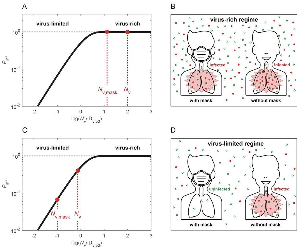
Airborne transmission by droplets and aerosols is important for the spread of viruses. Face masks are a well-established preventive measure, but their effectiveness for mitigating SARS-CoV-2 transmission is still under debate. We show that variations in mask efficacy can be explained by different regimes of virus abundance and related to population-average infection probability and reproduction number. For SARS-CoV-2, the viral load of infectious individuals can vary by orders of magnitude. We find that most environments and contacts are under conditions of low virus abundance (virus-limited) where surgical masks are effective at preventing virus spread. More advanced masks and other protective equipment are required in potentially virus-rich indoor environments including medical centers and hospitals. Masks are particularly effective in combination with other preventive measures like ventilation and distancing.

Airborne transmission is one of the main pathways for the transmission of respiratory viruses, including the severe acute respiratory syndrome coronavirus 2 (SARS-CoV-2) (1). Wearing face masks has been widely advocated to mitigate transmission. Masks are thought to protect people in two ways: source control reducing the emission and spread of respiratory viruses through airborne droplets and aerosols, and wearer protection reducing the inhalation of airborne respiratory viruses.

The effectiveness of masks, however, is still under debate. Compared to N95/FFP2 respirators which have very low particle penetration rates (around ~5%), surgical and similar masks exhibit higher and more variable penetration rates (around ~30–70%) (2, 3). Given the large number of particles emitted upon respiration and especially upon sneezing or coughing , the number of respiratory particles that may penetrate masks is substantial, which is one of the main reasons leading to doubts about their efficacy in preventing infections. Moreover, randomized clinical trials show inconsistent or inconclusive results, with some studies reporting only a marginal benefit or no effect of mask use (5, 6). Thus, surgical and similar masks are often considered to be ineffective. On the other hand, observational data show that regions or facilities with a higher percentage of the population wearing masks have better control of the coronavirus disease 2019 (COVID-19) (7–9).

Famed longevity pioneer Aubrey de Grey, Chief Science Officer of SENS Research Foundation, joins Geoffrey Woo, Founder and Chairman of Health Via Modern Nutrition Inc., for an enlightening conversation about advances in longevity, the investments and technologies that extend life, and the challenges and opportunities of a world in which people live longer. He walks us through his damage repair therapies with a focus on rejuvenation, prevention, and wearable technologies. Filmed on May 17, 2021. To continue the discussion with fellow Real Vision members on this interview, click here to visit the Exchange: https://rvtv.io/2T7nqZL

Key Learnings: Longevity escape velocity, which is a term de Grey coined, is the idea in which life expectancy is extended longer than the time that is passing, and he estimates a 50% chance that aging could be brought under medical control in as little as 15 years’ time. To learn more about SENS’ research and advancements, please visit their site here: https://www.sens.org/.

Scientists working at Canada’s highest-security infectious-disease laboratory have been collaborating with Chinese military researchers to study and conduct experiments on deadly pathogens.

Seven scientists in the special pathogens unit at the National Microbiology Laboratory (NML) in Winnipeg and Chinese military researchers have conducted experiments and co-authored six studies on infectious diseases such as Ebola, Lassa fever and Rift Valley fever. The publication dates of the studies range from early 2016 to early 2020.

The Globe and Mail has also learned that one of the Chinese researchers, Feihu Yan, from the People’s Liberation Army’s (PLA) Academy of Military Medical Sciences, worked for a period of time at the Winnipeg lab, a Level 4 facility equipped to handle some of the world’s deadliest diseases. This researcher is credited as a co-author on all six of the papers. However, on two of them, he is listed as being affiliated with both the Winnipeg lab and the military medical academy.

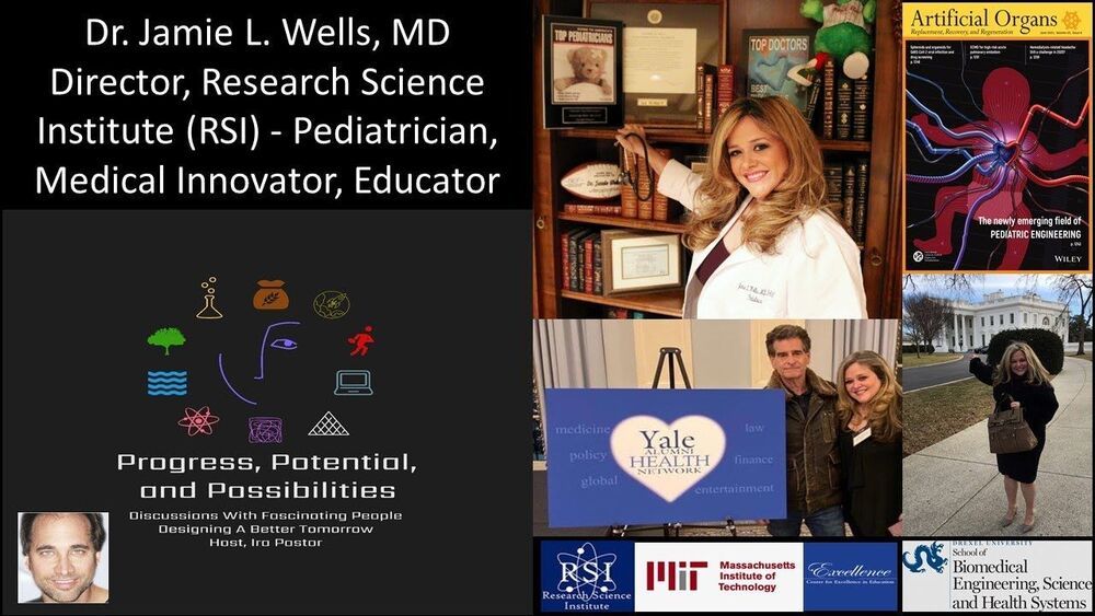
Pediatrician, Medical Innovator, Educator — Dr. Jamie Wells, MD, FAAP — Director, Research Science Institute (RSI), Center for Excellence in Education, Massachusetts Institute of Technology (MIT) — Professor, Drexel University School of Biomedical Engineering, Science and Health Systems.


Dr. Jamie L. Wells, MD, FAAP, is an Adjunct Professor at Drexel University’s School of Biomedical Engineering, Science and Health Systems, where she has been involved in helping to spearhead the nation’s first-degree program focused on pediatric engineering, innovation, and medical advancement.

Dr. Wells is an award-winning Board-certified pediatrician with many years of experience caring for patients. With her BA with Honors from Yale, and her MD from Jefferson Medical College, Philadelphia, PA, she has served as a Clinical Instructor/Attending at NYU Langone, Mt. Sinai-Beth Israel and St. Vincent’s Medical Centers in Manhattan.

Dr. Wells also serves as Director of the Research Science Institute (RSI), at Center for Excellence in Education (CEE), a non-profit, 501©(3) organization, collaboratively sponsored with MIT bring together top U.S. and international high school students for an intensive, six-week summer program that provides students with the opportunity to conduct original, cutting-edge research.

Dr. Wells is on the leadership council of the Wistar Institute (the USA’s first independent biomedical research facility and certified cancer center), Ambassador of the Healthcare Global Blockchain Business Council, was a grant reviewer for the Susan G. Komen Community Grants Program, judged both the local, district and world robotics championships for Dean Kamen’s F.I.R.S.T. (For Inspiration & Recognition of Science & Technology) nonprofit, as well as the Miss America’s Outstanding Teen scholarship competition (for which she is now a member of its Board of Directors), and is the Chair of the Yale Alumni Health Network (YAHN).

Dr. Wells has a proven track record of success in communications on various platforms (e.g. BBC, Reuters TV, Fox News Channel, Discovery Health), routinely appearing as a medical expert and has published over 400 articles as director of medicine for an educational advocacy nonprofit.

Dr. Wells has broad ranging interests in patient safety and optimizing care delivery, bioethics, public health and policy, science communications, med-tech innovation, diagnosis and management of disease, identifying healthcare trends, bridging interdisciplinary knowledge gaps to prevent medical error and expedite discovery, thought integration of clinical practice and biomedical engineering approaches with an emphasis on patient-centered design, debunking medical myths and pediatrics.

In a major breakthrough, researchers at Massachusetts General Hospital (MGH) have discovered how amyloid beta—the neurotoxin believed to be at the root of Alzheimer’s disease (AD)—forms in axons and related structures that connect neurons in the brain, where it causes the most damage. Their findings, published in Cell Reports, could serve as a guidepost for developing new therapies to prevent the onset of this devastating neurological disease.

Among his many contributions to research on AD, Rudolph Tanzi, Ph.D., vice chair of Neurology and co-director of the McCance Center for Brain Health at MGH, led a team in 1986 that discovered the first Alzheimer’s disease gene, known as APP, which provides instructions for making protein precursor (APP). When this protein is cut (or cleaved) by enzymes—first, beta secretase, followed by gamma secretase—the byproduct is amyloid beta (sometimes shortened to Abeta). Large deposits of amyloid beta are believed to cause neurological destruction that results in AD. Amyloid beta formed in the brain’s axons and nerve endings causes the worst damage in AD by impairing communication between nerve cells (or neurons) in the brain. Researchers around the world have worked intensely to find ways to block the formation of amyloid beta by preventing cleavage by beta secretase and gamma secretase. However, these approaches have been hampered by safety issues.

Despite years of research, a major mystery has remained. “We knew that Abeta is made in the axons of the brain’s nerve cells, but we didn’t know how,” says Tanzi. He and his colleagues probed the question by studying the brains of mice, as well as with a research tool known as Alzheimer’s in a dish, a three-dimensional cell culture model of the disease created in 2014 by Tanzi and a colleague, Doo Yeon Kim, Ph.D. Earlier, in 2013, several other MGH researchers, including neurobiologist Dora Kovacs, Ph.D. (who is married to Tanzi), and Raja Bhattacharyya, Ph.D., a member of Tanzi’s lab, showed that a form of APP that has undergone a process called palmitoylation (palAPP) gives rise to amyloid beta. That study indicated that, within the neuron, palAPP is transported in a fatty vesicle (or sac) known as a lipid raft. But there are many forms of lipid rafts.

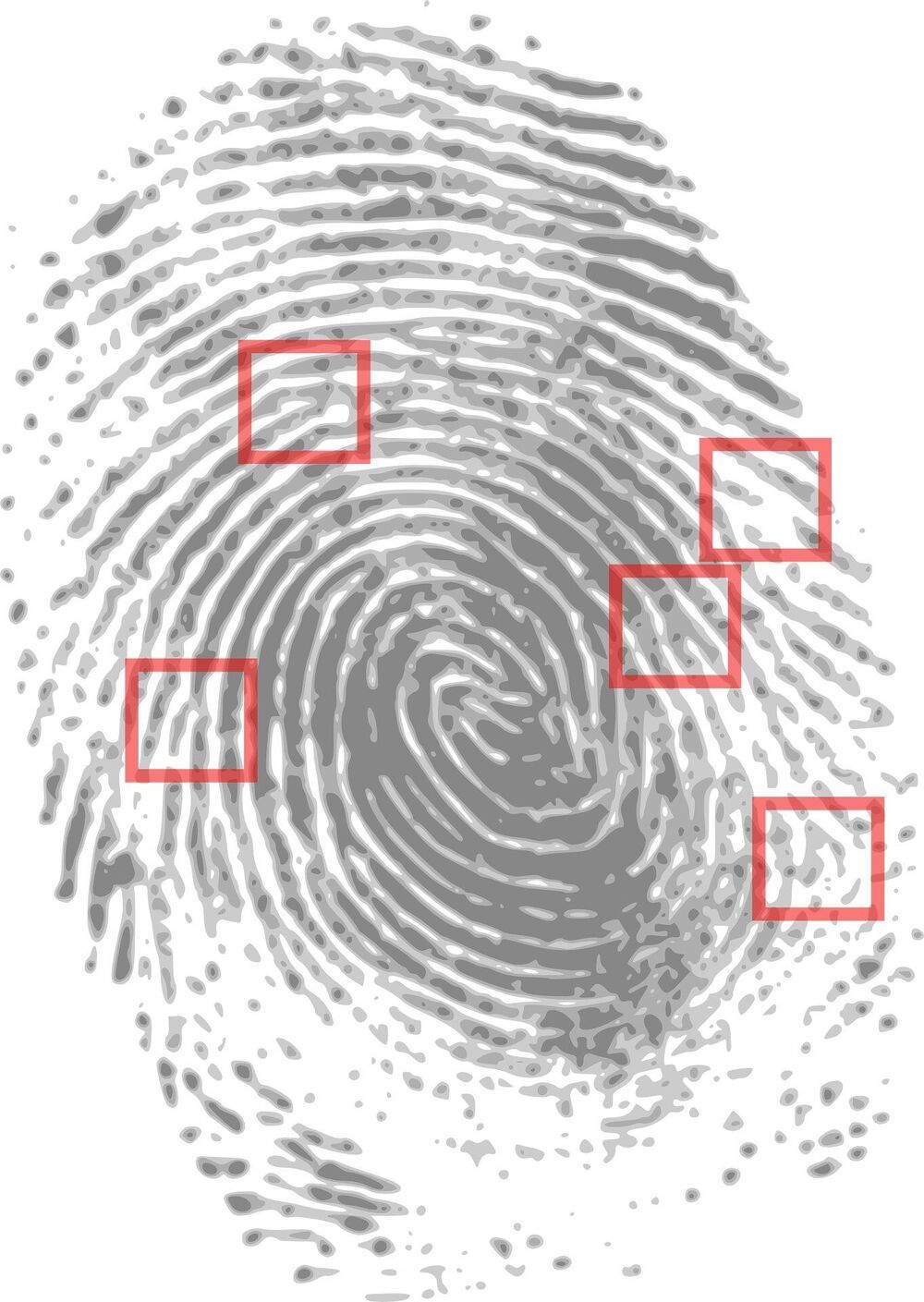
The National Center of Excellence in Mass Spectrometry Imaging at NPL, in collaboration with the University of Surrey and Ionoptika Ltd reveal latest findings showing how a single fingerprint left at a crime scene could be used to determine whether someone has touched or ingested class A drugs.

In a paper published in Royal Society of Chemistry’s Analyst journal, the consortium reveal how they have been able to identify the differences between the fingerprints of people who touched cocaine compared with those who have ingested the drug—even if the hands are not washed. The science behind the advance is the spectrometry imaging tools applied to the detection of cocaine and its metabolites in fingerprints.

In 2020 researchers were able to determine the difference between touch and ingestion if someone had washed their hands prior to giving a sample. Given that a suspect at a crime scene is unlikely to wash their hands before leaving fingerprints, these new findings are a significant advantage to crime forensics.