Toggle light / dark theme

A new study led by University of Maryland and UCLA researchers found that DNA from tissue samples can be used to accurately predict the age of bats in the wild. The study also showed age-related changes to the DNA of long-lived species are different from those in short-lived species, especially in regions of the genome near genes associated with cancer and immunity. This work provides new insight into causes of age-related declines.

This is the first research paper to show that animals in the wild can be accurately aged using an epigenetic clock, which predicts age based on specific changes to DNA. This work provides a new tool for biologists studying animals in the wild. In addition, the results provide insight into possible mechanisms behind the exceptional longevity of many bat species. The study appears in the March 12, 2021, issue of the journal Nature Communications.

“We hoped that these epigenetic changes would be predictive of age,” said Gerald Wilkinson, a professor of biology at UMD and co-lead author of the paper. “But now we have the data to show that instead of having to follow animals over their lifetime to be sure of their age, you can just go out and take a tiny sample of an individual in the wild and be able to know its age, which allows us to ask all kinds of questions we couldn’t before.”

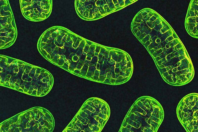
The inside of a mitochondria is made up of a folded membrane, which has evolved to produce the greatest surface area possible between two parts of the mitochondria known as the intermembrane space (the outer part) and the mitochondrial matrix (the inner part). To drastically oversimplify this entire process, the mitochondria uses glucose (and ethanol if it’s available) to pump hydrogen ions (with the occasional deuterium and tritium ion) across the membrane which separates these two compartments of the mitochondria (known as the cristae) into the intermembrane space. These hydrogen ions then flow back into the mitochondrial matrix through a very special protein called ATP synthase, which uses the electrostatic potential energy of the hydrogen ion to manufacture ATP.

Unfortunately, as we get older this inner membrane starts to decay and become smaller. As the cristae starts to shrink, there is less space for ATP synthase, which means there is less ATP produced, which ultimately means that our cells do not have enough energy to maintain all of our cellular functions. As you can imagine, this lack of energy is catastrophic for the health of the cell, and will eventually lead to either cell senescent (where the cell essentially becomes dormant), or complete cell death.

Numerous different suggestions have been put forward as to explain why exactly why mitochondria decay in this way, including mutations within the DNA of the mitochondria (they have their own chromosomes), as well as the build up of oxidative agents within the cell itself which cause direct damage to the mitochondria. However, a group of scientists lead by Dr Hazel Szeto have discovered that the decay of the mitochondrial cristae is linked to declining levels of a phospholipid (fat) called cardiolipin. It turns out that as we age, oxidative agents within our body destroy this phospholipid, which is essential for maintaining the folded inner membrane of the mitochondria.

Several different causes of aging have been discovered, but the question remains whether there are common underlying mechanisms that determine aging and lifespan. Researchers from the Max Planck Institute for Biology of Ageing and the CECAD Cluster of Excellence in Ageing research at the University Cologne have now come across folate metabolism in their search for such basic mechanisms. Its regulation underlies many known aging signaling pathways and leads to longevity. This may provide a new possibility to broadly improve human health during aging.

In recent decades, several cellular signaling pathways have been discovered that regulate the lifespan of an organism and are thus of enormous importance for aging research. When researchers altered these signaling pathways, this extended the lifespan of diverse organisms. However, the question arises whether these different signaling pathways converge on common metabolic pathways that are causal for longevity.

We probably cannot slow the rate at which we get older because of biological constraints, an unprecedented study of lifespan statistics in human and non-human primates has confirmed.

The study set out to test the ‘invariant rate of aging’ hypothesis, which says that a species has a relatively fixed rate of aging from adulthood. An international collaboration of scientists from 14 countries, including José Manuel Aburto from Oxford’s Leverhulme Centre for Demographic Science, analyzed age-specific birth and death data spanning centuries and continents. Led by Fernando Colchero, University of Southern Denmark and Susan Alberts, Duke University, North Carolina, the study was a huge endeavor requiring monitoring wild populations of primates over several decades.

Jose Manuel Aburto says, Our findings support the theory that, rather than slowing down death, more people are living much longer due to a reduction in mortality at younger ages. We compared birth and death data from humans and and found this general pattern of mortality was the same in all of them. This suggests that biological, rather than environmental factors, ultimately control longevity.

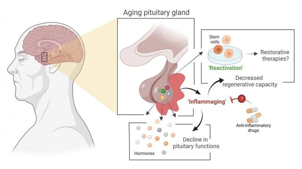
Stem cell biologist Hugo Vankelecom (KU Leuven) and his colleagues have discovered that the pituitary gland in mice ages as the result of an age-related form of chronic inflammation. It may be possible to slow down this process or even partially repair it. The researchers have published their findings in PNAS.

The pituitary is a small, globular gland located underneath the brain that plays a major role in the , explains Professor Hugo Vankelecom from the Department of Development and Regeneration at KU Leuven. “My research group discovered that the pituitary gland ages as a result of a form of chronic inflammation that affects tissue and even the organism as a whole. This usually goes unnoticed and is referred to as ‘inflammaging’—a contraction of inflammation and aging. Inflammaging has previously been linked to the aging of other organs.” Due to the central role played by the pituitary, its aging may contribute to the reduction of hormonal processes and hormone levels in our body—as is the case with menopause, for instance.

The study also provides significant insight into the stem cells in the aging . In 2012, Vankelecom and his colleagues showed that a prompt reaction of these stem cells to injury in the gland leads to repair of the tissue, even in adult animals. “As a result of this new study, we now know that stem cells in the pituitary do not lose this regenerative capacity when the organism ages. In fact, the stem cells are only unable to do their job because, over time, the pituitary becomes an ‘inflammatory environment’ as a result of the chronic inflammation. But as soon as the stem cells are taken out of this environment, they show the same properties as stem cells from a young pituitary.”

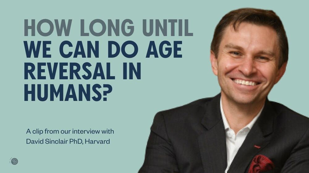
In a minute and 27 seconds we get the what from an eye regeneration for mice, to monkey trials to start later this year, to human trials by 2023, and full body in a decade.


David Sinclair—a world-leading biologist, Harvard Medical School Professor, and author of The New York Times best-selling book @Lifespan.

🧬 His work on understanding why we age and how to slow down the aging process has contributed significantly to getting the longevity science to where it is today. David’s numerous discoveries have been published in the most respected scientific journals. He co-founded many biotech companies, including Life Biosciences, MetroBiotech, and InsideTracker.

🧬 David has received more than 25 awards and honors for his research. He was included in TIME Magazine’s list of the “100 most influential people in the world” in 2014 and “50 Most Influential People in Health Care” in 2018.

🧬 David and his colleagues have recently published a Nature paper with extraordinary results of their epigenetic reprogramming therapy that has successfully restored vision in mice. The paper has become the most accessed paper in the past 12 months at the journal.

If you like this clip and want to watch the full episode be sure to join our Longevity Ambassador list. Every month, co-hosts Sourav Sinha and Sergey Young sit down with a longevity pioneer or healthcare experts to dive deep into the science, investing and technological breakthroughs of aging.

Sign up here: https://sergeyyoung.com/ama-list.

Follow me on Instagram: https://www.instagram.com/sergeyyoung200/

The resulting implant consists of cells attached to the scaffold, which permits the targeted delivery of therapeutic cells to the diseased region within the eye. A non-cryopreserved formulation of this cellular therapy is being employed in an ongoing Phase I/IIa clinical trial sponsored by RPT. The cryopreserved formulation enabled by the work of Pennington and colleagues will facilitate anticipated Phase IIb and Phase III clinical trials as well as ultimate commercialization and clinical application of the product.


Scientists at UC Santa Barbara, University of Southern California (USC), and the biotechnology company Regenerative Patch Technologies LLC (RPT) have reported new methodology for preservation of RPT’s stem cell-based therapy for age-related macular degeneration (AMD).

The new research, recently published in Scientific Reports, optimizes the conditions to cryopreserve, or freeze, an consisting of a single layer of ocular generated from supported by a flexible scaffold about 3×6 mm in size. This implant is currently in clinical trial for the treatment of AMD, the leading cause of blindness in aging populations. The results demonstrate that the implant can be frozen, stored for long periods and distributed in frozen form to clinical sites where it is designed to be thawed and immediately implanted into the eyes of patients with macular degeneration. The capacity to cryopreserve this and other cell-based therapeutics will extend and enable on-demand distribution to distant clinical sites, increasing the number of patients able to benefit from such treatments.

The report published by lead author Britney Pennington and colleagues achieves a milestone that brings ocular implants one step closer to the clinic. “This is the first published report that demonstrates high viability and function of adherent ocular cells following cryopreservation, even after long-term frozen storage,” said Pennington, head of process development at RPT and assistant project scientist at UC Santa Barbara.

The World Health Organization classifies processed meat as a Group 1 carcinogen. Processed meat includes ham, sausage, bacon, pepperoni; they’re meats that have been preserved with salt or smoke, meat that has been cured, and meat treated with chemical preserves. Other Group 1 carcinogens include formaldehyde, tobacco, and UV radiation. Group 1 carcinogens have ‘enough evidence to conclude that it can cause cancer in humans.’


There is no question whether or not our current meat production complex is inhumane, unsanitary, or bad for the environment. Almost all chickens (99.9%), turkeys (99.8%), and most cows (70.4%) eaten in the United States are raised on factory farms. There are horrific consequences to this practice.

For example, the EPA estimates agriculture is the biggest contaminator of rivers and streams, to the point where feedlots, crop production, and manure runoff have led almost half (46%) of the U.S.’s rivers to be “in poor biological condition.”

Scientific American also explains, “TDM-approved feed containing antibiotics [are] a necessity if [factory farm animals] were to stay healthy in their crowded, manure-gilded home. Antibiotics also help farm animals grow faster on less food, so their use has long been a staple of industrial farming.” Many scientists worry that antibiotics used at such a scale on farms create unstoppable, drug-resistant bacteria that can transfer to humans; think inconveniences like nose infections or UTIs turned deadly because of the lack of antibiotics available to treat them.

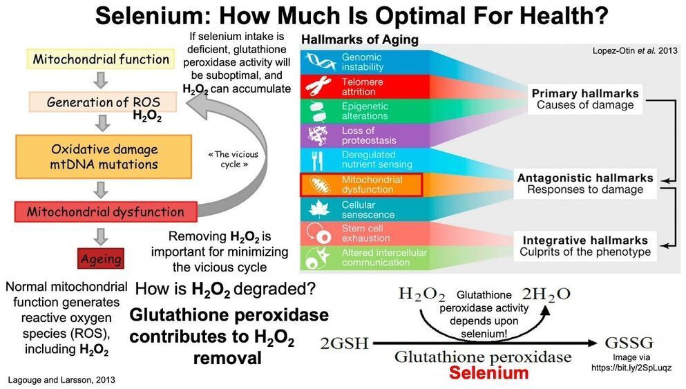
For those who track their diet, eating only the RDA for many nutrients may not optimize health. For example, the RDA for selenium is 55 micrograms per day, but is that amount optimal for reducing risk of death for all causes?


Papers referenced in the video:

The role of mitochondrial DNA mutations and free radicals in disease and ageing.
https://pubmed.ncbi.nlm.nih.gov/23432181/

The Hallmarks of Aging.
https://pubmed.ncbi.nlm.nih.gov/23746838/

Dietary Reference Intakes for Vitamin C, Vitamin E, Selenium, and Carotenoids.
https://www.ncbi.nlm.nih.gov/books/NBK225470/

Dietary and serum selenium in coronary heart disease and all-cause mortality: An international perspective.
https://pubmed.ncbi.nlm.nih.gov/33377378/

Association between selenium intake, diabetes, and mortality in adults: findings fromNational Health and Nutrition Examination Survey (NHANES) 2003–2014
https://pubmed.ncbi.nlm.nih.gov/34039451/

Dietary Antioxidants, Circulating Antioxidant Concentrations, Total Antioxidant Capacity, and Risk of All-Cause Mortality: A Systematic Review and Dose-Response Meta-Analysis of Prospective Observational Studies.
https://pubmed.ncbi.nlm.nih.gov/30239557/