Toggle light / dark theme

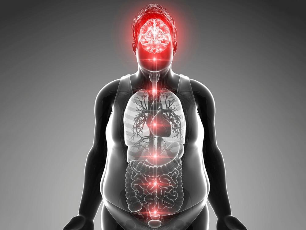
Thus, the researchers propose that disturbance in the DVC’s timekeeping leads to obesity, rather than being the result of excessive body weight.


When rats are fed a high fat diet, this disturbs the body clock in their brain that normally controls satiety, leading to over-eating and obesity. That’s according to new research published in The Journal of Physiology.

The number of people with obesity has nearly tripled worldwide since 1975.[1] In England alone, 28% of adults are obese and another 36% are overweight.[2] Obesity can lead to several other diseases such as Type 2 diabetes, heart disease, stroke, and some types of cancer.[3]

This new research may be a cornerstone for future clinical studies that could restore the proper functioning of the body clock in the brain, to avoid overeating.

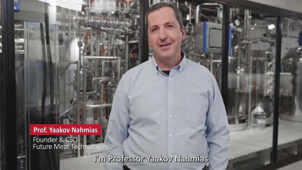
“After demonstrating that cultured meat can reach cost parity faster than the market anticipated, this production facility is the real game-changer,” said Yaakov Nahmias, Future Meat Technologies founder and chief scientific officer, in a press release. “This facility demonstrates our proprietary media rejuvenation technology in scale, allowing us to reach production densities 10-times higher than the industrial standard.”

Cultured meat is made by extracting cells from animal tissue and giving them nutrients, oxygen, and moisture while keeping them at the same temperature they’d be at inside an animal’s body. The cells divide and multiply then start to mature, with muscle cells joining to create muscle fibers and fat cells producing lipids. The resulting nuggets of meat can be used to make processed products like burgers or sausages. Structured cuts of meat with blood vessels and connective tissue, like steak or chicken breast, require scaffolds, and researchers are creating these with biomaterials, like cellulose from plants. Companies are working on several varieties of more elaborate cultured products, from bacon to salmon.

As reported by Bloomberg, Future Meat aims to start offering its products in US restaurants by the end of next year—but must get approval from the FDA first. On top of that approval, public opinion is another hurdle the company and its competitors will need to clear before they see widespread success; for every person who’s opposed to factory farming, there’s a person who’s squeamish about the idea of meat grown in a bioreactor, despite the avian (or bovine, or porcine) lives being spared. Getting these consumers to view cultured meat favorably will be a matter of education, taste/texture as compared to the ‘real thing,’ and cost competitiveness.

“I think this virus is here to stay with us and it will evolve like influenza pandemic viruses, it will evolve to become one of the other viruses that affects us,” Dr. Mike Ryan, executive director of the World Health Organization’s Health Emergencies Program, said at a press briefing.


Covid-19 could become endemic like the flu and circulate in the population at low levels.

Circa 23 March 2020


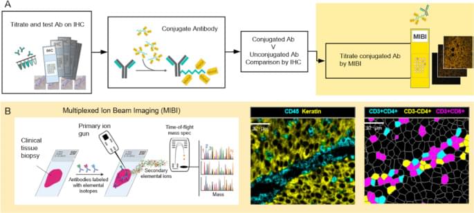
The ways in which a neoplastic cell arises and evades the immune system is the result of a departure from the systems biology that governs health. Understanding this biology requires methods that can resolve the heterogeneity of cell types, determine their states, whether they are activated (e.g., HLA-DR high) or suppressed (e.g., PD-1 high), and map their relationships or distances to one another. MIBI provides single cell resolution and sensitivity to phenotypically characterize the complex tissue environments including the TME. Executed similarly to IHC yet with the capability to profile 40+ markers simultaneously, MIBI is broadly applicable to a wide range of analyses performed in anatomic pathology including cell classification, spatial characterization, and assessment of marker expression. The MIBIscope produces data (multilayer TIFF files) that can be accessed by many analysis platforms currently available, such as those found in commercial software packages such as Fiji, Halo, and VisioPharm or freely available bioinformatic packages developed with open-source programming languages (e.g., R, Python).

All tumor types were stained, imaged, and analyzed using a single staining panel and standardized protocol. The workflow is flexible such that slides can be stained in batches and stored until imaged on the MIBIscope. Stained slides are typically stored under vacuum but protection from light is not necessary as the labels are stable metal isotopes rather than light-sensitive fluorophores. Once imaged it is possible to reimage the tissue as only a modest depth of the tissue is sputtered and analyzed during a single acquisition [16]. One limitation of the current project performed with an earlier version of the MIBIScope is the relatively small FOV size (500 μm by 500 μm) needed for images with 0.5 µm resolution. The current MIBIScope enables FOVs of 800 μm by 800 μm to be imaged in 70 min at fine resolution (650 nm). The resolution can be controlled at the instrument and acquisition at a slightly lower resolution than used in this study (1 μm) can be performed in 17 min. The 800 μm FOV captures 82% of a 1 mm TMA core. FOVs across cores of a TMA can be selected and then imaged in a single run. For whole sections it is possible to acquire adjacent images and stitch the images together using techniques commonly performed with other imaging technologies [22]. The need for tiling is particularly acute for imaging brain sections where multiple FOVs are collected to generate a larger image. Together with researchers at Stanford University, we are currently developing tiling methods to map large regions of brain tissue which will be described in a future publication. Because MIBI is still an early technology, the underlying methods for each stage of the processing pipeline are constantly evolving and improving, not just for accuracy but for generality. While the methods themselves are evolving, the pipeline tasks, at a high level, such as mass calibration, filtering, etc., are defined and have been automated through the MIBI/O software, and, as importantly, allows for appropriate user input when necessary. As more data becomes available, and the user base of MIBI grows, data processing should become more standardized.

The immediate utility of MIBI will be for understanding the biological mechanisms present in disease microenvironments. The results demonstrate the ability to detect a range of marker expression across many tumor types. The images can be segmented to define cell boundaries and then the expression of phenotypic markers used to classify cell instances into their cell class, such as proliferating tumor cells or nonproliferating tumor cells and various immune cells. Additional markers have been used on other sample sets to further define myeloid cell subsets, B cell subsets and stromal elements including vascular endothelial cells. This study also demonstrated the possibilities for calculating distances between different cell subsets including tumor and immune cells in addition to PD-1 and PD-L1 expressing immune cell subsets.
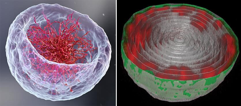
The groups also explained why in previous studies by other scientists, the chromatin appeared to fill the cell nuclei. “When scientists plate cells on a glass slide in order to study them under a microscope, they change their volume and physically flatten them. This may perturb some of the forces governing chromatin arrangement and reduce the distance between the upper part of the nucleus to its base,” Safran explains.


If you open a biology textbook and run through the images depicting how DNA is organized in the cell’s nucleus, chances are you’ll start feeling hungry; the chains of DNA would seem like a bowl of ramen: long strings floating in liquid. However, according to two new studies—one experimental and the other theoretical—that are the outcome of the collaboration between the groups of Prof. Talila Volk of the Molecular Genetics Department and Prof. Sam Safran of the Chemical and Biological Physics Department at the Weizmann Institute of Science, this image should be reconsidered. Clarifying it is essential since DNA’s spatial arrangement in the nucleus can affect the expression of genes contained within the DNA molecule, and hence the proteins found in the cell.

This story began when Volk was studying how mechanical forces influence cell nuclei in the muscle and found evidence that muscle contractions had an immediate effect on gene expression patterns. “We couldn’t explore this further because existing methods relied on imaging of chemically preserved cells, so they failed to capture what happens in the cell nuclei of an actual working muscle,” she says.

To address this issue, Dr. Dana Lorber, a research associate in Volk’s group, led the design of a device that makes it possible to study muscle nuclei in live fruit fly larvae. The device holds the tiny, translucent larva within a groove that allows it to contract and relax its muscles but keeps its movement constrained so that it can be scanned by a fluorescence microscope. Using the device, the researchers obtained images of the internal, linearly-organized complexes of DNA and its proteins (known as chromatin), surrounded by the membrane of the muscle nuclei.

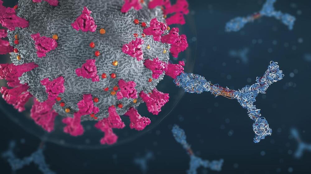
In fact, these antibodies could even fight off a virus engineered, on purpose, to be highly resistant to neutralization. This virus contained 20 mutations that are known to prevent SARS-CoV-2 antibodies from binding to it. Antibodies from people who were only vaccinated or only had prior COVID infections were essentially useless against this mutant virus. But antibodies in people with the “hybrid immunity” could neutralize it.


That’s how one scientist describes the findings of a series of studies looking at the antibodies created by individuals who were infected by the virus and then had an mRNA vaccine.

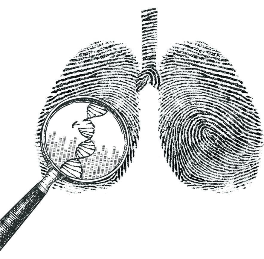
A genomic analysis of lung cancer in people with no history of smoking has found that a majority of these tumors arise from the accumulation of mutations caused by natural processes in the body. This study was conducted by an international team led by researchers at the National Cancer Institute (NCI), part of the National Institutes of Health (NIH), and describes for the first time three molecular subtypes of lung cancer in people who have never smoked.

These insights will help unlock the mystery of how lung cancer arises in people who have no history of smoking and may guide the development of more precise clinical treatments. The findings were published September 6 2021, in Nature Genetics.

“What we’re seeing is that there are different subtypes of lung cancer in never smokers that have distinct molecular characteristics and evolutionary processes,” said epidemiologist Maria Teresa Landi, M.D., Ph.D., of the Integrative Tumor Epidemiology Branch in NCI’s Division of Cancer Epidemiology and Genetics, who led the study, which was done in collaboration with researchers at the National Institute of Environmental Health Sciences, another part of NIH, and other institutions. “In the future we may be able to have different treatments based on these subtypes.”

Nanoengineers at the University of California San Diego have developed COVID-19 vaccine candidates that can take the heat. Their key ingredients? Viruses from plants or bacteria.

The new fridge-free COVID-19 vaccines are still in the early stage of development. In mice, the vaccine candidates triggered high production of neutralizing antibodies against SARS-CoV-2, the that causes COVID-19. If they prove to be safe and effective in people, the vaccines could be a big game changer for global distribution efforts, including those in rural areas or resource-poor communities.

“What’s exciting about our vaccine technology is that is thermally stable, so it could easily reach places where setting up ultra-low temperature freezers, or having trucks drive around with these freezers, is not going to be possible,” said Nicole Steinmetz, a professor of nanoengineering and the director of the Center for Nano-ImmunoEngineering at the UC San Diego Jacobs School of Engineering.