Toggle light / dark theme

“We think that this indicates that gut bacteria and fungi influence anti-tumor immune responses in many, if not all, types of cancer.”


Cedars-Sinai Cancer researchers have discovered that intestinal microorganisms help regulate anti-tumor immune responses to radiation treatments, and that fungi and bacteria have opposing effects on those responses. The study, conducted in laboratory mice 0 illuminates a path toward improving the effectiveness of radiation and immune-based treatments for patients with melanoma, breast and many other cancers.

The study, published on Aug. 13 in the peer-reviewed journal Cancer Cell, builds on prior studies that focused on the role of intestinal bacteria in influencing immune responses to chemotherapy and immunotherapy. Here the investigators sought to determine what role both bacteria and fungi in the gut might play in the response to radiation therapy.

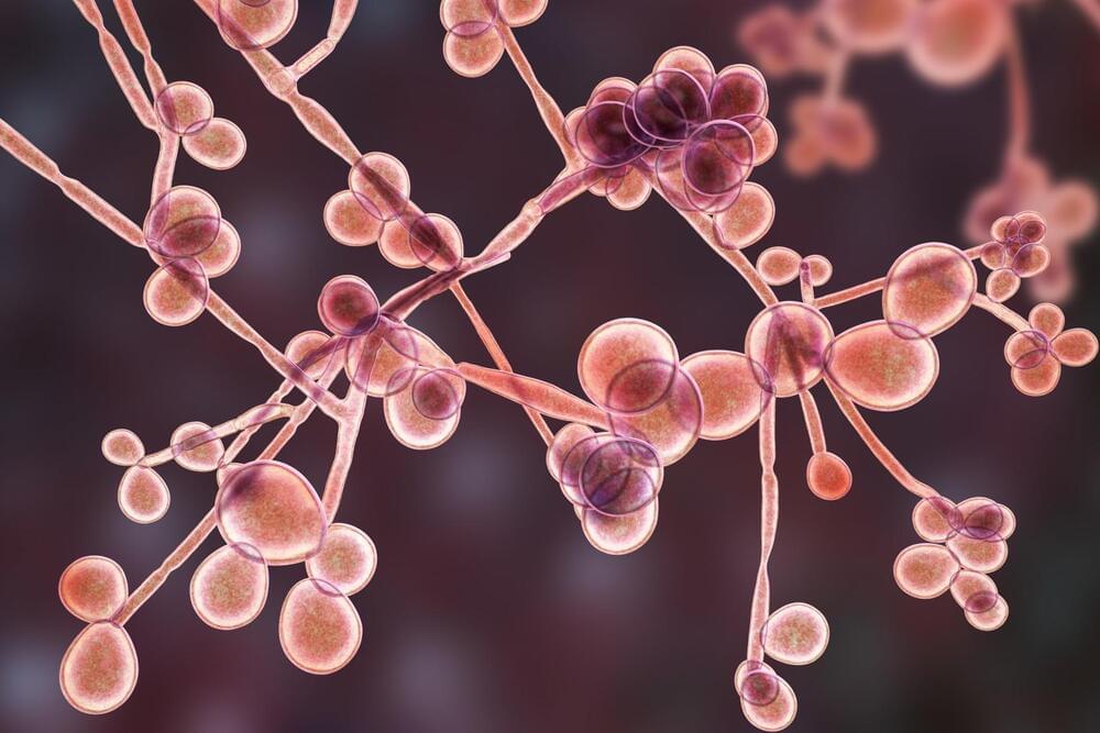
Trillions of microorganisms live in normal human intestines. These so-called commensal microorganisms are “friendly” bacteria and fungi that help process nutrients and play key roles in regulating the immune system in everything from infections to allergies. The research team found that reducing levels of commensal fungi in the intestines enhanced the anti-tumor immune response in the mice following radiation therapy. Conversely, they showed that depletion of commensal bacteria reduced the anti-tumor response.
Circa 2014


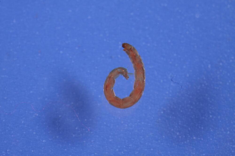
New collaborative research published in the journal Nature Communications by scientists from Japan, Russia and the US contains the genetic analysis on a species of African midge, which can survive a wide array of extreme conditions including large variations in temperature, extreme drought and even airless vacuums such as space. The team successfully deciphered the genetic mechanism that makes the midge invulnerable to these harsh conditions. Prof. Noriyuki Satoh and Dr. Takeshi Kawashima of Prof. Satoh’s Marine Genomics Unit, as well as Prof. Alexander Mikeyhev of the Ecology and Evolution Unit, and Mr. Manabu Fujie and Dr. Ryo Koyanagi of the DNA Sequencing Section at the Okinawa Institute of Science and Technology Graduate University have contributed to the collaboration.

The midge, Polypedilum vanderplanki, is capable of anhydrobiosis, a unique state that allows an organism to survive even after losing 97% of its body water. Anhydrobiotic organisms are also able to survive other severe conditions such as extreme temperatures ranging from 90°C to-270°C, vacuums and high doses of radiation; all of which would be lethal to most other life forms.

The midge found in northern Nigeria lives in an environment where the dry season lasts for at least six months and droughts can last up to eight months. By the time eggs have hatched and larvae have developed, the pools of water they breed in have dried up. However these dried larvae can survive in this dehydrated state for more than 17 years. “This is a very interesting kind of phenomena,” remarks Prof. Satoh. “The first descriptions of this midge were more than 60 years ago… But serious research started only ten years ago.”

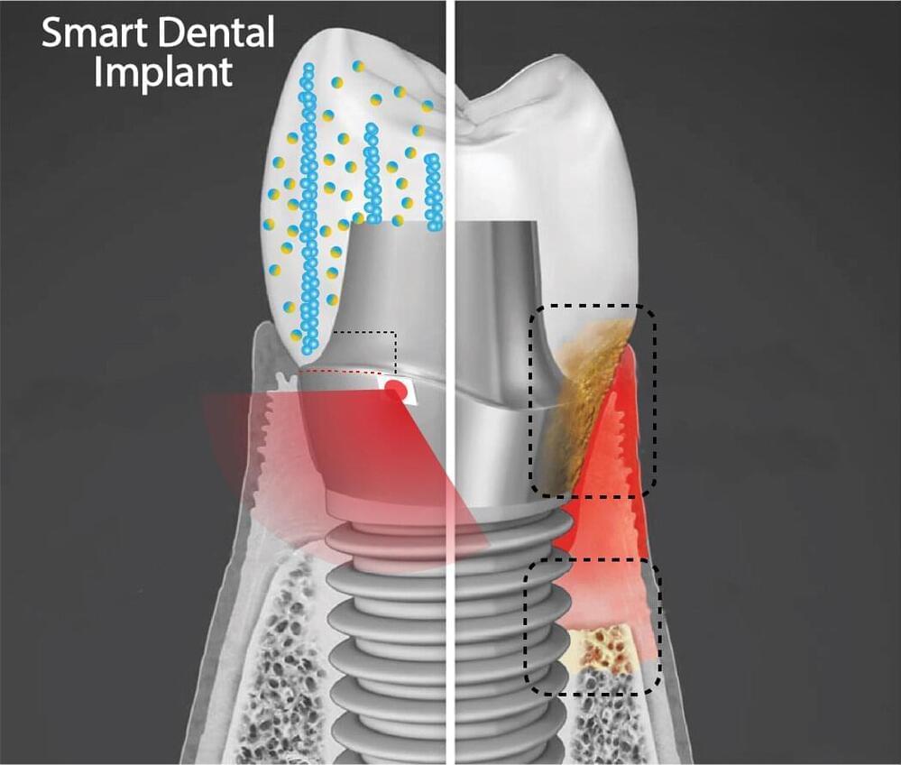
Hwang and colleagues lay out their platform, which could one day be integrated not only into dental implants but other technologies, such as joint replacements, as well.


More than 3 million people in America have dental implants, used to replace a tooth lost to decay, gum disease, or injury. Implants represent a leap of progress over dentures or bridges, fitting much more securely and designed to last 20 years or more.

But often implants fall short of that expectation, instead needing replacement in five to 10 years due to local inflammation or gum disease, necessitating a repeat of a costly and invasive procedure for patients.

“We wanted to address this issue, and so we came up with an innovative new ,” says Geelsu Hwang, an assistant professor in the University of Pennsylvania School of Dental Medicine, who has a background in engineering that he brings to his research on oral health issues.

What’s exciting about our findings is that we have identified a molecular pathway that is activated in normal acute wounds in humans, and altered in diabetic wounds in mice,” said Ghaidaa Kashgari, Ph.D., a postdoctoral researcher in the UCI School of Medicine Department of Medicine. “This finding strongly indicates clinical relevance and may improve our understanding of wound healing biology and could lead to new therapies.


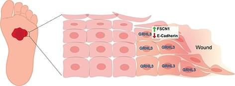
A University of California, Irvine-led study identifies a new molecular pathway that promotes the healing of wounds in the skin. Titled “GRHL3 activates FSCN1 to relax cell-cell adhesions between migrating keratinocytes during wound reepithelialization,” the study was published today in JCI Insight.

The molecular pathway identified is controlled by an evolutionary conserved gene called a Grainyhead like 3 (GRHL3), which is a gene required for mammalian development. Without this gene, several abnormalities may occur, including spina bifida, defective epidermal barrier, defective eyelid closure and soft-tissue syndactyly, a condition in which children are born with fused or webbed fingers.

The study reveals how during wound healing, GRHL3 works to activate a protein coding gene called Fascin Actin-Bundling Protein 1 (Fscn1) to loosen the adhesion between wounded skin cells so they can migrate efficiently to close the wound. Researchers also found that alterations in this process may result in chronic, non-healing wounds, such as diabetic ulcers that affect millions of patients every year.

Many human diseases can differ between males and females in their prevalence, manifestation, severity or age of onset. Examples include Lupus, where more than 80% of patients are females; Alzheimer’s disease, where females have higher incidence and tend to suffer quicker cognitive decline; and COVID-19 infections that are frequently more severe in males.

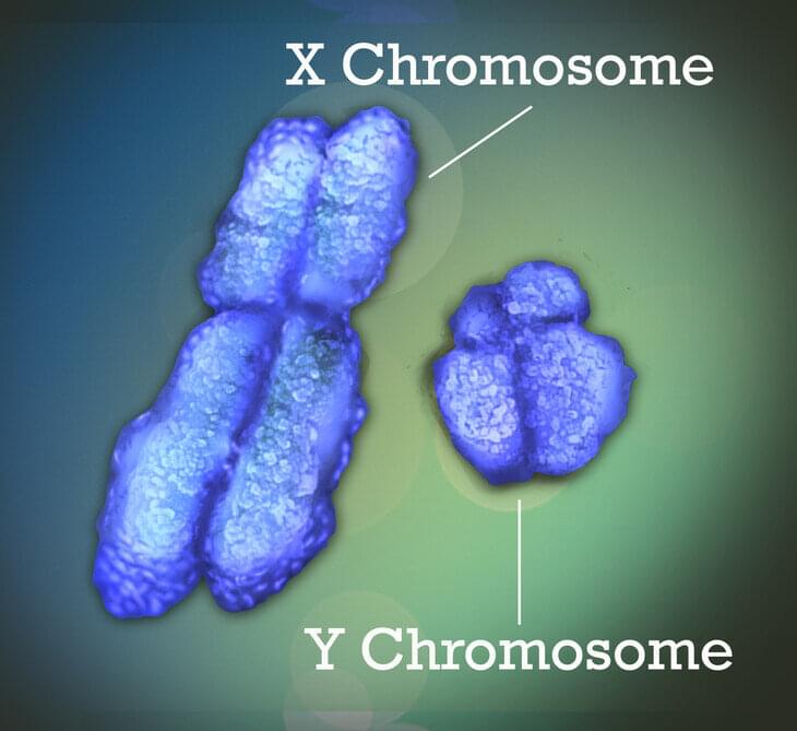
These sex differences may have a that is attributable to the sex . The X chromosome—one of the two sex chromosomes—is known to play an important role in human development and disease. New research led by Penn State College of Medicine reveals for the first time that sex-biased diseases can be attributable to that escape X chromosome inactivation (XCI), a process that ensures that females do not overexpress genes on their X-chromosomes.

The team developed a that can identify these XCI escape genes, and it may also help in determining whether a female will develop a sex-biased disease and if the disease will become progressively worse over time. The tool may even be useful in understanding the in immune responses to COVID-19, as the disease is thought to produce more severe symptoms and higher mortality in men than in women.

In this video Dr. Katcher reveals his thought on the future of aging if E5 is fulfils on its promise.

Dr Katcher’s book is on Amazon.
The Illusion of Knowledge: The paradigm shift in aging research that shows the way to human rejuvenation.
https://amzn.to/3jJ5deD

Dr Harold Katcher is one of the discovers of the human breast cancer gene BRCA1, and has thousands of citations in the scientific literature with publications ranging from protein structure to bacteriology, bioinformatics and biochemistry. He was the Academic Director for Natural Sciences of the University of Maryland Global Campus and is now the Chief Scientific Officer at Yuvan Research Inc, a company working on the development of rejuvenation treatments.

Dr Katcher’s new book, the Illusion of Knowledge, the paradigm shift in aging research that shows the way to human rejuvenation will be launched on 4th September 2021 and is already available in electronic form. The book launch will take place at The Book Passage in the Ferry Building in San Francisco at 3:00 pm Pacific Time.

************************************************************
Health claims Disclosure: Information provided on this video is not a substitute for direct, individual medical treatment or advice. Please consult with your doctor first. Products or services mentioned in this video are not a recommendation.

Audio Copyright Disclaimer:
Please note that we have full authorization to the music that we used in our videos as they were created using the service WeVideo which provides the rights to the music. The rights are detailed in the terms of use that can be reviewed here https://www.wevideo.com/terms-of-use and any following inquiries should be addressed to [email protected].

Summary: A new study on aging reveals a surprising discovery about the connection between protein shape and mitochondrial health.

Source: Buck Institute.

Every cell in the body goes through thousands of chemical reactions each day, and each reaction involves tiny protein molecules folded into precise shapes to perform their functions. Misfolded proteins underlie some of the most common and devastating diseases of aging, like Alzheimer’s and Parkinson’s. A major focus of aging research is discovering ways to maintain protein shape and prevent misfolded proteins from wreaking havoc on cellular function.

New photo-oxygenation catalyst targets amyloid structure, recruits brain immune system cells.

A small, light-activated molecule recently tested in mice represents a new approach to eliminating clumps of amyloid protein found in the brains of Alzheimer’s disease patients. If perfected in humans, the technique could be used as an alternative approach to immunotherapy and used to treat other diseases caused by similar amyloids.

Researchers injected the molecule directly into the brains of live mice with Alzheimer’s disease and then used a specialized probe to shine light into their brains for 30 minutes each day for one week. Chemical analysis of the mouse brain tissue showed that the treatment significantly reduced amyloid protein. Results from additional experiments using human brain samples donated by Alzheimer’s disease patients supported the possibility of future use in humans.