Toggle light / dark theme

“Identification of these compounds means that we are one step closer to being able to molecularly diagnose dementia,” said senior author of the study, Professor Mitsuhiro Yanagida, who leads the G0 Cell Unit at OIST.


Summary: Researchers identified 33 metabolic compounds in blood samples that differed between those with dementia and cognitively healthy older adults. 7 of the metabolites were elevated in dementia patients, while 26 were at lower levels compared to samples of those without dementia. Elevating levels of those metabolites could have a neuroprotective effect against dementia.

Source: OIST

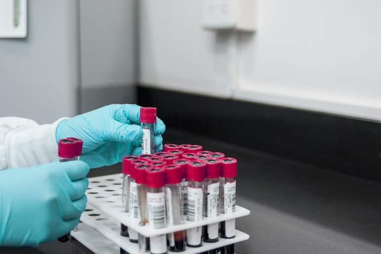
Scientists in Japan have identified metabolic compounds within the blood that are associated with dementia.

The study revealed that the levels of 33 metabolites differed in patients with dementia, compared to elderly people with no existing health conditions. Their findings, published this week in PNAS, could one day aid diagnosis and treatment of dementia.

Many human diseases can differ between males and females in their prevalence, manifestation, severity or age of onset. Examples include Lupus, where more than 80% of patients are females; Alzheimer’s disease, where females have higher incidence and tend to suffer quicker cognitive decline; and COVID-19 infections that are frequently more severe in males.

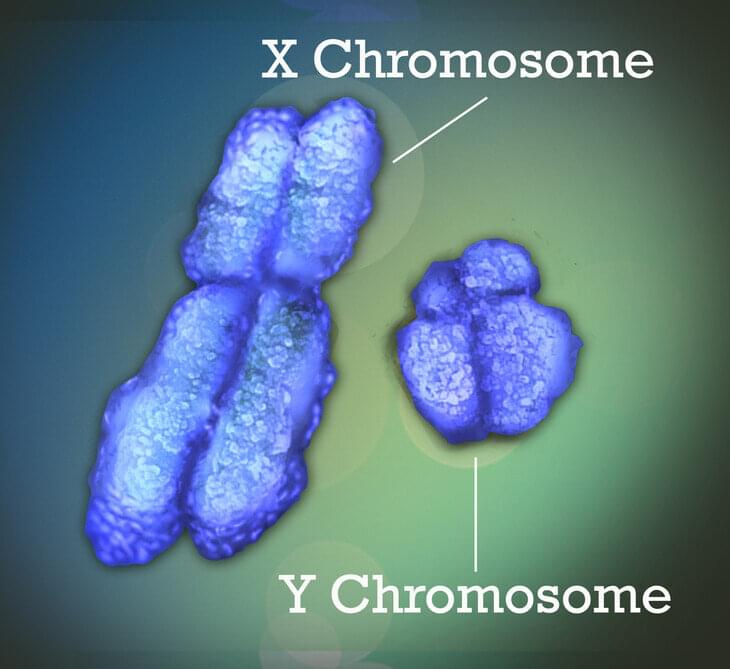
These sex differences may have a that is attributable to the sex . The X chromosome—one of the two sex chromosomes—is known to play an important role in human development and disease. New research led by Penn State College of Medicine reveals for the first time that sex-biased diseases can be attributable to that escape X chromosome inactivation (XCI), a process that ensures that females do not overexpress genes on their X-chromosomes.

The team developed a that can identify these XCI escape genes, and it may also help in determining whether a female will develop a sex-biased disease and if the disease will become progressively worse over time. The tool may even be useful in understanding the in immune responses to COVID-19, as the disease is thought to produce more severe symptoms and higher mortality in men than in women.

Summary: A new study on aging reveals a surprising discovery about the connection between protein shape and mitochondrial health.

Source: Buck Institute.

Every cell in the body goes through thousands of chemical reactions each day, and each reaction involves tiny protein molecules folded into precise shapes to perform their functions. Misfolded proteins underlie some of the most common and devastating diseases of aging, like Alzheimer’s and Parkinson’s. A major focus of aging research is discovering ways to maintain protein shape and prevent misfolded proteins from wreaking havoc on cellular function.

New photo-oxygenation catalyst targets amyloid structure, recruits brain immune system cells.

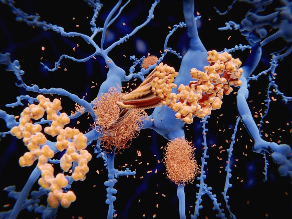
A small, light-activated molecule recently tested in mice represents a new approach to eliminating clumps of amyloid protein found in the brains of Alzheimer’s disease patients. If perfected in humans, the technique could be used as an alternative approach to immunotherapy and used to treat other diseases caused by similar amyloids.

Researchers injected the molecule directly into the brains of live mice with Alzheimer’s disease and then used a specialized probe to shine light into their brains for 30 minutes each day for one week. Chemical analysis of the mouse brain tissue showed that the treatment significantly reduced amyloid protein. Results from additional experiments using human brain samples donated by Alzheimer’s disease patients supported the possibility of future use in humans.

Exploring The Gut Microbiota-Brain Axis In Health, Disease, and Aging — Dr. Marina Ezcurra, Ph.D. University of Kent.


Dr. Marina Ezcurra (https://marinaezcurralab.com/) is a Lecturer in the Biology of Aging, and NeuroBiology, at the School of BioSciences, at the University of Kent, UK (https://www.kent.ac.uk/biosciences/people/2081/ezcurra-marina).

Dr. Ezcurra received her PhD from the Karolinska Institute in 2011. Her PhD research was a collaborative project between Karolinska and the Medical Research Council Laboratory of Molecular Biology at Cambridge, where she studied neural circuits and behavior using C. elegans in the lab of Dr. Bill Schafer.

During her PhD, Dr. Ezcurra identified extra-synaptic mechanisms by which nutritional status modulates nociception, involving neuro-peptidergic and dopaminergic signaling. She went on to do a postdoc working on aging with Dr. David Gems at University College London.

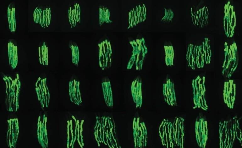
During her postdoc, Dr. Ezcurra developed methods to monitor the development of multiple age-related diseases in-vivo in C. elegans, leading to the discovery of a previously unknown process, Intestinal Biomass Conversion. This mechanism enables the C. elegans intestine to be broken down to produce vast amounts of yolk, resulting in poly-morbidity and mortality in aging nematodes. This work illustrates how aging and age-related diseases can be the result of run-on of wild-type gene function, rather than stochastic molecular damage.

Current research in Dr. Ezcurra’s group focuses on how host-microbiome interactions affect host aging, and is funded by The Wellcome Trust and Royal Society.

Dr. Ezcurra is a trustee board member of The British Society of Research on Aging.

Common medications can accumulate in gut bacteria, a new study has found, altering bacterial function and potentially reducing the effectiveness of the drug. These interactions—seen for a variety of medications, such as depression, diabetes, and asthma drugs—could help researchers to better understand individual differences in drug effectiveness and side-effects, according to the study published in Nature.

It is known that bacteria can chemically modify some drugs, a process known as biotransformation. This study, led by researchers from the Medical Research Council (MRC) Toxicology Unit at the University of Cambridge and the European Molecular Biology Laboratory (EMBL) in Germany, is the first to show that certain species of accumulate human drugs, altering the types of bacteria and their activity.

This could change the effectiveness of the both directly, as the accumulation could reduce the availability of the drug to the body, and indirectly, as altered bacterial function and composition could be linked to .

This makes sense, as we believe that inflammation in the central nervous system can start the autoimmune process (when a person’s immune system attacks part of their body) that causes MS.


Summary: A new study links viral infections including mononucleosis and pneumonia experienced during adolescence with an increased risk of developing multiple sclerosis.

Source: The Conversation

Multiple sclerosis (MS) is most often diagnosed between the ages of 20 and 50. Certain genes put a person at greater risk of getting this disease of the central nervous system, but scientists are still trying to understand the triggers.

My colleagues and I have been studying these triggers for many years. Our earlier research found that pneumonia in adolescence is associated with a raised risk of MS, so we decided to investigate whether other types of infection are associated with the condition.

Thus, the researchers propose that disturbance in the DVC’s timekeeping leads to obesity, rather than being the result of excessive body weight.


When rats are fed a high fat diet, this disturbs the body clock in their brain that normally controls satiety, leading to over-eating and obesity. That’s according to new research published in The Journal of Physiology.

The number of people with obesity has nearly tripled worldwide since 1975.[1] In England alone, 28% of adults are obese and another 36% are overweight.[2] Obesity can lead to several other diseases such as Type 2 diabetes, heart disease, stroke, and some types of cancer.[3]

This new research may be a cornerstone for future clinical studies that could restore the proper functioning of the body clock in the brain, to avoid overeating.

Circa 23 March 2020


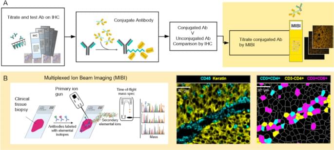
The ways in which a neoplastic cell arises and evades the immune system is the result of a departure from the systems biology that governs health. Understanding this biology requires methods that can resolve the heterogeneity of cell types, determine their states, whether they are activated (e.g., HLA-DR high) or suppressed (e.g., PD-1 high), and map their relationships or distances to one another. MIBI provides single cell resolution and sensitivity to phenotypically characterize the complex tissue environments including the TME. Executed similarly to IHC yet with the capability to profile 40+ markers simultaneously, MIBI is broadly applicable to a wide range of analyses performed in anatomic pathology including cell classification, spatial characterization, and assessment of marker expression. The MIBIscope produces data (multilayer TIFF files) that can be accessed by many analysis platforms currently available, such as those found in commercial software packages such as Fiji, Halo, and VisioPharm or freely available bioinformatic packages developed with open-source programming languages (e.g., R, Python).

All tumor types were stained, imaged, and analyzed using a single staining panel and standardized protocol. The workflow is flexible such that slides can be stained in batches and stored until imaged on the MIBIscope. Stained slides are typically stored under vacuum but protection from light is not necessary as the labels are stable metal isotopes rather than light-sensitive fluorophores. Once imaged it is possible to reimage the tissue as only a modest depth of the tissue is sputtered and analyzed during a single acquisition [16]. One limitation of the current project performed with an earlier version of the MIBIScope is the relatively small FOV size (500 μm by 500 μm) needed for images with 0.5 µm resolution. The current MIBIScope enables FOVs of 800 μm by 800 μm to be imaged in 70 min at fine resolution (650 nm). The resolution can be controlled at the instrument and acquisition at a slightly lower resolution than used in this study (1 μm) can be performed in 17 min. The 800 μm FOV captures 82% of a 1 mm TMA core. FOVs across cores of a TMA can be selected and then imaged in a single run. For whole sections it is possible to acquire adjacent images and stitch the images together using techniques commonly performed with other imaging technologies [22]. The need for tiling is particularly acute for imaging brain sections where multiple FOVs are collected to generate a larger image. Together with researchers at Stanford University, we are currently developing tiling methods to map large regions of brain tissue which will be described in a future publication. Because MIBI is still an early technology, the underlying methods for each stage of the processing pipeline are constantly evolving and improving, not just for accuracy but for generality. While the methods themselves are evolving, the pipeline tasks, at a high level, such as mass calibration, filtering, etc., are defined and have been automated through the MIBI/O software, and, as importantly, allows for appropriate user input when necessary. As more data becomes available, and the user base of MIBI grows, data processing should become more standardized.

The immediate utility of MIBI will be for understanding the biological mechanisms present in disease microenvironments. The results demonstrate the ability to detect a range of marker expression across many tumor types. The images can be segmented to define cell boundaries and then the expression of phenotypic markers used to classify cell instances into their cell class, such as proliferating tumor cells or nonproliferating tumor cells and various immune cells. Additional markers have been used on other sample sets to further define myeloid cell subsets, B cell subsets and stromal elements including vascular endothelial cells. This study also demonstrated the possibilities for calculating distances between different cell subsets including tumor and immune cells in addition to PD-1 and PD-L1 expressing immune cell subsets.