Toggle light / dark theme

Although we often think of knowledge as “knowing that” (for example, knowing that Paris is the capital of France), each of us also knows many procedures consisting of “knowing how,” such as knowing how to tie a knot or start a car. Now, a new study has found the brain programs that code the sequence of steps in performing a complex procedure.

In a just published paper in Psychological Science, researchers at Carnegie Mellon University have found a way to find decode the procedural information required to tie various knots with enough precision to identify which knot is being planned or performed. To reach this conclusion, Drs. Robert Mason and Marcel Just first trained a group of participants to tie seven types of knots, and then scanned their brains while they imagined tying, or actually tied the knots while they were in an MRI scanner. The main findings were that each knot had a distinctive neural signature, so the researchers could tell which knot was being tied from the sequence of brain images collected. Furthermore, the neural signatures were very similar for imagining tying a particular knot and planning to tie it.

Dr. Just said, “Tying a knot is an ancient and frequently performed that is the epitome of everyday procedural knowledge, making it an excellent target for investigation.”
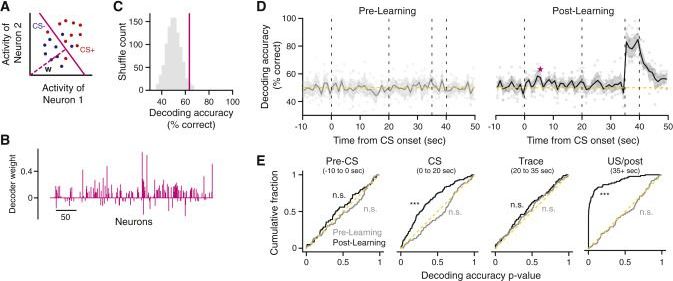
Episodic memory requires linking events in time, a function dependent on the hippocampus. In “trace” fear conditioning, animals learn to associate a neutral cue with an aversive stimulus despite their separation in time by a delay period on the order of tens of seconds. But how this temporal association forms remains unclear. Here we use two-photon calcium imaging of neural population dynamics throughout the course of learning and show that, in contrast to previous theories, hippocampal CA1 does not generate persistent activity to bridge the delay. Instead, learning is concomitant with broad changes in the active neural population. Although neural responses were stochastic in time, cue identity could be read out from population activity over longer timescales after learning. These results question the ubiquity of seconds-long neural sequences during temporal association learning and suggest that trace fear conditioning relies on mechanisms that differ from persistent activity accounts of working memory.

Eric Klien


Simulation theory points out that we might be living in a giant computer simulation. Exponential technological growth and how far we’ve already come in so little time are big indicators that we can’t possibly imagine what the future of humanity would look like in 100 years, or better yet, 1,000 years!
Will we be able to create simulations so indistinguishable from reality that the characters will not be aware that they are being simulated? Today on Cognitive Culture, you’ll learn about why you’re not real!

PLEASE SUBSCRIBE! Let’s get the channel to 5K subs! I love you guys so much and your support means all of the things to me!!

Follow me on Instagram @cognitive.culture for inspiration!

Here are more of my videos and my sources of information for this video:

How To Evolve With Technology

How To Experience Something Again For The First Time

How To Make A Successful New Year Resolution

“It’s not just about the smell,” said Adrian Cheok, one of the scientists behind the experiments. “It is part of a whole, integrated virtual reality or augmented reality. So, for example, you could have a virtual dinner with your friend through the internet. You can see them in 3D and also share a glass of wine together.”

In real life, odors are transmitted when airborne molecules waft into the nose, prompting specialized nerve cells in the upper airway to fire off impulses to the brain. In the recent experiments, performed on 31 test subjects at the Imagineering Institute in the Malaysian city of Nusajaya, researchers used electrodes in the nostrils to deliver weak electrical currents above and behind the nostrils, where these neurons are found.

The researchers were able to evoke 10 different virtual odors, including fruity, woody and minty.

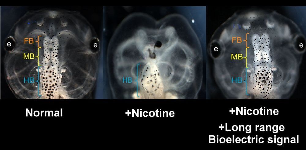
Researchers led by biologists at Tufts University have discovered that the brains of developing frog embryos damaged by nicotine exposure can be repaired by treatment with certain drugs called “ionoceuticals” that drive the recovery of bioelectric patterns in the embryo, followed by repair of normal anatomy, gene expression and brain function in the growing tadpole. The research, published today in Frontiers in Neuroscience, introduces intervention strategies based on restoring the bioelectric “blueprint” for embryonic development, which the researchers suggest could provide a roadmap for the exploration of therapeutic drugs to help repair birth defects.

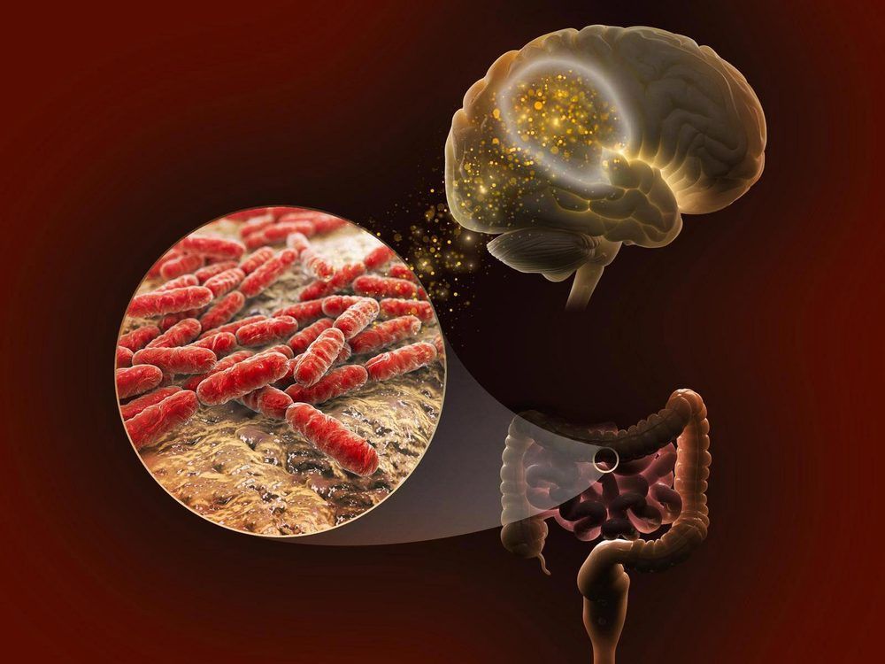
A molecule commonly produced by gut microbes appears to improve memory in mice.

A new study is among the first to trace the molecular connections between genetics, the gut microbiome, and memory in a mouse model bred to resemble the diversity of the human population.

While tantalizing links between the gut microbiome and brain have previously been found, a team of researchers from two U.S. Department of Energy national laboratories found new evidence of tangible connections between the gut and the brain. The team identified lactate, a molecule produced by all species of one gut microbe, as a key memory-boosting molecular messenger. The work was published recently in the journal BMC Microbiome.

“An international study involving Monash physicists has cornered a new approach to measure consciousness, potentially changing our understanding of complex neurological problems.

The study published today in Physical Review Research describes how tools from physics and complexity theory were used to determine the level of consciousness in fruit flies.

“This is a major problem in neuroscience, where it is crucial to differentiate between unresponsive vegetative patients and those suffering from a condition in which a patient is aware but cannot move or communicate verbally because of complete paralysis of nearly all voluntary muscles in the body,” said study author Dr. Kavan Modi, from the Monash University School of Physics and Astronomy.”


An international study involving Monash physicists has conrmed a new approach to measure consciousness, potentially changing our understanding complex neurological problems.

DOI: 10.1103/PhysRevResearch.2.023219

Organisation/s: Monash University.

An MRI in each other’s arms shows how physical contact alters the brains of couples.

Researchers at Aalto University and Turku PET Centre have developed a new method for simultaneous imaging brain activity from two people, allowing them to study social interaction.

In a recent study, the researchers scanned brain activity from 10 couples. Each couple spent 45 minutes inside the MRI scanner in physical contact with each other. The objective of the study was to examine how social contact activates the brain. The results were published in the theme issue Social Interaction in Neuropsychiatry of the journal Frontiers in Psychiatry.

Memory is the natural extension of attention and learning. The act of memory facilitates the formation, activation, and retention of circuits that contribute to the brain’s optimal functioning. Dr. Restak explains how we are the sum total of the memory we retain. Without memory, we wouldn’t know who we are.


The hippocampus, a portion of the brain located in the temporal lobe of each cerebral cortex, is the entry portal for information to be remembered. If the hippocampus is damaged, we have difficulty forming new memories.

This was demonstrated by Patient H. M., whose real name was Henry Molaison. He started having seizures when he was 10 years old. By age 20, he was completely incapacitated.

Since he could be felled with one of the sudden seizures at any time, he couldn’t work or form relationships, and lived at home with his mother. At age 27, in 1953, he underwent a new type of operation.

DNA damage is common to our cells, but when we’re young our bodies can fix it pretty easily. Unfortunately we lose that ability over time, leading to many of the symptoms of aging that we know all too well. A new study from MIT has found that reactivating a certain enzyme improves repair of DNA damage in neurons, which helps Alzheimer’s patients and others with cognitive decline.

Previous studies by the team have shown that an enzyme called HDAC1 seems to be involved in DNA repair in neurons. For the new study, the researchers examined what happens when HDAC1 doesn’t do its job.

The team engineered mice to be deficient in HDAC1, and monitored their health compared to normal mice. Things looked good during the animals’ youth – there were no differences in DNA damage or behavior between the two groups. But as they aged, the decline became clear.