Toggle light / dark theme

A research study in mice by investigators at the University of Rochester Medical Center (URMC) suggests it would be possible to repair the brain cell damage caused by multiple sclerosis (MS). The research was published in the journal Cell Reports.

The research, led by Steve Goldman, professor of Neurology and Neuroscience at URMC and co-director of the Center for Translational Neuromedicine, manipulated embryonic and induced pluripotent stem cells to create glia, a type of brain cell. Glial progenitor cells, a subtype of these cells, eventually form the primary support cells of the brain, astrocytes and oligodendrocytes, which play essential roles in the health and signaling behavior of nerve cells.

MS is an autoimmune disorder where the body’s immune system attacks oligodendrocytes. Oligodendrocytes manufacture myelin, which makes the insulation that allows nerve cells to communicate with each other. As myelin decreases in MS, the signaling between nerve cells is interrupted, which causes the loss of function that leads to problems with sensation, motor function and cognitive problems.
Interesting articles on theranostic iron nanowires. I’m interested in watching all aspects of development of nanobots, because I think it may lead to new forms of treatments for superlongevity and superintelligence.

Phys.org: Iron nanorobots go undercover to do surveillance on living cells in real time:

https://phys.org/…/2020-05-iron-nanorobots-undercover-surve…


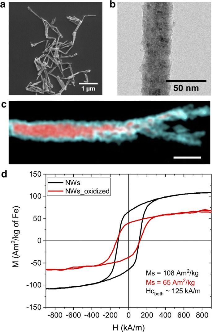
Identifying the precise location of cells and their migration dynamics is of utmost importance for achieving the therapeutic potential of cells after implantation into a host. Magnetic resonance imaging is a suitable, non-invasive technique for cell monitoring when used in combination with contrast agents.

This work shows that nanowires with an iron core and an iron oxide shell are excellent materials for this application, due to their customizable magnetic properties and biocompatibility. The longitudinal and transverse magnetic relaxivities of the core–shell nanowires were evaluated at 1.5 T, revealing a high performance as T2 contrast agents. Different levels of oxidation and various surface coatings were tested at 7 T. Their effects on the T2 contrast were reflected in the tailored transverse relaxivities. Finally, the detection of nanowire-labeled breast cancer cells was demonstrated in T2-weighted images of cells implanted in both, in vitro in tissue-mimicking phantoms and in vivo in mouse brain. Labeling the cells with a nanowire concentration of 0.8 μg of Fe/mL allowed the detection of 25 cells/µL in vitro, diminishing the possibility of side effects.

In 2015 Francesco Greco, head of the Laboratory of Applied Materials for Printed and Soft electronics (LAMPSe) at the Institute of Solid State Physics at Graz University of Technology, developed so-called “tattoo electrodes” together with Italian scientists.

These are conductive polymers that are printed using an inkjet printer on standard tattoo paper and then stuck to the skin like transfers to measure heart or muscle activity.

This type of electrode, optimized in 2018, opened up completely new paths in electrophysiological examinations, such as electrocardiography (ECG) or electromyography (EMG). Thanks to a thickness of 700 to 800 nanometres — that is about 100 times thinner than a human hair — the tattoos adapt to uneven skin and are hardly noticeable on the body.

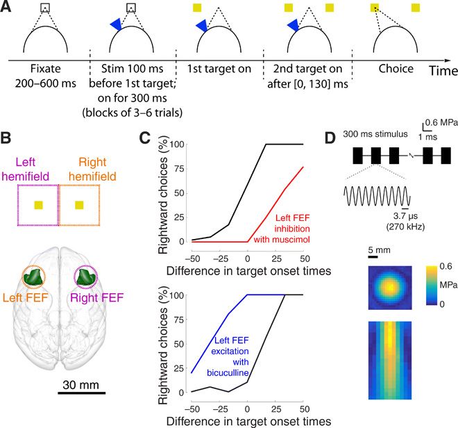
The ability to modulate neural activity in specific brain circuits remotely and systematically could revolutionize studies of brain function and treatments of brain disorders. Sound waves of high frequencies (ultrasound) have shown promise in this respect, combining the ability to modulate neuronal activity with sharp spatial focus. Here, we show that the approach can have potent effects on choice behavior. Brief, low-intensity ultrasound pulses delivered noninvasively into specific brain regions of macaque monkeys influenced their decisions regarding which target to choose. The effects were substantial, leading to around a 2:1 bias in choices compared to the default balanced proportion. The effect presence and polarity was controlled by the specific target region. These results represent a critical step towards the ability to influence choice behavior noninvasively, enabling systematic investigations and treatments of brain circuits underlying disorders of choice.

Noninvasive and reversible modulation of neuronal activity in specific brain circuits may allow us to diagnose and treat brain disorders in, targeted ways. Low-intensity ultrasound, applied to the brain noninvasively, can be used to modulate neural activity with spatial specificity superior to other noninvasive methods such as transcranial electrical or magnetic stimulation (15). The neuromodulatory potential of ultrasound has been highlighted in studies that targeted peri-motor regions of anesthetized rodents or rabbits. Brief, low-intensity stimuli lead to observable movements of the limbs or other body parts (613).

However, the enthusiasm about the neuromodulatory potential of ultrasound has recently been dampened by studies that called these effects into question (14, 15). In addition, such overt effects have not been observed in large mammals including humans. Only small changes in neural signals (1623) or small changes in reaction time or other metrics (2426) have been found. Yet, to make it truly useful, the approach would ideally provide neuromodulatory effects that are strong enough to manifest in behavior. For example, if clinicians are to determine which brain circuits drive a patient’s craving for an addictive drug, the neuromodulatory effects on a particular neural circuit should be potent enough to yield measurable changes in the subject’s choice behavior, i.e., whether the subject decides to use the drug or not.

Revealing yet another super-power in the skillful squid, scientists have discovered that squid massively edit their own genetic instructions not only within the nucleus of their neurons, but also within the axon — the long, slender neural projections that transmit electrical impulses to other neurons. This is the first time that edits to genetic information have been observed outside of the nucleus of an animal cell.

The study, led by Isabel C. Vallecillo-Viejo and Joshua Rosenthal at the Marine Biological Laboratory (MBL), Woods Hole, is published this week in Nucleic Acids Research.

A visual cortical prosthesis (VCP) has long been proposed as a strategy for restoring useful vision to the blind, under the assumption that visual percepts of small spots of light produced with electrical stimulation of visual cortex (phosphenes) will combine into coherent percepts of visual forms, like pixels on a video screen. We tested an alternative strategy in which shapes were traced on the surface of visual cortex by stimulating electrodes in dynamic sequence. In both sighted and blind participants, dynamic stimulation enabled accurate recognition of letter shapes predicted by the brain’s spatial map of the visual world. Forms were presented and recognized rapidly by blind participants, up to 86 forms per minute. These findings demonstrate that a brain prosthetic can produce coherent percepts of visual forms.

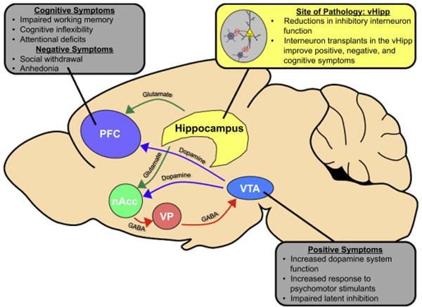
Schizophrenia is a devastating psychiatric disorder characterized by positive, negative and cognitive symptoms. While aberrant dopamine system function is typically associated with the positive symptoms of the disease, it is thought that this is secondary to pathology in afferent regions. Indeed, schizophrenia patients show dysregulated activity in the hippocampus and prefrontal cortex, two regions known to regulate dopamine neuron activity. These deficits in hippocampal and prefrontal cortical function are thought to result, in part, from reductions in inhibitory interneuron function in these brain regions. Therefore, it has been hypothesized that restoring interneuron function in the hippocampus and/or prefrontal cortex may be an effective treatment strategy for schizophrenia. In this article, we will discuss the evidence for interneuron pathology in schizophrenia and review recent advances in our understanding of interneuron development. Finally, we will explore how these advances have allowed us to test the therapeutic value of interneuron transplants in multiple preclinical models of schizophrenia.

Schizophrenia is devastating psychiatric disorder that affects approximately 1% of the population1. Positive symptoms, such as paranoia, grandiosity, delusions, and hallucinations, are often the most striking features of the disorder; however, schizophrenia patients also display characteristic negative and cognitive symptoms, which can be severely debilitating. Negative symptoms, such as blunted affect, emotional withdrawal, and social avoidance and cognitive symptoms, including disruptions in working memory, attentional deficits, disorganized thought, and cognitive inflexibility, can negatively influence social and occupational functioning and diminish quality of life2–4. Currently prescribed antipsychotic medications, which act as antagonists at the dopamine D2 receptor5, have been somewhat effective in treating the positive symptoms of schizophrenia6.

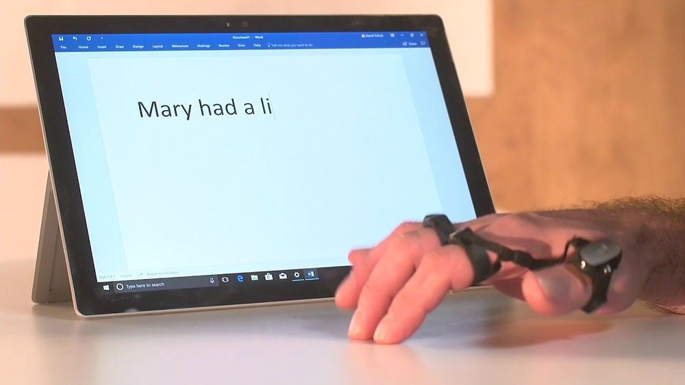
How will we interact with our electronic #devices in the #future? https://bit.ly/2Tm59F6

Touchscreens, keyboards and mice are the three dominant ways for us to interact with our devices in modern times. However, with the development of some new technologies (including #VoiceControl, #BrainComputerInterface, #brainwaves control, #gesture control, muscular signals interpretation and so forth), many start to ask: What will be the next way for us to talk to our machines?

In this article, we will first introduce some potential alternatives of touchscreens, keyboards and mice. Then, a picture about what a future device should be like will be provided.

#technology #innovation #ConsumerElectronics


Smartphone is arguably one of the most successful electronic devices in modern times, and it should be safe to say that its triumph has a lot to do with the appearance of the first iPhone announced on June 29, 2007, in which the way how people interact with their mobile devices has been completely reinvented. As a matter of fact, such way based on our index finger and a touchscreen is so intuitive and works so efficiently that the design of the smartphones today remains essentially unchanged – a thin and tiny rectangular pad with most of its area covered by a touchscreen.

Temporary tattoo electrodes are the most recent development in the field of cutaneous sensors. They have successfully demonstrated their performances in the monitoring of various electrophysiological signals on the skin. These epidermal electronic devices offer a conformal and imperceptible contact with the wearer while enabling good quality recordings over time. Evaluations of brain activity in clinical practice face multiple limitations, where such electrodes can provide realistic technological solutions and increase diagnostics efficiency. Here we present the performance of inkjet-printed conducting polymer tattoo electrodes in clinical electroencephalography and their compatibility with magnetoencephalography. The working mechanism of these dry sensors is investigated through the modeling of the skin/electrode impedance for better understanding of the biosignals transduction at this interface. Furthermore, a custom-made skin phantom platform demonstrates the feasibility of high-density recordings, which are essential in localizing neuropathological activities. These evaluations provide valuable input for the successful application of these ultrathin electronic tattoos sensors in multimodal brain monitoring and diagnosis.