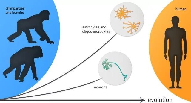
Oligodendrocytes and astrocytes displayed more differences in the human evolutionary lineage than neurons as compared to similar cells in other primates. Credit: Pavel Odinev/ Skoltech.

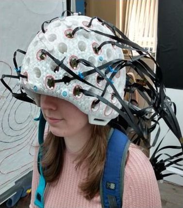
When it comes to monitoring electrical activity in the brain, patients typically have to lie very still inside a large magnetoencephalography (MEG) machine. That could be about to change, though, as scientists have developed a new version of a wearable helmet that does the same job.
Back in 2018, researchers at Britain’s University of Nottingham revealed the original version of their “MEG helmet.”
The 3D-printed device was fitted with multiple sensors that allowed it to read the tiny magnetic fields created by brain waves, just like a regular MEG machine. Unlike the case with one of those, however, wearers could move around as those readings were taking place.
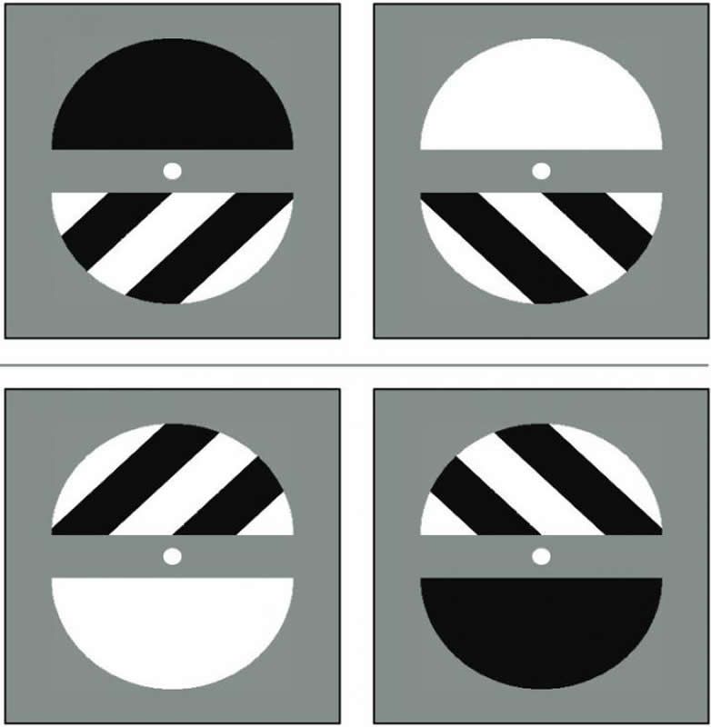
Summary: Optical illusions are helping researchers better understand attention and visual perception. Findings suggest attention operates periodically on the perceptual binding of visual information.
Source: University of Tokyo.
Rhythmic waves of brain activity cause us to see or not see complex images that flash before our eyes. An image can become practically invisible if it flashes before our eyes at the same time as a low point of those brain waves. We can reset that brain wave rhythm with a simple voluntary action, like choosing to push a button.
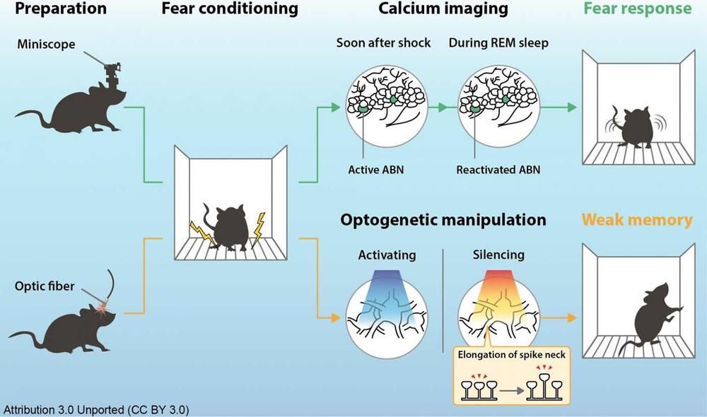
Summary: Hippocampal adult-born neurons are responsible for memory consolidation during REM sleep.
Source: University of Tsukuba.
The presence of dreaming during rapid-eye-movement (REM) sleep indicates that memory formation may occur during this sleep stage. But now, researchers from Japan have found that activity in a specific group of neurons is necessary for memory consolidation during REM sleep.
#Eternal life might not be attainable in the near future, but genetic engineers and doctors are working on new life extension technology. The research could lead to keeping our bodies young, and scientists are developing ways of downloading our brain’s consciousness onto digital media once the body is at the end of its life cycle.
#RT #Documentary offers you in-depth documentary films on topics that will leave no one indifferent. It’s not just front-page stories and global events, but issues that extend beyond the headlines. Social and environmental issues, shocking traditions, intriguing personalities, history, sports and so much more – we have documentaries to suit every taste. RTD’s film crews travel far and wide to bring you diverse and compelling stories. Discover the world with us!
SUBSCRIBE TO RTD Channel to get documentaries firsthand! http://bit.ly/1MgFbVy
FOLLOW US
RTD WEBSITE: https://RTD.rt.com/
RTD ON TWITTER: http://twitter.com/RT_DOC
RTD ON FACEBOOK: http://www.facebook.com/RTDocumentary
RTD ON INSTAGRAM https://www.instagram.com/rt_documentary_/
RTD LIVE https://rtd.rt.com/on-air/

· 24 mins ·
Safety was the reason the WHO stopped clinical trials of a drug that is not even an amphetamine. This happened before the racial divide, distraction, and mass confusion.
So let US think with a clear head. If Hydroxychloroquine is unsafe because of heart concerns, why give children amphetamines for ADHD, when marijuana and other natural measures offer many more safer alternatives? I know of them, why don’t the WHO and FDA, who know more than I do know as well? I can start a w… See More.
First-line stimulant class medications, such as methylphenidate and amphetamine formulations are FDA approved for the treatment of Attention Deficit Hyperactivity Disorder (ADHD) and narcolepsy. It is estimated that 4.4% of US adults experience some symptoms and disabilities of ADHD. However adults receive 32% of all issued stimulant prescriptions.1 Off-label treatment for conditions including weight management, fatigue related to depression, stroke, traumatic brain injury, or hyper-somnolence due to Obstructive Sleep Apnea (OSA) may account for the high prevalence of stimulant use in adults. Such conditions are frequently associated with history or risk of cardiovascular disease. Of note, OSA and other forms of sleep-disordered breathing have unfavorable effects on cardiovascular physiology, predisposing affected individuals to cardiovascular disease and cardiac arrhythmias.2,3
Due to reports of cardiovascular adverse events and observed physiological effects, the package inserts for stimulant drugs warn against use in patients with preexisting heart disease or cardiac structural abnormalities due to risk of sudden death, stroke, and myocardial infarction (MI).4–6 Furthermore, the FDA issued a safety announcement in 2011 stating that stimulant products and atomoxetine should not be used in patients with serious heart problems, or for whom an increase in blood pressure (BP) or heart rate (HR) would be problematic.7 Table 1 summarizes the available stimulant and stimulant-like medications, including those referenced by the FDA. Debate remains on the safety of stimulants in the cardiovascular population. Specifically, the use of stimulants in patients with history of or susceptible to arrhythmias has not been studied. In an effort to elucidate risk, interpretation of current evidence surrounding stimulant and stimulant-like drugs is offered.
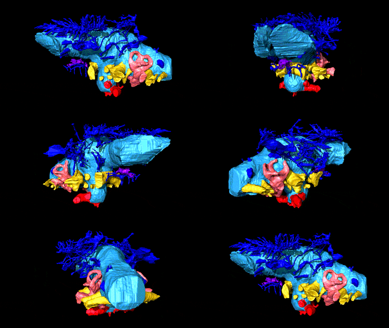
Paleontologists at St Petersburg University created the most detailed virtual 3D-model of the endocranial cast and blood vessels of the head of an ankylosaurian.
Paleontologists from St Petersburg University have been the first to study in detail the structure of the brain and blood vessels in the skull of the ankylosaur Bissektipelta archibaldi. It was a herbivorous dinosaur somewhat similar in appearance to a modern armadillo. The first three-dimensional computer reconstruction of a dinosaur endocast made in Russia — a digital cast of its braincase — was of help to the scientists. It made it possible to find out that ankylosaurs, and Bissektipelta in particular, were capable of cooling their brains, had an extremely developed sense of smell, and heard low-frequency sounds. However, their brain was one and a half times smaller than that of modern animals of the same size.
Ankylosaurs appeared on Earth in the middle of the Jurassic — about 160 million years ago — and existed until the end of the dinosaur era, which ended 65 million years ago. These herbivorous animals were somewhat reminiscent of modern turtles or armadillos, were covered with thick armor, and sometimes even had a bony club on the tail. The researchers became interested in the uniquely-preserved remains of ankylosaurs from Uzbekistan. Although these fossils have been known for 20 years, only now have the scientists had a unique opportunity to study the specimens from the inside using cutting-edge methods.

When Plato set out to define what made a human a human, he settled on two primary characteristics: We do not have feathers, and we are bipedal (walking upright on two legs). Plato’s characterization may not encompass all of what identifies a human, but his reduction of an object to its fundamental characteristics provides an example of a technique known as principal component analysis.
Now, Caltech researchers have combined tools from machine learning and neuroscience to discover that the brain uses a mathematical system to organize visual objects according to their principal components. The work shows that the brain contains a two-dimensional map of cells representing different objects. The location of each cell in this map is determined by the principal components (or features) of its preferred objects; for example, cells that respond to round, curvy objects like faces and apples are grouped together, while cells that respond to spiky objects like helicopters or chairs form another group.
The research was conducted in the laboratory of Doris Tsao (BS ‘96), professor of biology, director of the Tianqiao and Chrissy Chen Center for Systems Neuroscience and holder of its leadership chair, and Howard Hughes Medical Institute Investigator. A paper describing the study appears in the journal Nature on June 3.

Chronic stress has long been associated with the pathogenesis of psychological disorders such as depression and anxiety. Recent studies have found chronic stress can cause neuroinflammation: activation of the resident immune cells in the brain, microglia, to produce inflammatory cytokines. Numerous studies have implicated the inflammatory cytokine, interleukin-1 (IL-1), a master regulator of immune cell recruitment and activity in the brain, as the key mediator of psychopathology. However, how IL-1 disrupts neural circuits to cause behavioral and emotional problems seen in psychological disorders has not been determined.
…
The research team previously detailed how psychosocial stress results in peripheral immune activation, increased levels of circulating monocytes, and robust neuroimmunological responses in the brain. These responses include increases in IL-1 and other inflammatory cytokines, activation of brain glial cells and movements of peripheral immune cells to the brain, along with enhanced activity of specific neuronal pathways. The work makes it clear that inflammatory-related effects of stress are not just global effects, but are associated with increased IL-1 signaling within specific brain circuits.
The study shows for the first time that neuronal IL-1Rs in the hippocampus, a brain structure connected to learning and memory, is necessary and sufficient to mediate some of the behavioral deficits caused by chronic stress, pointing to a critical neuroimmune mechanism for the etiology of these types of disorders. Findings from the study augment the understanding of IL-1R signaling in physiological and behavioral responses to stress and also suggest that it may be possible to develop better medications to treat the consequences of chronic stress by limiting inflammatory signaling not just generally, which may not be beneficial in the long run, but to specific brain circuits.
“We created and validated a unique genetic mouse model to restrict IL-1R1 expression to different cell types to visualize and control IL-1Rs,” said Ning Quan, Ph.D., lead author, a professor of biomedical science in FAU’s Schmidt College of Medicine, and a member of the FAU Brain Institute (I-BRAIN). “We demonstrated that chronic social stress caused the mice to show social withdrawal and working memory deficits. These changes could be prevented if the neuronal IL-1R1 was deleted and restored if IL-1R1 was only allowed to be expressed on hippocampal neurons.”
For the study, researchers wanted to determine the degree to which IL-1 acts directly on hippocampal neurons to influence cognitive and mood changes with stress. To define the IL-1R-mediated neuronal response, they used novel and comprehensive IL-1R transgenic/reporter lines in which one can selectively delete IL-1R or restore IL-1R on specific cell types, including glutamatergic neurons. They also used modified viruses to manipulate hippocampal neurons and investigate the role of IL-1R in eliciting behavioral responses to stress. Their data show that social defeat-induced IL-1R signaling in hippocampal neurons perpetuated inflammation and promoted deficits in social interaction and working memory.