Toggle light / dark theme

Current optical techniques can image neuron activity only near the brain’s surface, but integrated neurophotonics could unlock circuits buried deep in the brain. Credit: Roukes et. al.

But current optogenetic studies of the brain are constrained by a significant physical limitation, says Laurent Moreaux, Caltech senior research scientist and lead author on the paper. Brain tissue scatters light, which means that light shone in from outside the brain can travel only short distances within it. Because of this, only regions less than about two millimeters from the brain’s surface can be examined optically. This is why the best-studied brain circuits are usually simple ones that relay sensory information, such as the sensory cortex in a mouse—they are located near the surface. In short, at present, optogenetics methods cannot readily offer insight into circuits located deeper in the brain, including those involved in higher-order cognitive or learning processes.

Integrated neurophotonics, Roukes and colleagues say, circumvents the problem. In the technique, the microscale elements of a complete imaging system are implanted near complex neural circuits located deep within the brain, in regions such as the hippocampus (which is involved in memory formation), striatum (which controls cognition), and other fundamental structures in unprecedented resolution. Consider the similar technology of functional magnetic resonance imaging (fMRI), the scanning technique currently used to image entire brains. Each voxel, or three-dimension pixel, in an fMRI scan is typically about a cubic millimeter in volume and contains roughly 100,000 neurons. Each voxel, therefore, represents the average activity of all of these 100,000 cells.

Portable sequencing is making it possible for biologists to perform DNA analysis anywhere in the world. How is this technology reshaping the way they work?

Thanks to nanopore technology, scientists can now collect samples and sequence them anywhere. It is the concept of backpacking applied to scientific research.

French molecular biologist Anne-Lise Ducluzeau has experienced this first hand during her research in the freezing environment of Alaska. “I remember driving back home with my sequencing station on the passenger seat, it was −20ºF (−29ºC) but the car was warm and reads kept coming,” ‪relates Ducluzeau, who has been using a portable sequencer for her research for the past four years.

Knowing which proteins are key to protection from disease, and the deficiencies in expression or activity that are hallmarks of disease, can inform individualized medicine and the development of new therapies.


Twenty years after the release of the human genome, the genetic “blueprint” of human life, an international research team, including the University of British Columbia’s Chris Overall, has now mapped the first draft sequence of the human proteome.

Their work was published Oct. 16 in Nature Communications and announced today by the Human Proteome Organization (HUPO).

“Today marks a in our overall understanding of human life,” says Overall, a professor in the faculty of dentistry and a member of the Centre for Blood Research at UBC. “Whereas the provides a complete ‘blueprint’ of , the human proteome identifies the individual building blocks of life encoded by this blueprint: proteins. ” Proteins interact to shape everything from life-threatening diseases to cellular structure in our bodies.”

Article. The research/article indicates that childhood trauma can not only impact the current generation, but future generations. Biochemical signals are sent to the germ cells, modifying the expression of some genes and/or the DNA structure.


Traumatic experiences can have a lasting impact, so children that suffer through them can feel their effects for a lifetime. Work has also shown that trauma can change the way genes are expressed, through epigenetics. Epigenetic changes do not alter the sequence of genes but they alter the biochemistry of DNA, and these changes are sometimes passed down to future generations through germ cells. Scientists have been working to learn more about how traumatic events get embedded in the genetic code of germ cells.

Image credit: Pkist

New research reported in The EMBO Journal has used a mouse model to suggest that childhood trauma can influence the composition of blood, and this is the conduit for passing the impact down to offspring.

Researchers from the University of Seville, in collaboration with colleagues from the Universities of Murcia and Marburg (Germany) have identified a new protein that makes it possible to repair DNA. The protein in question, called cryptochrome, has evolved to acquire this and other functions within the cell.

Ultraviolet radiation can damage the DNA, leading to mutations that disrupt cell function and can allow cancer cells to grow out of control. Our cells have DNA repair systems to defend themselves against this sort of damage. One of these systems is based on a protein, photolyase, which uses to repair DNA damage before it leads to mutations.

Over the course of evolution, the genes for photolyase duplicated and became specialized, creating new proteins, cryptochromes, which have honed their ability to perceive blue light and now perform other functions in cells. For example, cryptochromes use blue light as a signal to regulate and the rhythm that controls daily activity (the circadian rhythm) in fungi and animals.

We ask students to login via google as we share a lot of our content over google drive. To access the same, a google account is a must.


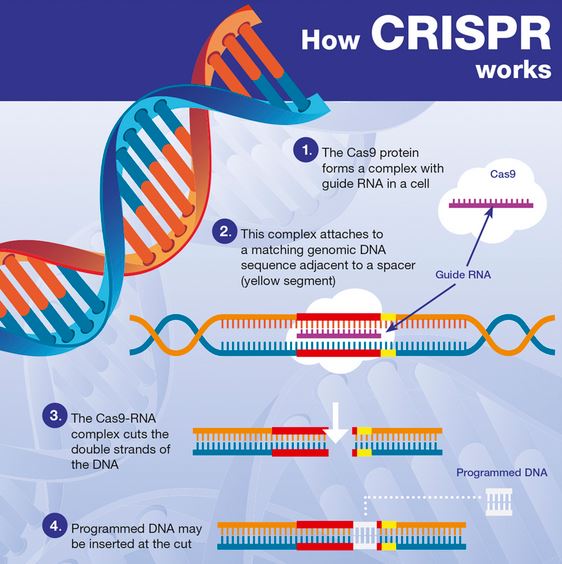
The CRISPR-Cas9 system has revolutionized genetic manipulations and made gene editing simpler, faster and easily accessible to most laboratories.

To its recognition, this year, the French-American duo Emmanuelle Charpentier and Jennifer Doudna have been awarded the prestigious Nobel Prize for chemistry for CRISPR.

The research, out today from the University of Colorado Anschutz Medical Campus and published in * Evolution and Human Behavior*, presents a hypothesis supporting a role for fructose, a component of sugar and high fructose corn syrup, and uric acid (a fructose metabolite), in increasing the risk for these behavioral disorders.

Johnson outlines research that shows a foraging response stimulates risk taking, impulsivity, novelty seeking, rapid decision making, and aggressiveness to aid the securing of food as a survival response. Overactivation of this process from excess sugar intake may cause impulsive behavior that could range from ADHD, to bipolar disorder or even aggression.” “Johnson notes, “We do not blame aggressive behavior on sugar, but rather note that it may be one contributor.”” “The identification of fructose as a risk factor does not negate the importance of genetic, familial, physical, emotional and environmental factors that shape mental health,” he adds.


Huh, want to know more.

“New research suggests that conditions such as attention deficit hyperactivity syndrome (ADHD), bipolar disorder, and even aggressive behaviors may be linked with sugar intake, and that it may have an evolutionary basis.

The research, out today from the University of Colorado Anschutz Medical Campus and published in Evolution and Human Behavior, presents a hypothesis supporting a role for fructose, a component of sugar and high fructose corn syrup, and uric acid (a fructose metabolite), in increasing the risk for these behavioral disorders.

“We present evidence that fructose, by lowering energy in cells, triggers a foraging response similar to what occurs in starvation,” said lead author Richard Johnson, MD, professor at the University of Colorado School of Medicine on the CU Anschutz Medical Campus.

Summary: New artificial intelligence technology will analyze clinical data, brain images, and genetic information from Alzheimer’s patients to look for new biomarkers associated with the neurodegenerative disease.

Source: University of Pennsylvania

As the search for successful Alzheimer’s disease drugs remains elusive, experts believe that identifying biomarkers — early biological signs of the disease — could be key to solving the treatment conundrum. However, the rapid collection of data from tens of thousands of Alzheimer’s patients far exceeds the scientific community’s ability to make sense of it.

Controlling brains with light.


Thanks to optogenetics, in just ten years we’ve been able to artificially incept memories in mice, decipher brain signals that lead to pain, untangle the neural code for addiction, reverse depression, restore rudimentary sight in blinded mice, and overwrite terrible memories with happy ones. Optogenetics is akin to a universal programming language for the brain.

But it’s got two serious downfalls: it requires gene therapy, and it needs brain surgery to implant optical fibers into the brain.

This week, the original mind behind optogenetics is back with an update that cuts the cord. Dr. Karl Deisseroth’s team at Stanford University, in collaboration with the University of Minnesota, unveiled an upgraded version of optogenetics that controls behavior without the need for surgery. Rather, the system shines light through the skulls of mice, and it penetrates deep into the brain. With light pulses, the team was able to change how likely a mouse was to have seizures, or reprogram its brain so it preferred social company.