Toggle light / dark theme

Canadian firm says it could make 10 million doses per month — if its innovative production method wins FDA approval.

A Canadian company says that it has produced a COVID −19 vaccine just 20 days after receiving the coronavirus’s genetic sequence, using a unique technology that they soon hope to submit for FDA approval.

Medicago CEO Bruce Clark said his company could produce as many as 10 million doses a month. If regulatory hurdles can be cleared, he said in a Thursday interview, the vaccine could start to become available in November 2021.

First wave 🌊.


Your questions answered — an update (11−03−2020): Professor Neil Ferguson on the current status of the COVID-19 Coronavirus outbreak, case numbers, intervention measures and challenges countries are currently facing.

Read all reports including estimates of epidemic size, transmissibility, severity, phylogenetics, undetected cases, prevalence and symptom progression here: https://www.imperial.ac.uk/mrc-gida

The Abdul Latif Jameel Institute for Disease and Emergency Analytics (J-IDEA) brings together global health researchers in the School of Public Health at Imperial College London. Drawing on Imperial’s expertise in data analytics, epidemiology and economics, J-IDEA improves our understanding of diseases and health emergencies in the most vulnerable populations across the globe. The Institute links governments, research institutions and communities to develop practical and effective long-term solutions, shape health policy and deliver better quality of life for all.

Abdul Latif Jameel Institute for Disease and Emergency Analytics (J-IDEA)
Website: https://www.imperial.ac.uk/JIDEA
Twitter: @Imperial_JIDEA, https://twitter.com/Imperial_JIDEA

MRC Centre for Global Infectious Disease Analysis (MRC GIDA)
Website: https://www.imperial.ac.uk/mrc-gida
Twitter: @MRC_Outbreak, https://twitter.com/MRC_Outbreak

Interviewee: Professor Neil Ferguson, Imperial College London, Director of J-IDEA and MRC GIDA
Interviewer and Producer: Dr Sabine van Elsland, Imperial College London, J-IDEA, MRC GIDA
Producer: Dr Katharina Hauck, Imperial College London, Deputy Director of J-IDEA, MRC GIDA
Associate Producer: Oliver Geffen Obregon, School of Public Health, Imperial College London.
Director and A Cam Operator: Tiago Melo, Digital Learning Hub
B Cam Operator: Jack Lowe, Digital Learning Hub
C Cam Operator: Erdvilas Abukevicius, Digital Learning Hub
Editor: Anne Marie Rützou Bruntse, Digital Learning Hub
Assistant editor: Tiziana Mangiaratti, Digital Learning Hub.

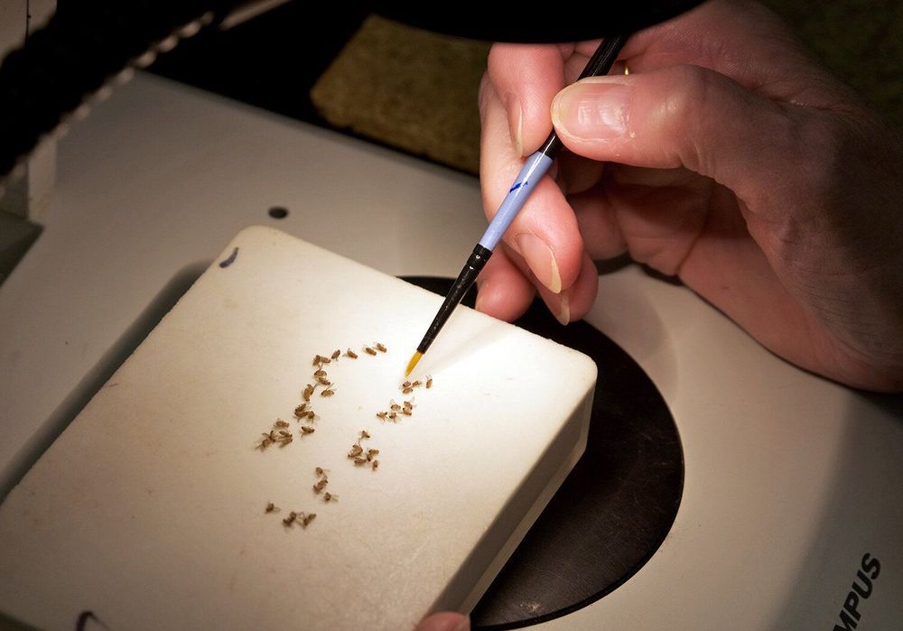
Just as there is a mysterious dark matter that accounts for 85 percent of our universe, there is a “dark” portion of the human genome that has perplexed scientists for decades. A study published March 9, 2020, in Genome Research identifies new portions of the fruit fly genome that, until now, have been hidden in these dark, silent areas.

The collaborative paper titled “Gene Expression Networks in the Drosophila Genetic Reference Panel” is the culmination of years of research by Clemson University geneticists Trudy Mackay and Robert Anholt. Their groundbreaking findings could significantly advance science’s understanding of a number of genetic disorders.

The “dark” portion refers to the approximate 98 percent of the genome that doesn’t appear to have any obvious function. Only 2 percent of the human genome codes for proteins, the building blocks of our bodies and the catalysts of the chemical reactions that allow us to thrive. Scientists have been puzzled by this notion since the 1970s when gene sequencing technologies were first developed, revealing the proportion of coding to noncoding regions of the genome.

Circa 2015 o.o take their Gene’s and could make immunity greater for stomaches.


Vultures have a unique genetic make-up allowing them to digest carcasses and guard themselves against constant exposure to pathogens in their diet, according to the first Eurasian vulture genome published in the open access journal Genome Biology. The study also finds that this species of Asian vulture is more closely related to the North American bald eagle than previously thought.

The cinereous vulture or black vulture, Aegypius monachus, is the largest bird of prey, and an iconic bird in the Far East. The species plays a key role in the ecosystem by removing rotting carcasses, thus preventing the spread of disease.

As their feeding habits involve constant exposure to pathogens, vultures are suspected to have strong immune systems, having evolved mechanisms to prevent infection by the microbes found in their diet. Despite the potential interest in the immune system of scavengers, little is known about the genetic variations involved in vultures’ immune processes.

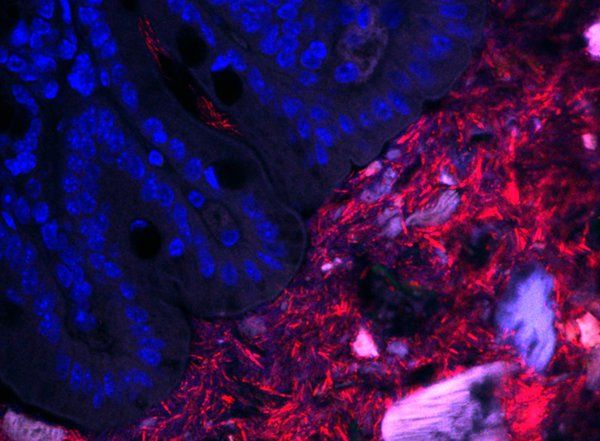
Over many years, the Mazmanian laboratory has described how Bacteroides fragilis in the gut produces beneficial molecules that protect mice from inflammatory bowel disease and autism-like symptoms. Like a densely populated city, a vast majority of the B. fragilis in the gut live within the central part of the intestinal tube, called the lumen. However, the Mazmanian laboratory discovered in 2013 that some B. fragilis reside in the bacterial equivalent of small towns, nestled into microscopic pockets within the tissue walls lining the tube. These sparse populations are protected by mucus and are largely unaffected by antibiotics, suggesting that they act as population reservoirs that ensure long-term colonization.

“For humans, where we live can dictate how we behave—for example, a person living in a city likely has a different everyday life than a person living in a small rural community,” says former graduate student Gregory Donaldson (PhD ‘18), the first author on the new paper. “For the bacteria that we study, the intestines represent their entire world, so we wanted to know how differently they behave depending on how far away from the intestinal surface they are.”

Though they may live in different habitats within the gut, these B. fragilis populations all have the same genetic code. What may differ, however, is how they express those genes—is a bacterium expressing a gene for replication and division, for example, or perhaps for an enzyme that digests food? Donaldson aimed to measure and compare gene expression in these two populations (intestinal wall tissue and lumen of the gut) to determine what, if any, differences were seen.

This posed a technical challenge. Because the population of bacteria living in the tissue lining is so small, their genetic material becomes obscured during sequencing by the genetic material of the mouse cells, which is far more abundant than that of the bacteria. Though mice and bacteria are distinctly different genetically, sifting through the mouse RNA to find the bacterial RNA is like finding a needle in a haystack.

Here, a crucial collaboration with Ashlee Earl of the Broad Institute made the research possible. Earl and her team led the development of a new technique, called hybrid selection RNA-sequencing, designed to fish out the elusive strands of bacterial RNA like using a magnet to search for the needles in a haystack.

“Inspired by a previous approach to sequence small populations of parasites in human blood, we developed a technique that could boost the amount of bacterial RNA we could detect in these host-rich samples by orders of magnitude,” says Earl. “This technique not only helped to reveal a new aspect of the Bacteroides-host relationship, but now provides us with a more general tool for listening in on conversations between humans and their rarest inhabitants.”

This technique revealed that B. fragilis residing in the gut tissue are surprisingly metabolically active and thriving despite their sparser population. Donaldson and his team found that one particular gene in these bacteria help them to establish a foothold in the tissue lining. Without this gene, they are unable to colonize this habitat, which is detrimental to their long-term colonization of the animal.

In contrast, B. fragilis living in the lumen are busy digesting food and nutrients, but they appear to be more focused on surviving than growing. This suggests that the epithelial surface is actually the preferred habitat for these bacteria.

As B. fragilis in the gut is known to provide beneficial protections for the host, this work suggests that a spatially intimate relationship is necessary for these positive effects. Ultimately, understanding how bacteria establish residency and persist in the gut may lead to new strategies to reinforce a healthy human microbiome to prevent or treat disease.

“While we know a great deal about which bacteria reside in humans through DNA sequencing techniques and how microbial community membership changes as a result of disease or other factors, we know far less what bacteria are doing while living inside of us,” says Mazmanian. “For the first time, this work gives us a glimpse into the lifestyle and behavior of an important human gut bacteria, while colonizing mice. Knowing what key bacteria are actually doing in the gut may help develop rationale and robust therapies from the microbiome.”


The microscopic populations of bacteria in our intestines are, in some ways, just like us: They live in communities, eat, work, reproduce, and eventually die. Many of these bacterial species live in harmony with our bodies, providing benefits to us in exchange for nutrients and shelter. When the right kinds of bacteria cannot establish the proper symbiotic relationship with our bodies, we may be at a greater risk for a variety of immune, neurological, and metabolic diseases.

Now, in collaboration with the Broad Institute of MIT and Harvard, Caltech researchers have discovered that a certain species of bacteria behaves differently depending on where in the gut it takes up residence. The work was done in mouse models, but mapping the geography of microbial populations in the gut—collectively referred to as the gut microbiome—may be crucial for one day being able to treat and reshape a human microbiome gone awry.

The research was conducted primarily in the laboratory of Sarkis Mazmanian, Luis B. and Nelly Soux Professor of Microbiology and Heritage Medical Research Institute Investigator. A paper describing the research appears on March 9 in the journal Nature Microbiology.

In a new publication in Nature Plants, assistant professor of Plant Science at the University of Maryland Yiping Qi has established a new CRISPR genome engineering system as viable in plants for the first time: CRISPR-Cas12b. CRISPR is often thought of as molecular scissors used for precision breeding to cut DNA so that a certain trait can be removed, replaced, or edited. Most people who know CRISPR are likely thinking of CRISPR-Cas9, the system that started it all. But Qi and his lab are constantly exploring new CRISPR tools that are more effective, efficient, and sophisticated for a variety of applications in crops that can help curb diseases, pests, and the effects of a changing climate. With CRISPR-Cas12b, Qi is presenting a system in plants that is versatile, customizable, and ultimately provides effective gene editing, activation, and repression all in one system.

“This is the first demonstration of this new CRISPR-Cas12b system for plant genome engineering, and we are excited to be able to fill in gaps and improve systems like this through new technology,” says Qi. “We wanted to develop a full package of tools for this system to show how useful it can be, so we focused not only on editing, but on developing gene repression and activation methods.”

It is this complete suite of methods that has ultimately been missing in other CRISPR systems in . The two major systems available before this paper in plants were CRISPR-Cas9 and CRISPR-Cas12a. CRISPR-Cas9 is popular for its simplicity and for recognizing very short DNA sequences to make its cuts in the genome, whereas CRISPR-Cas12a recognizes a different DNA targeting sequence and allows for larger staggered cuts in the DNA with additional complexity to customize the system. CRISPR-Cas12b is more similar to CRISPR-Cas12a as the names suggest, but there was never a strong ability to provide gene activation in plants with this system. CRISPR-Cas12b provides greater efficiency for gene activation and the potential for broader targeting sites for , making it useful in cases where genetic expression of a trait needs to be turned on/up (activation) or off/down (repression).

Keep it Clean. Please avoid obscene, vulgar, lewd, racist or sexually-oriented language. PLEASE TURN OFF YOUR CAPS LOCK. Don’t Threaten. Threats of harming another person will not be tolerated. Be Truthful. Don’t knowingly lie about anyone or anything. Be Nice. No racism, sexism or any sort of –ism that is degrading to another person. Be Proactive. Use the ‘Report’ link on each comment to let us know of abusive posts. Share with Us. We’d love to hear eyewitness accounts, the history behind an article.

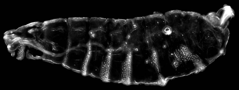
Researchers at Princeton University have revealed the inner workings of a gene repression mechanism in fruit fly embryos, adding insight to the study of human diseases.

Led by graduate student Shannon Keenan, the team used light to activate in developing and traced the effects on a protein called Capicua, or Cic. Located in a cell’s nucleus, Cic binds to DNA and performs the specialized task of silencing . The study, published in Developmental Cell and made available online March 5, reveals the dynamics of gene repression by this protein.

In a complex piece of music, the silences running through the melody contribute as much to the score’s effect as the sounded notes. The that control development rely on highly sophisticated temporal patterns of gene activation and repression to create life’s beautiful symphonies. When a pattern is disrupted, it’s like a wrong note in the music. In this case, Cic is a repressor protein that silences certain parts of the genome, allowing other genes to express in harmony with one another. Understanding how repressors like Cic work allows researchers to better conduct the orchestra.