During the pandemic, the National Museum of Mathematics found new ways to build human connections.

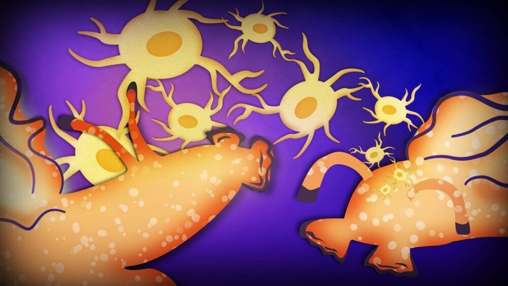
LOS ANGELES — UCLA neuroscientists reported Monday that they have transferred a memory from one animal to another via injections of RNA, a startling result that challenges the widely held view of where and how memories are stored in the brain.
The finding from the lab of David Glanzman hints at the potential for new RNA-based treatments to one day restore lost memories and, if correct, could shake up the field of memory and learning.
“It’s pretty shocking,” said Dr. Todd Sacktor, a neurologist and memory researcher at SUNY Downstate Medical Center in Brooklyn, N.Y. “The big picture is we’re working out the basic alphabet of how memories are stored for the first time.” He was not involved in the research, which was published in eNeuro, the online journal of the Society for Neuroscience.
Many scientists are expected to view the research more cautiously. The work is in snails, animals that have proven a powerful model organism for neuroscience but whose simple brains work far differently than those of humans. The experiments will need to be replicated, including in animals with more complex brains. And the results fly in the face of a massive amount of evidence supporting the deeply entrenched idea that memories are stored through changes in the strength of connections, or synapses, between neurons.

The world’s most powerful laser is scheduled for a slate of experiments next year.
The laser, in Romania, managed to fire at 10 petawatts — that’s one-tenth the power of all the sunlight that reaches Earth concentrated into a single laser beam — during a test run in March. Now, according to ExtremeTech, the scientists behind it intend to discover new high-energy cancer treatments and simulate supernovas to reveal how the stellar explosions form heavy metals.
The laser is part of the European Union’s Extreme Light Infrastructure project. The hope is that lasers will lead to new medical techniques, a better understanding of how the universe works, and improved nuclear safety.

Covid-19, a virus that many experts believe came to us from bats, has been transmitted on from humans to pets and other animals. Here’s why some scientists are worried that so-called spillbacks could potentially perpetuate a cycle of infection. Photo: Markus Scholz/Zuma Press.
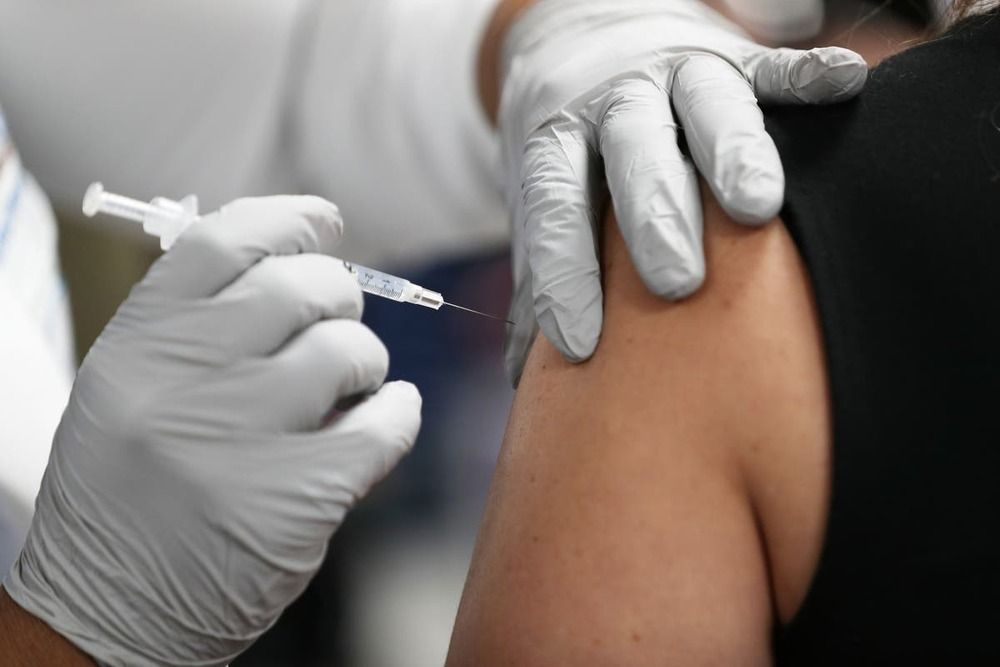
New study looked at the effects of the vaccine on 237 healthcare workers.
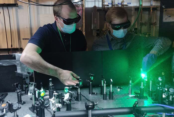
A new class of quantum dots deliver a stable stream of single, spectrally tunable infrared photons under ambient conditions and at room temperature, unlike other single photon emitters. This breakthrough opens a range of practical applications, including quantum communication, quantum metrology, medical imaging and diagnostics, and clandestine labeling.
“The demonstration of high single-photon purity in the infrared has immediate utility in areas such as quantum key distribution for secure communication,” said Victor Klimov, lead author of a paper published today in Nature Nanotechnology by Los Alamos National Laboratory scientists.
The Los Alamos team has developed an elegant approach to synthesizing the colloidal-nanoparticle structures derived from their prior work on visible light emitters based on a core of cadmium selenide encased in a cadmium sulfide shell. By inserting a mercury sulfide interlayer at the core/shell interface, the team turned the quantum dots into highly efficient emitters of infrared light that can be tuned to a specific wavelength.
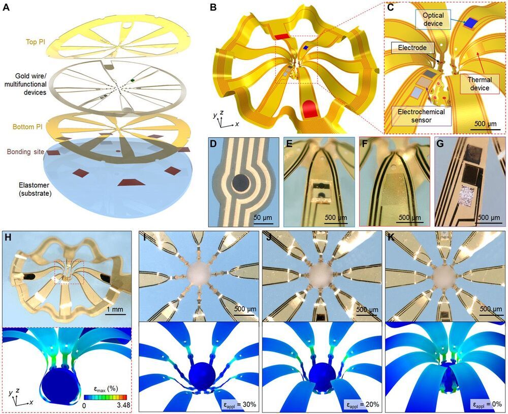
Three-dimensional (3D), submillimeter-scale constructs of neural cells, known as cortical spheroids, are of rapidly growing importance in biological research because these systems reproduce complex features of the brain in vitro. Despite their great potential for studies of neurodevelopment and neurological disease modeling, 3D living objects cannot be studied easily using conventional approaches to neuromodulation, sensing, and manipulation. Here, we introduce classes of microfabricated 3D frameworks as compliant, multifunctional neural interfaces to spheroids and to assembloids. Electrical, optical, chemical, and thermal interfaces to cortical spheroids demonstrate some of the capabilities. Complex architectures and high-resolution features highlight the design versatility. Detailed studies of the spreading of coordinated bursting events across the surface of an isolated cortical spheroid and of the cascade of processes associated with formation and regrowth of bridging tissues across a pair of such spheroids represent two of the many opportunities in basic neuroscience research enabled by these platforms.
Progress in elucidating the development of the human brain increasingly relies on the use of biosystems produced by three-dimensional (3D) neural cultures, in the form of cortical spheroids, organoids, and assembloids (1–3). Precisely monitoring the physiological properties of these and other types of 3D biosystems, especially their electrophysiological behaviors, promises to enhance our understanding of the interactions associated with development of the nervous system, as well as the evolution and origins of aberrant behaviors and disease states (4–8). Conventional multielectrode array (MEA) technologies exist only in rigid, planar, and 2D formats, thereby limiting their functional interfaces to small areas of 3D cultures, typically confined to regions near the bottom contacting surfaces.
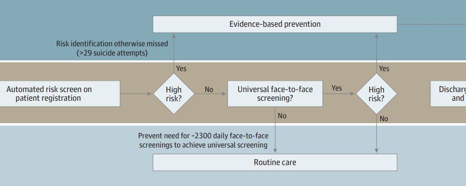
Importance Numerous prognostic models of suicide risk have been published, but few have been implemented outside of integrated managed care systems.
Objective To evaluate performance of a suicide attempt risk prediction model implemented in a vendor-supplied electronic health record to predict subsequent suicidal ideation and suicide attempt.
Design, Setting, and Participants This observational cohort study evaluated implementation of a suicide attempt prediction model in live clinical systems without alerting. The cohort comprised patients seen for any reason in adult inpatient, emergency department, and ambulatory surgery settings at an academic medical center in the mid-South from June 2019 to April 2020.

LONDON (AP) — The rainbow flag flew proudly Thursday above the Bank of England in the heart of London’s financial district to commemorate World War II codebreaker Alan Turing, the new face of Britain’s 50-pound note.
The design of the bank note was unveiled before it is being formally issued to the public on June 23, Turing’s birthday. The 50-pound note is the most valuable denomination in circulation but is little used during everyday transactions, especially during the coronavirus pandemic as digital payments increasingly replaced the use of cash.
The new note, which is laden with high-level security features and is made of longer-lasting polymer, completes the bank’s rejig of its paper currencies over the past few years. Turing’s image joins that of Winston Churchill on the five-pound note, novelist Jane Austen on the 10-pound note and artist J. M. W. Turner on the 20-pound note.
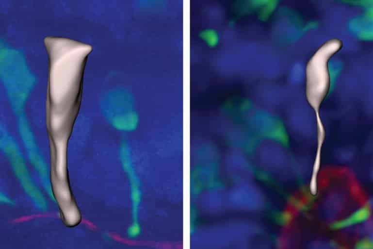
Summary: Using brain organoid models, researchers have identified how the brain grows much larger and has three times as many neurons, as the brains of chimpanzees and gorillas.
Source: UK Research and Innovation.
A new study is the first to identify how human brains grow much larger, with three times as many neurons, compared with chimpanzee and gorilla brains. The study, led by researchers at the Medical Research Council (MRC) Laboratory of Molecular Biology in Cambridge, UK, identified a key molecular switch that can make ape brain organoids grow more like human organoids, and vice versa.