Toggle light / dark theme

Perhaps better than medication.


Summary: Inexpensive, commercial video games may help those suffering from anxiety and depression to combat their symptoms. Researchers found that, in addition to conventional games, virtual reality games could help people overcome mental health problems.

Source: Lero

Popular video games have the potential to provide low-cost, easy access, effective and stigma-free support for some mental health issues, researchers at Lero, the Science Foundation Ireland Research Centre for Software, have found.

The team at Lero, a world leader in connected-health research, said video games could be used where conventional therapies are not available because of cost or location, or as an addition to traditional therapeutic treatments for depression or anxiety.

Summary: Researchers found an increased inflammatory signal in patients with the C90rf72 subtype of ALS. The increased inflammatory biomarkers could be found in peripheral serum tests.

Source: Thomas Jefferson University.

Amyotrophic lateral sclerosis (ALS), also known as Lou Gehrig’s disease, is a neurodegenerative disease that strikes nearly 5000 people in the U.S. every year.

Summary: A new imaging study reveals how the MFSD2A transporter protein provides a gateway for omega-3 fatty acids to enter the brain.

Source: Columbia University.

Spectacular images of a molecule that shuttles omega-3 fatty acids into the brain may open a doorway for delivering neurological therapeutics to the brain.

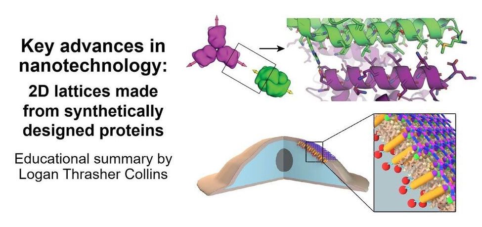
Check out my short video in which I explain some super exciting research in the area of nanotechnology: de novo protein lattices! I specifically discuss a journal article by Ben-Sasson et al. titled “Design of biologically active binary protein 2D materials”.


Here, I explain an exciting nanotechnology paper “Design of biologically active binary protein 2D materials” (https://doi.org/10.1038/s41586-020-03120-8).

Though I am not involved in this particular research myself, I have worked in adjacent areas such as de novo engineering of aggregating antimicrobial peptides, synthetic biology, nanotechnology-based tools for neuroscience, and gene therapy. I am endlessly fascinated by this kind of computationally driven de novo protein design and would love to incorporate it in my own research at some point in the future.

I am a PhD candidate at Washington University in St. Louis and the CTO of the startup company Conduit Computing. I am also a published science fiction writer and a futurist. To learn more about me, check out my website: https://logancollinsblog.com/.

Stimulation of the nervous system with neurotechnology has opened up new avenues for treating human disorders, such as prosthetic arms and legs that restore the sense of touch in amputees, prosthetic fingertips that provide detailed sensory feedback with varying touch resolution, and intraneural stimulation to help the blind by giving sensations of sight.

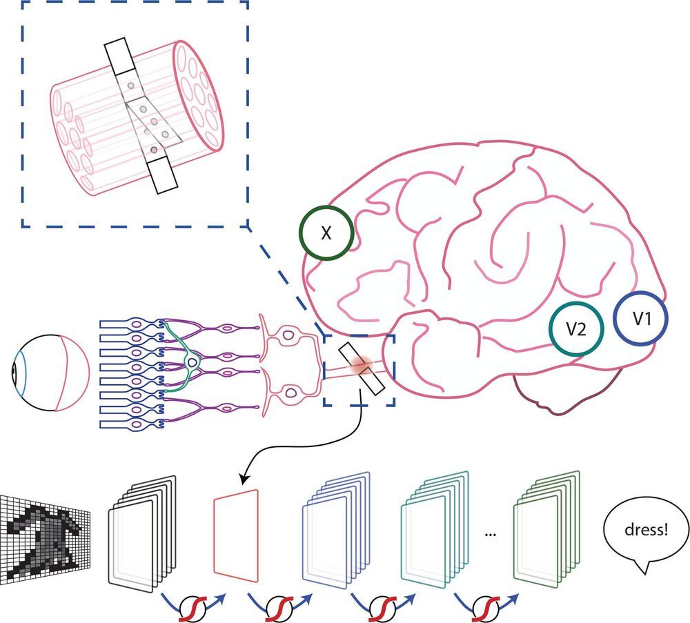
Scientists in a European collaboration have shown that optic nerve stimulation is a promising neurotechnology to help the blind, with the constraint that current technology has the capacity of providing only simple visual signals.

Nevertheless, the scientists’ vision (no pun intended) is to design these simple visual signals to be meaningful in assisting the blind with daily living. Optic nerve stimulation also avoids invasive procedures like directly stimulating the brain’s visual cortex. But how does one go about optimizing stimulation of the optic nerve to produce consistent and meaningful visual sensations?

Now, the results of a collaboration between EPFL, Scuola Superiore Sant’Anna and Scuola Internazionale Superiore di Studi Avanzati, published today in Patterns, show that a new stimulation protocol of the optic nerve is a promising way for developing personalized visual signals to help the blind–that also take into account signals from the visual cortex. The protocol has been tested for the moment on artificial neural networks known to simulate the entire visual system, called convolutional neural networks (CNN) usually used in computer vision for detecting and classifying objects. The scientists also performed psychophysical tests on ten healthy subjects that imitate what one would see from optic nerve stimulation, showing that successful object identification is compatible with results obtained from the CNN.

“We are not just trying to stimulate the optic nerve to elicit a visual perception,” explains Simone Romeni, EPFL scientist and first author of the study. “We are developing a way to optimize stimulation protocols that takes into account how the entire visual system responds to optic nerve stimulation.”

“The research shows that you can optimize optic nerve stimulation using machine learning approaches. It shows more generally the full potential of machine learning to optimize stimulation protocols for neuroprosthetic devices,” continues Silvestro Micera, EPFL Bertarelli Foundation Chair in Translational Neural Engineering and Professor of Bioelectronics at the Scuola Superiore Sant’Anna.

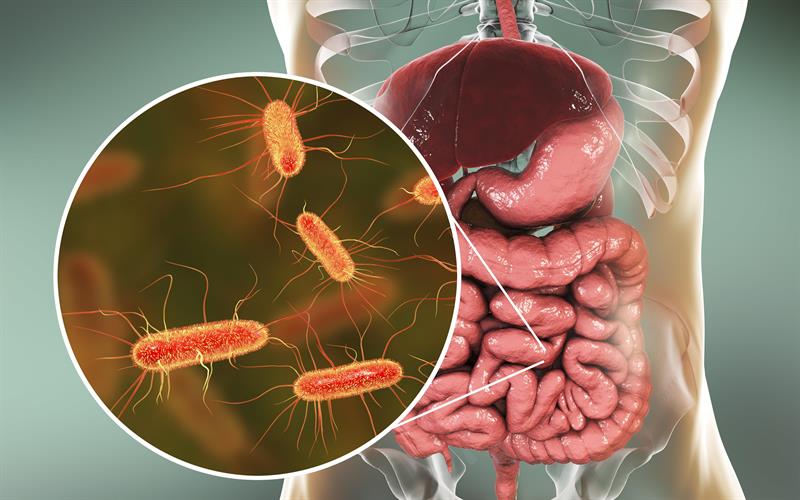
Possibly one of the most surprising ways in which our mind and body are interlinked with one another is the gut-brain axis, which is a collection of bidirectional biochemical signals which are transmitted between the nervous system of the body and the digestive system. This is understandably surprising, as the functions of these two distinct parts of the body are completely different to one another. The gut is unlike most other parts of the body, because a large part of its function and health is dictated by cells which are not part of the body, but are instead bacteria cells which colonise the inner lining of the gut.

It has been known for a while now that the makeup of the gut flora changes as we age, which has in turn been linked to cognitive decline through the disruption of the aforementioned gut-brain axis. It has even been shown that faecal transplants can help to correct this cognitive decline in mice, and has been shown to be able to generate a direct positive effect on cognitive function.

Further research into this phenomenon has revealed that the graduate degradation of the gut flora, or more commonly referred to as the ‘good’ bacteria inside the gut has revealed that these bacteria play an important role at keeping unwanted bacteria in check. Researchers at the University Of Florida have found that certain types of ‘good’ bacteria inside the gut produce a chemical known as butyrate, which supresses the growth of pathogenic bacteria such as Enterobacteriaceae. These pathogenic, or ‘bad’ bacteria effect the body in numerous ways, such as interfering with the protein folding, resulting in a build up of toxic and mis-formed proteins within the body. This disruption to protein folding causes problems all across the body, including in the muscles, intestines, gonads, and most notably the brain and central nervous system.

Summary: Neuroinflammation may be a key player in the pathological brain changes produced as a result of chronic opioid use. Microglia is likely responsible for the majority of the changes.

Source: boston university school of medicine.

Prevalence rates of opioid use disorder (OUD) have increased dramatically, accompanied by a surge of overdose deaths–nearly 50000 in the U.S. in 2019. While opioid dependence has been extensively studied in preclinical models, an understanding of the biological alterations that occur in the brains of people who chronically use opioids and who are diagnosed with OUD remains limited.

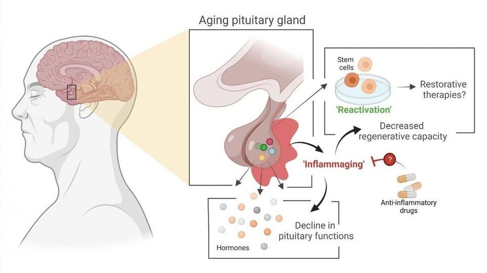
Stem cell biologist Hugo Vankelecom (KU Leuven) and his colleagues have discovered that the pituitary gland in mice ages as the result of an age-related form of chronic inflammation. It may be possible to slow down this process or even partially repair it. The researchers have published their findings in PNAS.

The pituitary is a small, globular gland located underneath the brain that plays a major role in the , explains Professor Hugo Vankelecom from the Department of Development and Regeneration at KU Leuven. “My research group discovered that the pituitary gland ages as a result of a form of chronic inflammation that affects tissue and even the organism as a whole. This usually goes unnoticed and is referred to as ‘inflammaging’—a contraction of inflammation and aging. Inflammaging has previously been linked to the aging of other organs.” Due to the central role played by the pituitary, its aging may contribute to the reduction of hormonal processes and hormone levels in our body—as is the case with menopause, for instance.

The study also provides significant insight into the stem cells in the aging . In 2012, Vankelecom and his colleagues showed that a prompt reaction of these stem cells to injury in the gland leads to repair of the tissue, even in adult animals. “As a result of this new study, we now know that stem cells in the pituitary do not lose this regenerative capacity when the organism ages. In fact, the stem cells are only unable to do their job because, over time, the pituitary becomes an ‘inflammatory environment’ as a result of the chronic inflammation. But as soon as the stem cells are taken out of this environment, they show the same properties as stem cells from a young pituitary.”

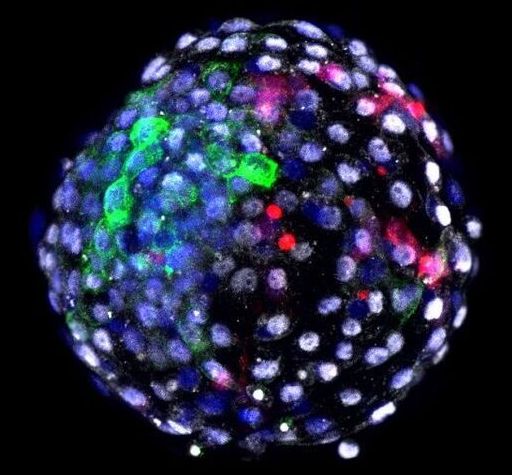
The way the team made the human–monkey embryo is similar to previous attempts at half-human chimeras.

Here’s how it goes. They used de-programmed, or “reverted,” human stem cells, called induced pluripotent stem cells (iPSCs). These cells often start from skin cells, and are chemically treated to revert to the stem cell stage, gaining back the superpower to grow into almost any type of cell: heart, lung, brain…you get the idea. The next step is preparing the monkey component, a fertilized and healthy monkey egg that develops for six days in a Petri dish. By this point, the embryo is ready for implantation into the uterus, which kicks off the whole development process.

This is where the chimera jab comes in. Using a tiny needle, the team injected each embryo with 25 human cells, and babied them for another day. “Until recently the experiment would have ended there,” wrote Drs. Hank Greely and Nita Farahany, two prominent bioethicists who wrote an accompanying expert take, but were not involved in the study.