We will need to tap into our brains and enter the era of neuroreality if we want to reach the next level of truly immersive worlds.



Scientists at Albert Einstein College of Medicine have found that stem cells in the brain’s hypothalamus govern how fast aging occurs in the body. The finding, made in mice, could lead to new strategies for warding off age-related diseases and extending lifespan. The paper was published online today in Nature.
The hypothalamus was known to regulate important processes including growth, development, reproduction and metabolism. In a 2013 Nature paper, Einstein researchers made the surprising finding that the hypothalamus also regulates aging throughout the body. Now, the scientists have pinpointed the cells in the hypothalamus that control aging: a tiny population of adult neural stem cells, which were known to be responsible for forming new brain neurons.
“Our research shows that the number of hypothalamic neural stem cells naturally declines over the life of the animal, and this decline accelerates aging,” says senior author Dongsheng Cai, M.D., Ph.D., (professor of molecular pharmacology at Einstein. “But we also found that the effects of this loss are not irreversible. By replenishing these stem cells or the molecules they produce, it’s possible to slow and even reverse various aspects of aging throughout the body.”

Cory Doctorow has made several careers out of thinking about the future, as a journalist and co-editor of Boing Boing, an activist with strong ties to the Creative Commons movement and the right-to-privacy movement, and an author of novels that largely revolve around the ways changing technology changes society. From his debut novel, Down And Out In The Magic Kingdom (about rival groups of Walt Disney World designers in a post-scarcity society where social currency determines personal value), to his most acclaimed, Little Brother (about a teenage gamer fighting the Department of Homeland Security), his books tend to be high-tech and high-concept, but more about how people interface with technologies that feel just a few years into the future.
But they also tend to address current social issues head-on. Doctorow’s latest novel, Walkaway, is largely about people who respond to the financial disparity between the ultra-rich and the 99 percent by walking away and building their own networked micro-societies in abandoned areas. Frightened of losing control over society, the 1 percent wages full-on war against the “walkaways,” especially after they develop a process that can digitize individual human brains, essentially uploading them to machines and making them immortal. When I talked to Doctorow about the book and the technology behind it, we started with how feasible any of this might be someday, but wound up getting deep into the questions of how to change society, whether people are fundamentally good, and the balance between fighting a surveillance state and streaming everything to protect ourselves from government overreach.
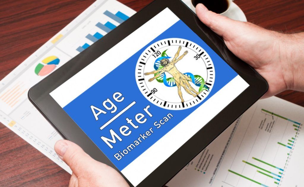
Chronological age has been typically used as a way to gauge how someone is aging, however this is a poor measure indeed. People tend to age at different rates due to a variety of reasons, environment, diet, diseases in earlier life, stress, exercise and lifestyle all play a role in how a person ages.
Clearly a better way to measure aging is needed if we are to accurately assess how someone is aging for the purposes of health monitoring and research. One way to do this is to use functional aging as a way to determine how someone is aging.
Functional aging is defined as a combination of the chronological, physiological, mental, and emotional ages of a person that give an overall measure of their rate of aging.

Researchers in the US have reported what they believe is a first-of-its-kind reversal of brain damage, after treating a drowned and resuscitated toddler with a combination of oxygen therapies.
The little girl, whose heart didn’t beat on her own for 2 hours after drowning, showed deep grey matter injury and cerebral atrophy with grey and white matter loss after the incident, and could no longer speak, walk, or respond to voices – but would uncontrollably squirm around and shake her head.
Amazingly, thanks to a course of oxygen treatments – including hyperbaric oxygen therapy (HBOT) – administered by a team from LSU Health New Orleans and the University of North Dakota, doctors were able to significantly reverse the brain damage experienced by the toddler.
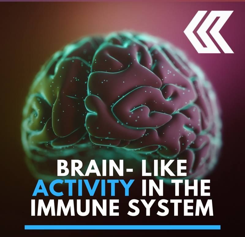

Electric fields can be used to guide transplanted human neural stem cells — cells that can develop into various brain tissues — to repair brain damage in specific areas of the brain, scientists at the University of California, Davis have discovered.
It’s well known that electric fields can locally guide wound healing. Damaged tissues generate weak electric fields, and research by UC Davis Professor Min Zhao at the School of Medicine’s Institute for Regenerative Cures has previously shown how these electric fields can attract cells into wounds to heal them.
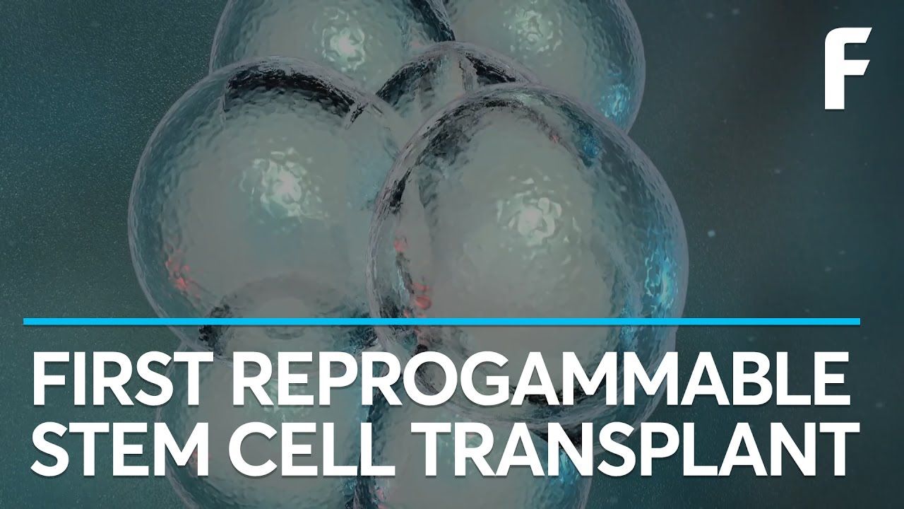
“Microglia play an important role in Alzheimer’s and other diseases of the central nervous system. Recent research has revealed that newly discovered Alzheimer’s-risk genes influence microglia behavior,” Jones said in an interview for a UCI press release. “Using these cells, we can understand the biology of these genes and test potential new therapies.”
A Renewable Method
The skin cells had been donated by patients from UCI’s Alzheimer’s Disease Research Center. These were first subjected to a genetic process to convert them into induced pluripotent stem (iPS) cells — adult cells modified to behave as an embryonic stem cell, allowing them to become other kinds of cells. These iPS cells were then exposed to differentiation factors designed to imitate the environment of developing microglia, which transformed them into the brain cells.
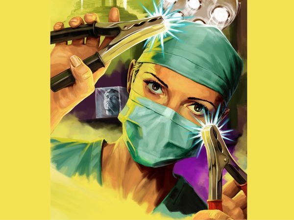
One afternoon in February 2011, Kelly Dwyer strapped on a pair of snowshoes and set out to hike a beaver pond trail near her home in Hooksett, New Hampshire. When the sun dropped below the horizon hours later, the 46-year-old environmental educator still hadn’t returned home. Her husband, David, was worried. Grabbing his cellphone and a flashlight, he told their two daughters he was going to look for Mom. As he made his way toward the pond, sweeping his flashlight beam across the darkening winter landscape, he called out for Kelly. That’s when he heard the moans.
Running toward them, David phoned their daughter Laura, 14, and told her to call 911. His flashlight beam soon settled on Kelly, submerged up to her neck in a hole of dark water in the ice. As David clutched her from behind to keep her head above water, Kelly slumped into unconsciousness. By the time rescue crews arrived, her body temperature was in the 60s and her pulse was almost too faint to register. Before she could reach the ambulance, Kelly’s heart stopped. The EMTs attempted CPR—a process doctors continued for three hours at a hospital in nearby Manchester. They warmed her frigid body. Nothing. Even defibrillation wouldn’t restart her heart. Kelly’s core temperature hovered in the 70s. David assumed he’d lost her for good.

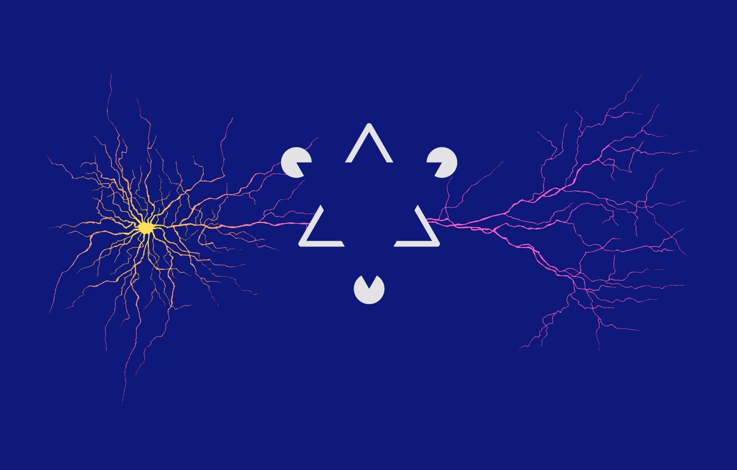
The research team of Prof. Sonja Hofer at the Biozentrum, University of Basel, has discovered why our brain might be so good at perceiving edges and contours. Neurons that respond to different parts of elongated edges are connected and thus exchange information. This can make it easier for the brain to identify contours of objects. The results of the study are now published in the journal Nature.
Individual visual stimuli are not processed independently by our brain. Rather neurons exchange incoming information to form a coherent perceptual image from the myriad of visual details impinging on our eyes. How our visual perception arises from these interactions is still unclear. This is partly due to the fact that we still know relatively little about the rules that determine which neurons in the brain are connected to each other, and what information they exchange. The research team of Prof. Sonja Hofer at the Biozentrum, University Basel studies neuronal networks in the brain. She has now investigated in the mouse model what information individual neurons in the visual cortex receive from other neurons about the wider visual field.