Great discuss of time travel by Dr. Brain greene.


Great discuss of time travel by Dr. Brain greene.
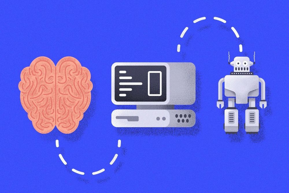
Machine interfaces today can link up brains to play tetris together. Like it’s not hard enough to find a place for the L-shaped block without another cerebrum trying to overrule you.
Let’s go farther: What if we could create a digital replica of your brain and upload and download it like a piece of software?
This feat, aka whole brain emulation (WBE), is still decades, perhaps more than a century away. Outside of the pure science challenge, it could make us confront some of the most daunting questions about what it means to be human, and where man ends and machine begins.

No one needs intelligence more than the military. That’s why the U.S. armed forces and intelligence services are working on a stunning array of pioneering brain development techniques that could one day make their way into civilian life. “The sophistication of our weapons and communications technologies in the Navy and elsewhere is growing dramatically,” says Harold Hawkins, a cognitive psychologist and the director of a program at the Office of Naval Research studying brain training. “To have intellectually stronger people to deal with these new systems is going to be critical.”
The Army, Navy and Air Force are all funding substantial research programs, but a $12 million program approved in January by the Intelligence Advanced Research Projects Activity (IARPA) is one of the largest. It will pay for the first year of a planned three-and-a-half-year program called Strengthening Human Adaptive Reasoning and Problem-solving (SHARP).
The SHARP program is studying techniques both ancient and avant-garde, from meditation to low-dose electrical stimulation of the brain, with an aim toward making intelligence analysts, well, more intelligent. Also on the drawing board are large-scale studies of computerized games that have shown promise in smaller studies for strengthening “working memory” — the critical-thinking ability to not simply remember facts and figures but to juggle and manipulate them. “If these interventions are actually doing what we think they’re doing,” says Adam Russell, a neuroscientist and the SHARP program’s manager at IARPA, “we should be able to demonstrate that with large numbers of participants, strong metrics and a real-world test battery.”
A team of scientists at UC San Francisco and the National Institutes of Health have achieved another CRISPR first, one which may fundamentally alter the way scientists study brain diseases.
In a paper published August 15 in the journal Neuron, the researchers describe a technique that uses a special version of CRISPR developed at UCSF to systematically alter the activity of genes in human neurons generated from stem cells, the first successful merger of stem cell-derived cell types and CRISPR screening technologies.
Though mutations and other genetic variants are known to be associated with an increased risk for many neurological diseases, technological bottlenecks have thwarted the efforts of scientists working to understand exactly how these genes cause disease.
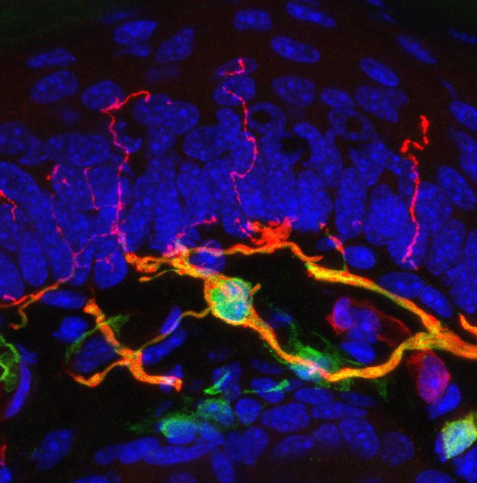
Most people who’ve been jabbed by a needle know the drill: First the pierce, then the sharp, searing pain and an urge to pull away, or at least wince. While the exact circuitry behind this nearly universal reaction is not fully understood, scientists may have just found an important piece of the puzzle: a previously unknown sensory organ inside the skin.
Dubbed the nociceptive glio-neural complex, this structure is not quite like the typical picture of a complex organ like the heart or the spleen. Instead, it’s a simple organ made up of a network of cells called glial cells, which are already known to surround and support the body’s nerve cells. In this case, the glial cells form a mesh-like structure between the skin’s outer and inner layers, with filament-like protrusions that extend into the skin’s outer layer. (Also find out about a type of simple organ recently found in humans, called the interstitium.)
As the study team reports today in the journal Science, this humble organ seems to play a key role in the perception of mechanical pain—discomfort caused by pressure, pricking, and other impacts to the skin. Until now, individual cells called nociceptive fibers were thought to be the main starting points for this kind of pain.
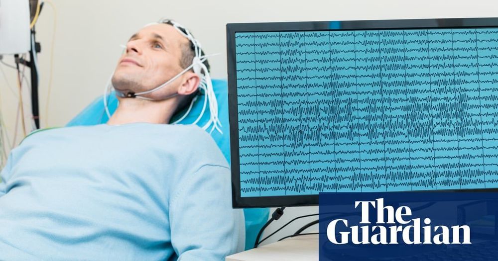
When Stephen Hawking wanted to speak, he chose letters and words from a synthesiser screen controlled by twitches of a muscle in his cheek.
But the painstaking process the cosmologist used might soon be bound for the dustbin. With a radical new approach, doctors have found a way to extract a person’s speech directly from their brain.
The breakthrough is the first to demonstrate how a person’s intention to say specific words can be gleaned from brain signals and turned into text fast enough to keep pace with natural conversation.
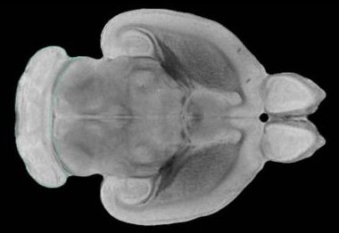
New research, published today in Nature, reveals how increasing brain stiffness as we age causes brain stem cell dysfunction, and demonstrates new ways to reverse older stem cells to a younger, healthier state.
The results have far reaching implications for how we understand the ageing process, and how we might develop much-needed treatments for age-related brain diseases.
As our bodies age, muscles and joints can become stiff, making everyday movements more difficult. This study shows the same is true in our brains, and that age-related brain stiffening has a significant impact on the function of brain stem cells.
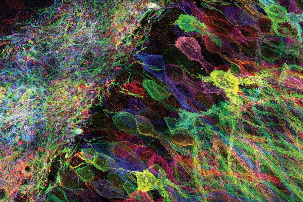
A long-standing controversy in neuroscience centers on a simple question: How do neurons in the brain share information? Sure, it’s well-known that neurons are wired together by synapses and that when one of them fires, it sends an electrical signal to other neurons connected to it. But that simple model leaves a lot of unanswered questions—for example, where exactly in neurons’ firing is information stored? Resolving these questions could help us understand the physical nature of a thought.
Two theories attempt to explain how neurons encode information: the rate code model and the temporal code model. In the rate code model, the rate at which neurons fire is the key feature. Count the number of spikes in a certain time interval, and that number gives you the information. In the temporal code model, the relative timing between firings matters more—information is stored in the specific pattern of intervals between spikes, vaguely like Morse code. But the temporal code model faces a difficult question: If a gap is “longer” or “shorter,” it has to be longer or shorter relative to something. For the temporal code model to work, the brain needs to have a kind of metronome, a steady beat to allow the gaps between firings to hold meaning.
Every computer has an internal clock to synchronize its activities across different chips. If the temporal code model is right, the brain should have something similar. Some neuroscientists posit that the clock is in the gamma rhythm, a semiregular oscillation of brain waves. But it doesn’t stay consistent. It can speed up or slow down depending on what a person experiences, such as a bright light. Such a fickle clock didn’t seem like the full story for how neurons synchronize their signals, leading to ardent disagreements in the field about whether the gamma rhythm meant anything at all.
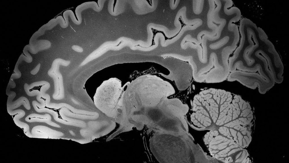
S cientists are very careful about claiming that no one else has ever done something before — the last thing they need is some overlooked lab saying, um, right here! — but researchers at Massachusetts General Hospital are confident they’re on solid ground. Their high-resolution MRIs of a complete, intact human brain, they say, are “unprecedented.”
Other labs have sliced up brains and seen features down to 80 or even 50 microns. (One micron is a 10,000th of a centimeter, and 75 of them is about the width of a human hair.) The MGH team got 100-micron resolution in a whole brain, producing the most detailed three-dimensional images of an intact brain ever seen.
The scientists started with an MRI machine with a 7-tesla magnet, a significantly stronger magnetic field than the 0.5-to-3 teslas of most MRIs in clinical use, which optimized the signal-to-noise ratio. But they also built custom state-of-the-art software that, depending which physics parameters it directs the MRI to optimize, reveals particular features of the tissue, from tiny bleeds to swelling to white and gray matter.