Toggle light / dark theme

Researchers reverse stroke damage in animal model using stem cell exosomes.


Expanding upon previous work that developed a treatment using a type of extracellular vesicles known as exosomes—small fluid-filled structures that are created by stem cells—investigators at the University of Georgia (UGA) present brain-imaging data for a new stroke treatment that supported full recovery in swine, modeled with the same pattern of neurodegeneration as seen in humans with severe stroke. Findings from this new study were published recently in Translational Stroke Research through an article titled “Neural Stem Cell Extracellular Vesicles Disrupt Midline Shift Predictive Outcomes in Porcine Ischemic Stroke Model.”

Amazingly, it’s been almost a quarter-century since the first drug was approved for stroke. Yet, what’s even more striking is that only a single drug remains approved today, so having a greater understanding of the molecular mechanisms that underlie stroke cases should lead to new therapies that could provide dramatic improvements in patient outcomes.

The researchers at UGA’s Regenerative Bioscience Center report the first observational evidence during a midline shift—when the brain is being pushed to one side—to suggest that a minimally invasive and nonoperative exosome treatment can now influence the repair and damage that follow a severe stroke.

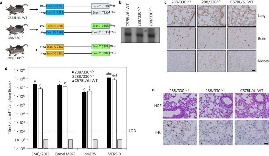
A, C57BL/6J mice were genetically engineered using CRISPR–Cas9 genomic editing to encode 288L and 330R in mDPP4 on one chromosome (heterozygous, 288/330+/−) or on both chromosomes (homozygous, 288/330+/+). b, Northern blot of mDPP4 mRNA expression. c, Immunohistochemistry (IHC) of mDPP4 protein in the lungs, brain and kidneys of individual C57BL/6J wild-type (WT), 288/330+/− and 288/330+/+ mice. d, Viral titres for MERS-CoV at 3 days post-infection from C57BL/6J WT, 288/330+/− and 288/330+/+ (all n = 4) mice infected with 5 × 10⁵ plaque-forming units (p.f.u.) of the indicated viruses. Bar graphs show means + s.d.

A molecule produced by the brain that activates the same receptors as marijuana is protective against stress by reducing anxiety-causing connections between two brain regions, Vanderbilt University Medical Center researchers report.

This finding, published today in Neuron, could help explain why some people use marijuana when they’re anxious or under stress. It could also mean that pharmacologic treatments that increase levels of this molecule, known as “2-AG,” in the brain could regulate anxiety and depressive symptoms in people with stress-related anxiety disorders, potentially avoiding a reliance on medical marijuana or similar treatments.

When mice are exposed to acute stress, a break in an anxiety-producing connection between the amygdala and the frontal cortex caused by 2-AG temporarily disappears, causing the emergence of anxiety-related behaviors.

The information-processing capabilities of the brain are often reported to reside in the trillions of connections that wire its neurons together. But over the past few decades, mounting research has quietly shifted some of the attention to individual neurons, which seem to shoulder much more computational responsibility than once seemed imaginable.

The latest in a long line of evidence comes from scientists’ discovery of a new type of electrical signal in the upper layers of the human cortex. Laboratory and modeling studies have already shown that tiny compartments in the dendritic arms of cortical neurons can each perform complicated operations in mathematical logic. But now it seems that individual dendritic compartments can also perform a particular computation — “exclusive OR” — that mathematical theorists had previously categorized as unsolvable by single-neuron systems.

“I believe that we’re just scratching the surface of what these neurons are really doing,” said Albert Gidon, a postdoctoral fellow at Humboldt University of Berlin and the first author of the paper that presented these findings in Science earlier this month.

The discovery marks a growing need for studies of the nervous system to consider the implications of individual neurons as extensive information processors. “Brains may be far more complicated than we think,” said Konrad Kording, a computational neuroscientist at the University of Pennsylvania, who did not participate in the recent work. It may also prompt some computer scientists to reappraise strategies for artificial neural networks, which have traditionally been built based on a view of neurons as simple, unintelligent switches.

The Limitations of Dumb Neurons

In the 1940s and ’50s, a picture began to dominate neuroscience: that of the “dumb” neuron, a simple integrator, a point in a network that merely summed up its inputs. Branched extensions of the cell, called dendrites, would receive thousands of signals from neighboring neurons — some excitatory, some inhibitory. In the body of the neuron, all those signals would be weighted and tallied, and if the total exceeded some threshold, the neuron fired a series of electrical pulses (action potentials) that directed the stimulation of adjacent neurons.

At around the same time, researchers realized that a single neuron could also function as a logic gate, akin to those in digital circuits (although it still isn’t clear how much the brain really computes this way when processing information). A neuron was effectively an AND gate, for instance, if it fired only after receiving some sufficient number of inputs.


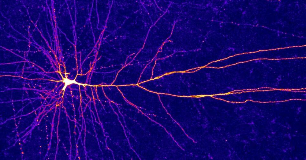
The dendritic arms of some human neurons can perform logic operations that once seemed to require whole neural networks.
https://www.usf.edu/…/neuroscientists-discover-brain-pressu…

If you found this article interesting please like and share it from our Facebook page: https://m.facebook.com/story.php?story_fbid=511500699478279&id=383136302314720


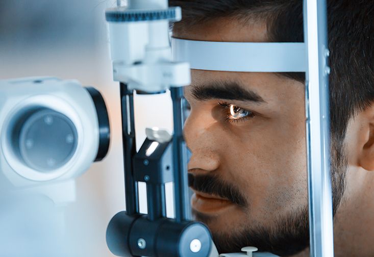
Researchers at the University of South Florida have discovered a novel feedback pathway from the brain to the eye that modulates eye pressure – a significant advancement in the effort to diagnose and treat glaucoma. Glaucoma is associated with increased pressure in the eye due to a reduce ability of the eye to maintain proper fluid drainage. The heightened pressure applies mechanical strain to the optic nerve as the nerve exits the eye, resulting in vision loss and potential blindness.

It has long been hypothesized that brain pressure might also play a role in glaucoma because the amount of strain on the optic nerve depends not just on eye pressure, but the difference in pressure between the eye and brain. The groundbreaking study published in the Journal of Physiology shows, for the first time, that eye and brain pressure are physiologically connected. The neuroscientists came to this conclusion by altering brain pressure in animal models and noting changes in the fluid drainage properties of the eye that could be blocked by chemicals that eliminate feedback signals from the brain. Interestingly, the eye’s ability to clear fluid changed in a manner that restored a healthy pressure difference across the optic nerve.

“The drainage control system may service to protect the optic nerve from swings in eye or brain pressure,” said Chris Passaglia, PhD, professor in the USF Department of Medical Engineering. “Its discovery offers a new target for glaucoma treatment, wherein the modulatory mechanisms of the system might be exploited to help lower eye pressure and impede disease progression in glaucoma patients.”

Neuroregeneration entails not only neurogenesis, but also regrowth of lost connections and birth of non-neuronal cells. While adult neurogenesis in humans is only known to occur definitively in a few precisely circumscribed regions of the brain, work in other species suggests that science has only scratched the surface of the full regenerative potential of our own nervous systems.

The serotonergic system has widely been shown to control many aspects of neuroregeneration. In some regions, it facilitates neurogenesis, while in others, it seems to inhibit it. In the case of inhibition, a recent example has been published in PLOS Biology. The authors used a zebrafish model of Alzheimer’s disease to show that amyloid-induced interleukin-4 (IL4) promotes neurogenic stem cell proliferation by suppressing the production of serotonin. In these animals, there is a unique neuro-immune interaction through which IL4 secreted by dying neurons activates microglia. In turn, microglia reciprocate by revving up neural stem cell proliferation.

“Tim Crow must be proud to see his theory being tested at a complex level.” That’s how I tweeted the news on a recent Brain article by van den Heuvel et al (2019). Tim Crow’s theory on schizophrenia as a possible by-product of human brain evolution was quite inspiring and led to many fruitful discussions in our evolutionary psychiatry group when I was a junior trainee (which I wrote about a while ago: EPSIG Newsletter, June 2018). And here it was, the theory was tested by using novel methodology. Now I am pleased to say that the article did not disappoint, so I can enjoy the initial thrill and share my take with the Mental Elf World.

Tim Crow’s original question was intriguing: “Is schizophrenia the price that Homo sapiens pay for language?” (Crow, 1997). He argued that schizophrenia may be considered an extreme variation of brain systems which are relatively new in evolutionary timescale. Brain structures that are mostly implicated in schizophrenia were also unique to humans as mediators of language and higher cognitive functions. Those relatively new (in evolutionary timescale) brain systems may be more vulnerable to insults (e.g. stress, trauma, neurodevelopmental conditions) and manifest as dysfunctional brain circuits in schizophrenia.

The prevalence of schizophrenia is fairly constant across human populations (Jablensky et al. 1992), and the prevalence does not change despite low fecundity rates of people with schizophrenia. This can only be possible in the case of overall genetic predisposition across the population.

“A new model based on the blood-vessel network in a rat brain shows that the vessel position within its circulatory network does not influence the blood flow nor how nutrients are transported. Instead, transport is controlled mostly by the dilation of vessels. As well as providing new insights into the circulatory system, the model could lead to better artificial tissues and brain-scanning techniques – and might even improve the performance of solar panels.”

Nutrient flow in the brain is controlled by blood-vessel dilation, reveals network model

If you enjoyed this article, please like and follow our Facebook page for the latest news on neuroscience, psychology, and artificial intelligence:

https://www.facebook.com/The-Neuro-Network-383136302314720/


New understanding of blood transport could lead to better solar panels.

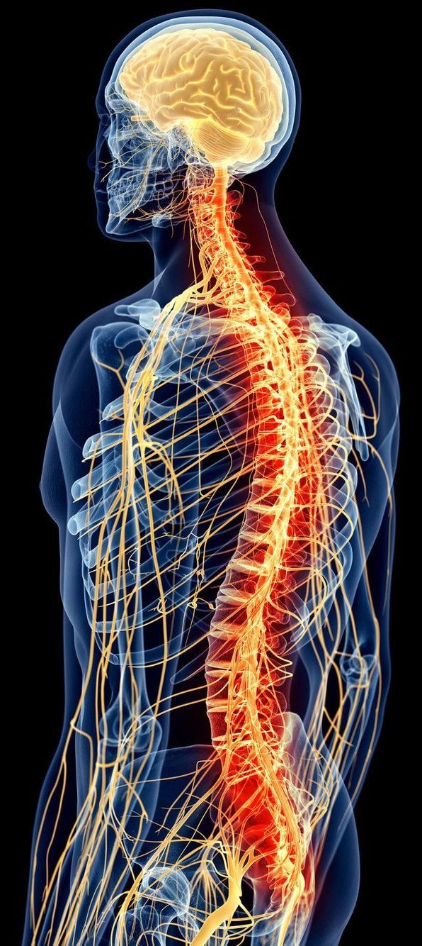
In neurological diseases such as Alzheimer’s disease, Parkinson’s disease or Multiple Sclerosis, brain neurons are constantly being lost, resulting in memory lapses, speech disorders, mood swings and movement disorders, for example, as well as muscle tremors in the case of Parkinson’s. After six years of development, MedUni Vienna researchers from the Department of Neurology (Head: Thomas Berger), led by Roland Beisteiner, have developed a new method of treatment that represents a world first. Using a non-invasive ultrasound technique, it is now possible to reach all areas of the brain and activate neurons that can help to regenerate brain functions. The preliminary data, which have been prominently published on the international stage, show that this can improve brain performance. This has positioned Vienna as a world leader in an important sector of medicine.

The new method is called transcranial pulse stimulation with ultrasound (TPS) and was developed in collaboration with Swiss commercial partner Storz Medical and its project leader, Ernst Marlinghaus. “For the first time in the world, TPS enables us to penetrate into all areas of the brain by means of an ultrasound pulse delivered directly to the skull in a non-invasive, painless procedure, during which the patient is fully conscious, and to specifically target particular areas of the brain and stimulate them,” explains Beisteiner. The study was part of the inter-university cluster led by Roland Beisteiner and Tecumseh Fitch, which is attempting to improve patients’ brain functions by means of brain stimulation and is being jointly run by MedUni Vienna and the University of Vienna. Such clinical procedures must be carried out with great precision and must be tailored to the individual patient. However, the existing electromagnetic techniques such as e.g.

The insulin pumps diabetics currently rely on do a great job of delivering the hormone as needed, but need regular replacing due to what are known as fibrils. These form over a day or two as insulin compounds accumulate into clumps and create the risk of blockages, but scientists in Australia have engineered what they say is a safer alternative, with egg yolks serving as their starting point.

The formation of fibrils means that diabetics need to replace their insulin pumps every 24 to 72 hours to avoid the risk of dangerous blockages, which bring with them a risk of life-threatening under-dosing. Beyond the dangers to the patient’s well-being, the need to regularly replace the pump increases the workload needed to manage their disease and means that portions of the medicine often go to waste.

So, there is considerable interest in developing synthetic insulin that doesn’t behave in this way. Researchers at Melbourne’s Florey Institute of Neuroscience and Mental Health approached this problem through a new technique it developed with scientists in Japan, whereby the insulin is engineered from egg yolks to allow for greater freedom over the final design.