Toggle light / dark theme

Neurological conditions or injuries that result in the inability to communicate can be devastating. Patients with such speech loss often rely on alternative communication devices that use brain–computer interfaces (BCIs) or nonverbal head or eye movements to control a cursor to spell out words. While these systems can enhance quality-of-life, they can only produce around 5–10 words per minute, far slower than the natural rate of human speech.

Researchers from the University of California San Francisco today published details of a neural decoder that can transform brain activity into intelligible synthesized speech at the rate of a fluent speaker (Nature 10.1038/s41586-019‑1119-1).

“It has been a longstanding goal of our lab to create technology to restore communication for patients with severe speech disabilities,” explains neurosurgeon Edward Chang. “We want to create technologies that can generate synthesized speech directly from human brain activity. This study provides a proof-of-principle that this is possible.”

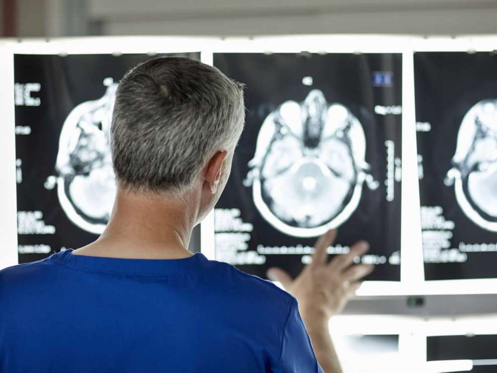
Utilizing a recently developed brain imaging technique new research suggests that measuring accumulated levels of a protein called tau may predict future neurodegeneration associated with Alzihemer’s disease. The discovery promises to accelerate clinical trial research offering a novel way to predict the progression of the disease before major symptoms appear.

Exactly what occurs in the human brain during the earliest stages of Alzheimer’s disease remains quite a mystery for dementia researchers. While studies have homed in on several pathological signs signaling moderate to severe cases of Alzheimer’s, it’s still unclear what the initial triggers for the disease are, and without this vital information scientists are struggling to generate effective drugs and treatments to slow or prevent the disease.

The two big pathological signs of Alzheimer’s most researchers agree on are accumulations of amyloid and tau proteins in the brain. Abnormal aggregations of amyloid proteins, into what are referred to as plaques, are generally considered to be the primary causative mechanism behind Alzheimer’s. Masses of misfolding tau proteins, forming what are known as neurofibrillary tangles, are also seen in the disease.

Brain organoids are made from human pluripotent stem cells, which are cells that can become any kind of cell in the adult body. When the stem cells are introduced to certain chemicals, they can be coaxed into becoming brain cells, then put into a liquid with the nutrients they need to survive.

“The amazing thing is that, after this, they pretty much do everything alone,” says Alysson Muotri, a molecular biologist at UC San Diego. The cells self-assemble into spheres that contain neural progenitor cells, or cells that will become brain cells. Over the course of a few weeks, those cells turn into different kinds of neurons that can act just like neurons in the human brain.

In a study preprint published on bioRxiv and presented at the Society for Neuroscience conference last month, Muotri and his colleagues reported that they recorded spontaneous and complex electrical activity from their lab-grown mini brains. It’s the first time that brain organoids have spontaneously produced brain waves similar to human brain activity, Nature reported.

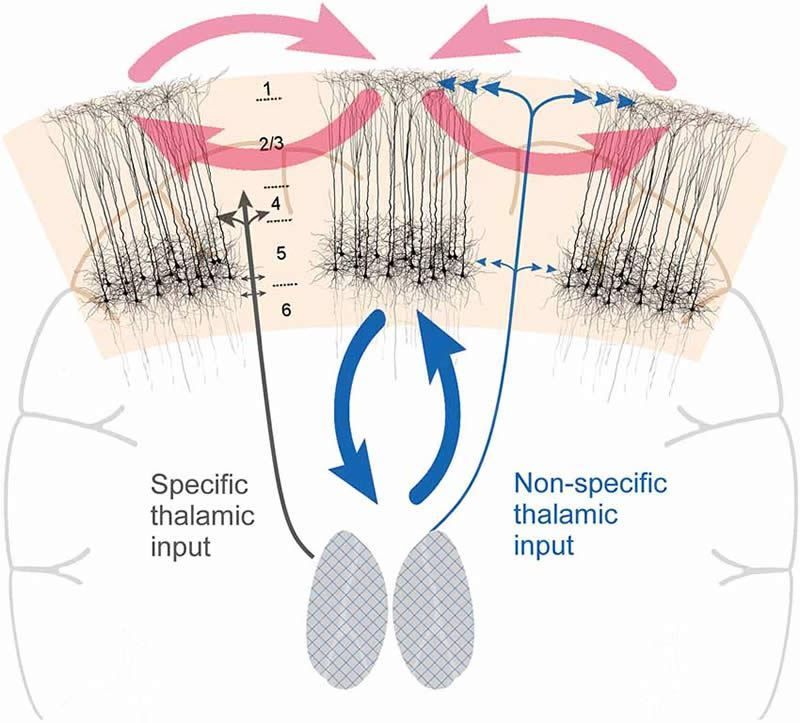
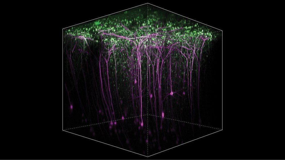
Researchers believe they have identified specific neurons that are responsible for conscious awareness. Previous studies have implicated both thalamocortical circuits and cortico-cortico circuits in consciousness. The new study reports these networks intersect via L5p neurons. Directly activating L5p neurons made mice react to weaker sensory stimuli. The researchers say if consciousness requires L5p neurons, all brain activity without them must be unconscious.

Researchers at Johns Hopkins Medicine have successfully used a laser-assisted imaging tool to “see” what happens in brain cells of mice learning to reach out and grab a pellet of food. Their experiments, they say, add to evidence that such motor-based learning can occur in multiple areas of the brain, even ones not typically associated with motor control.

“Scientists should be looking at the entire brain to understand specific types of learning,” says Richard Huganir, Ph.D., Bloomberg Distinguished Professor and Director of the Solomon H. Snyder Department of Neuroscience at the Johns Hopkins University School of Medicine. “Different parts of the brain contribute to learning in different ways, and studying brain cell receptors can help us decipher how this works.”

The work, say the researchers, may ultimately inform efforts to develop treatments for learning-based and neurocognitive disorders.

Scientists have uncovered a new kind of electrical process in the human brain that could play a key role in the unique way our brains compute.

Our brains are computers that work using a system of connected brain cells, called neurons, that exchange information using chemical and electric signals called action potentials. Researchers have discovered that certain cells in the human cortex, the outer layer of the brain, transmit signals in a way not seen in corresponding rodent cells. This process might be important to better understanding our unique brains and to improving programs that are based on a model of the human brain.

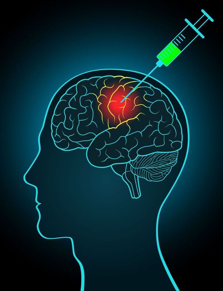
A newly published study has described the successful results in mice of a novel vaccine designed to prevent neurodegeneration associated with Alzheimer’s disease. The researchers suggest this “dementia vaccine” is now ready for human trials, and if successful could become the “breakthrough of the next decade.”

The new study, led by the Institute for Molecular Medicine and University of California, Irvine, describes the effect of a vaccine designed to generate antibodies that both prevent, and remove, the aggregation of amyloid and tau proteins in the brain. The accumulation of these two proteins is thought to be the primary pathological cause of neurodegeneration associated with Alzheimer’s disease.

The research revealed the vaccine led to significant decreases in both tau and amyloid accumulation in the brains of bigenic mice engineered to exhibit aggregations of these toxic proteins. Many prior failed Alzheimer’s treatments over the past few years have focused individually on either amyloid or tau protein reductions, but growing evidence suggests a synergistic relationship between the two toxic proteins may be driving neurodegeneration. Hence the hypothesis a combination therapy may be the most effective way to prevent this kind of dementia.