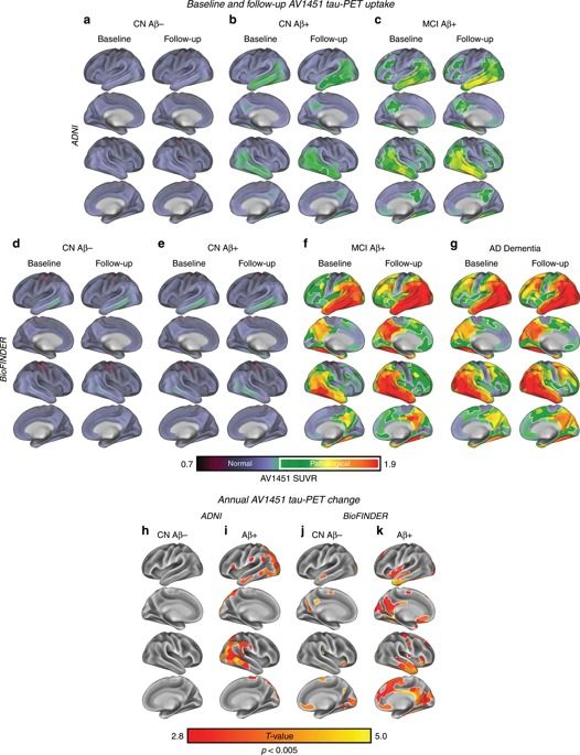
Tau accumulation is associated with disease progression in Alzheimer’s disease. Here the authors use resting state fMRI and tau-PET to demonstrate that baseline connectivity in Alzheimer’s disease is associated with tau spreading.


This article is reprinted by permission from NextAvenue.org.
During the last Alzheimer’s disease support meeting I attended at my mother’s assisted living center, I sheepishly asked if anyone else was worried about their own risk for the disease.
A lot of hands went up.
Humans might not be so special after all.
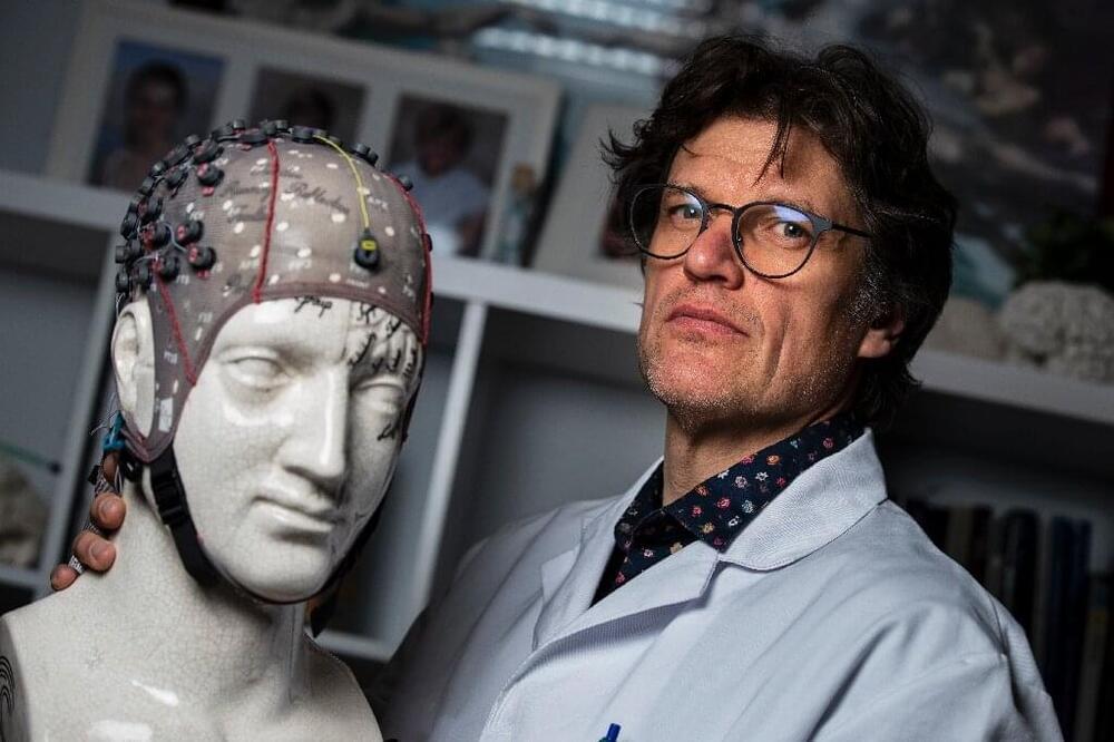
Not all patients who fall into a coma return, and when they do it can mark a moment of joy for their loved ones—but their troubles are rarely over.
Often, brain damage leaves them paralysed or unable to communicate.
Belgian neurologist Steven Laureys has dedicated himself to the question of how to improve the lives of the formerly comatose, and of their families.

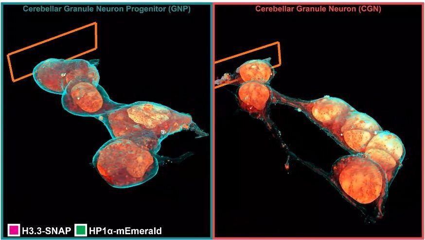
Inside a cell, tentacled vesicles shuttle cargo for sorting. DNA rearranges in the nucleus as stem cells differentiate into neurons. Neighboring neurons cling to one another through a web-like interface. And a new microscopy technique shows it all, in exquisite detail.
The technique, called cryo-SR/EM, melds images captured from electron microscopes and super-resolution light microscopes, resulting in brilliant, clear detailed views of the inside of cells—in 3D.
For years, scientists have probed the microscopic world inside cells, developing new tools to view these basic units of life. But each tool comes with a tradeoff. Light microscopy makes it simple to identify specific cellular structures by tagging them with easy-to-see fluorescent molecules. With the development of super-resolution (SR) fluorescence microscopy, these structures can be viewed with even greater clarity. But fluorescence can reveal only a few of the more than 10,000 proteins in a cell at a given time, making it difficult to understand how these few relate to everything else. Electron microscopy (EM), on the other hand, reveals all cellular structures in high-resolution pictures—but delineating one feature from all others by EM alone can be difficult because the space inside of cells is so crowded.


Boston, Mass. — Interoception is the awareness of our physiological states; it’s how animals and humans know they’re hungry or thirsty, and how they know when they’ve had enough to eat or drink. But precisely how the brain estimates the state of the body and reacts to it remains unclear. In a paper published in the journal Neuron, neuroscientists at Beth Israel Deaconess Medical Center (BIDMC) shed new light on the process, demonstrating that a region of the brain called the insular cortex orchestrates how signals from the body are interpreted and acted upon. The work represents the first steps toward understanding the neural basis of interoception, which could in turn allow researchers to address key questions in eating disorders, obesity, drug addiction, and a host of other diseases.
Using a mouse model his lab developed at BIDMC, Mark Andermann, PhD, principal investigator in the Division of Endocrinology, Diabetes and Metabolism at BIDMC and Associate Professor of Medicine at Harvard Medical School, and colleagues recorded the activity of hundreds of individual brain cells in the insular cortex to determine exactly what is happening as hungry animals ate.
The team observed that when mice hadn’t eaten for many hours, the activity pattern of insular cortex neurons reflected current levels of hunger. As the mice ate, this pattern gradually shifted over hours to a new pattern reflecting satiety. When mice were shown a visual cue predicting impending availability of food — akin to a person seeing a food commercial or a restaurant logo — the insular cortex appeared to simulate the future sated state for a few seconds, and then returned to an activity pattern related to hunger. These findings provided direct support for studies in humans that hypothesized that the insular cortex is involved in imagining or predicting how we will feel after eating or drinking.