Toggle light / dark theme

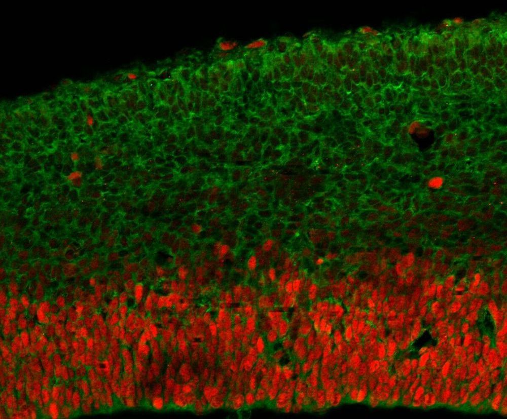
An international team of researchers that pooled genetic samples from developmentally disabled patients from around the world has identified dozens of new mutations in a single gene that appears to be critical for brain development.

“This is important because there are a handful of that are recognized as ‘hot spots’ for causing ,” said lead author Debra Silver, an associate professor of molecular genetics and microbiology in the Duke School of Medicine. “This gene, DDX3X, is going to be added to that list now.”

An analysis led by the Elliott Sherr lab at the University of California-San Francisco found that half of the DDX3X mutations in the 107 children studied caused a loss of function that made the gene stop working altogether, but the other half caused changes predicted to disrupt the function of the gene.
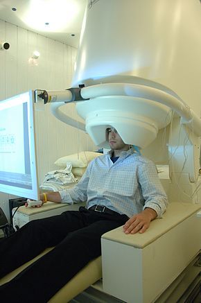
In a recent study, scientists at Baycrest’s Rotman Research Institute (RRI) found that research participants moved their eyes to determine whether they had seen an image before, and that their eye movement patterns could predict mistakes in memory. They obtained these results using an innovative new eye tracking technique they developed.

“Our findings indicate that eye movements play a functional role in retrieval,” says Dr. Jennifer Ryan, senior scientist at the RRI and Canada Research Chair in Cognitive Neuroscience of Memory. “They can tell us a lot about someone’s memory.”

This study builds on previous Baycrest research examining the link between eye movements and memory, including the role of our eye movements in memorization and the weakening connection between our eye movements and our brain activity as we age.

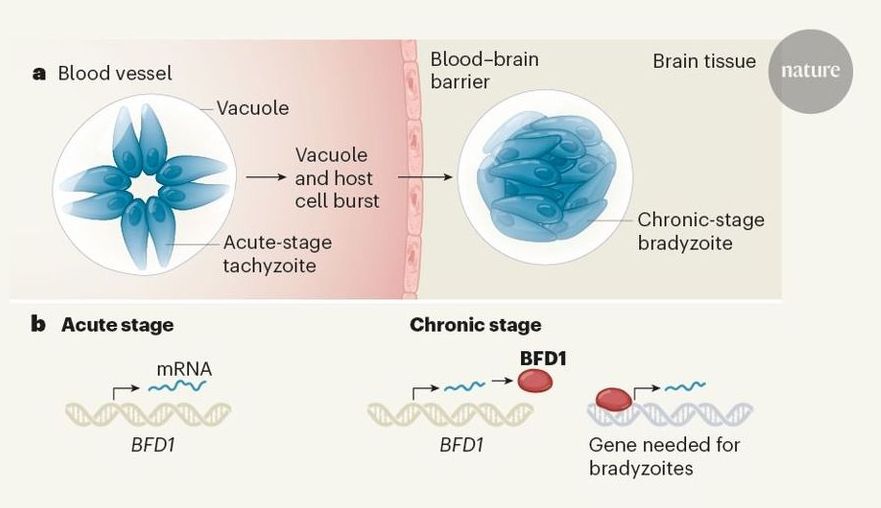
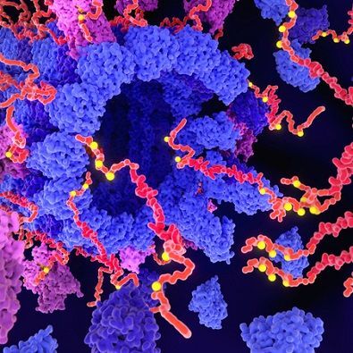
A gene has been found that controls the conversion of the parasite Toxoplasma gondii into a form that chronically infects the human brain. The discovery could aid the design of therapies to eliminate this currently untreatable infection. A transcription factor controls Toxoplasma gondii differentiation.

CRISPR Used To Edit Genes Inside A Patient With A Rare Form Of Blindness : Shots — Health News Doctors used CRISPR to edit genes of cells inside a patient’s eye, hoping to restore vision to a person blinded by a rare genetic disorder. A similar strategy might work for some brain diseases.

My most recent post, “Living in a Computer Simulation,” elicited some insightful comments from a reader skeptical of the possibility of mind uploading. Here is his argument with my own brief response to it below.

My comment concerns a reductive physicalist theory of the mind, which is the view that all mental states and properties of the mind will eventually be explained by scientific accounts of physiological processes and states … Basically, my argument is that for this view of the mind, mind uploading into a computer is completely impractical due to accumulation of errors.

In order to replicate the functioning of a “specific” human mind within a computer, one needs to replicate the functioning of all parts of that specific brain within the computer. [In fact, the whole human body needs to be represented because the mind is a product of all sensations of all parts of the body coalescing within the brain. But, for the sake of argument, let’s just consider replicating only the brain.] In order to represent a specific human brain in the computer, each neuron in the brain would need a digital or analog representation, instantiated in hardware, software or a combination of the two. Unless this representation is an exact biological copy (clone), it will have some inherent “error” associated with it. So, let’s do a sort of “error analysis” (admittedly non-rigorous).

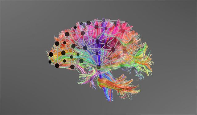
Researchers in Europe and the UK have managed to connect biological and artificial neurons together – and allow them to communicate long distances through the internet. The biological neurons were grown in one country, sent signals through an artificial synapse located in another to electronic neurons in a third country.

As advanced as supercomputers get, the human brain still utterly leaves them in the dust. It’s made up of neurons that communicate with each other through pulses of electrical signals, passed across tiny gaps known as synapses. These neurons can both process and store information, unlike computers that require separate types of memory for each task.

Artificial versions of neurons and synapses have shown to be far more powerful than traditional computer chip designs, but they’re still in the experimental stage. And now, a team of researchers has taken the next step and connected the artificial and biological versions between three different countries.

What happens to people who suffer severe injuries that make it impossible for them to communicate? They are often left at the mercy of doctors and families who are obligated to make vital decisions for them. According to New Scientist, however, now there are new mind-reading brain scanners that may remedy this situation.


The new scanners use functional near-infrared spectroscopy.

Researchers report an advance in the development of a blood test that could help detect pathological Alzheimer’s disease in people who are showing signs of dementia. This approach could be less invasive and less costly than current brain imaging and spinal fluid tests. The blood test detects the abnormal accumulation of a form of tau protein known as phosphorylated-tau-181 (ptau181), which is a biomarker that suggests brain changes from Alzheimer’s. The study, funded by the National Institutes of Health, was published on March 2 in Nature Medicine.

Over the past 15 years, research advances in the development of biomarkers like tau protein have enabled investigators to more accurately diagnose Alzheimer’s disease, select research participants, and measure response to investigational therapies. Tau and other biomarkers can be detected with PET scans of the brain and lab tests of spinal fluid. However, PET imaging is expensive and involves radioactive agents, and spinal fluid tests require spinal taps, which are invasive, complex and time-consuming. Simpler biomarker tests are still needed.

“The considerable time and resources required for screening research participants with PET scans and spinal taps slow the pace of enrollment for Alzheimer’s disease treatment studies,” said Richard J. Hodes, M.D., director of NIH’s National Institute on Aging (NIA), which funded much of the study. “The development of a blood test would enable us to rapidly screen a much larger and more diverse group of volunteers who wish to enroll in studies.”