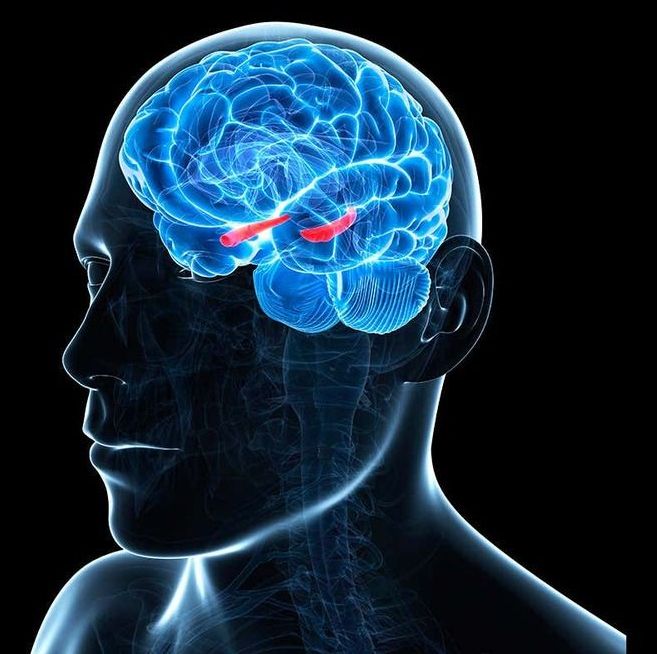
By Emma Young. Researchers recorded participants’ brain activity and emotions while they watched Forrest Gump.

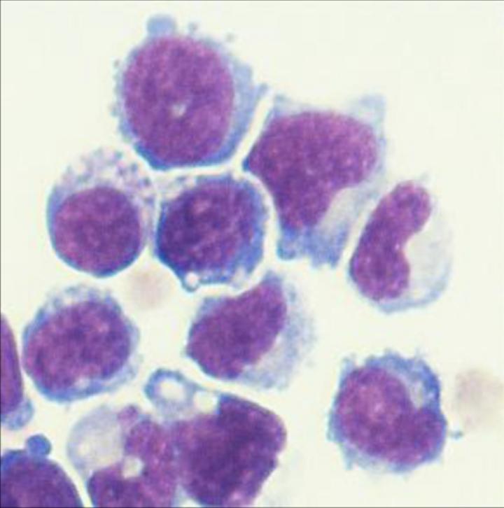
Immunotherapy is an increasingly powerful form of cancer treatment where the patient’s own immune system is equipped with heightened abilities to take down the disease, and one promising arm of this is known as adoptive cell therapy. This involves using altered versions of a patient’s own cells to trigger a more strong-handed response from their own immune system. Scientists at Johns Hopkins Kimmel Cancer Center are reporting an exciting advance in this area, demonstrating that engineered bone marrow cells can slow the growth of prostate and pancreatic cancers in mice.
The study builds on previous research where scientists demonstrated that a range of cancers, including melanomas, colon cancer and brain cancer, grow much more slowly in mice that are lacking a certain gene, known as p50, which seems to activate a stronger immune response. The Johns Hopkins researchers sought to further validate these earlier findings, while expanding the utility of a promising form of cancer therapy.
To do this, the team worked with what are known as immature myeloid cells, a type of white blood cell, which previous research had indicated could help switch on immune responses that fight tumors. In this case, the immature myeloid cells were taken from the bone marrow of mice engineered to lack the p50 gene, as a way of comparing them to the behavior of cells taken from mice who had the p50 gene in tact.

In a study of epilepsy patients, researchers at the National Institutes of Health monitored the electrical activity of thousands of individual brain cells, called neurons, as patients took memory tests. They found that the firing patterns of the cells that occurred when patients learned a word pair were replayed fractions of a second before they successfully remembered the pair. The study was part of an NIH Clinical Center trial for patients with drug-resistant epilepsy whose seizures cannot be controlled with drugs.
“Memory plays a crucial role in our lives. Just as musical notes are recorded as grooves on a record, it appears that our brains store memories in neural firing patterns that can be replayed over and over again,” said Kareem Zaghloul, M.D., Ph.D., a neurosurgeon-researcher at the NIH’s National Institute of Neurological Disorders and Stroke (NINDS) and senior author of the study published in Science.
Dr. Zaghloul’s team has been recording electrical currents of drug-resistant epilepsy patients temporarily living with surgically implanted electrodes designed to monitor brain activity in the hopes of identifying the source of a patient’s seizures. This period also provides an opportunity to study neural activity during memory. In this study, his team examined the activity used to store memories of our past experiences, which scientists call episodic memories.
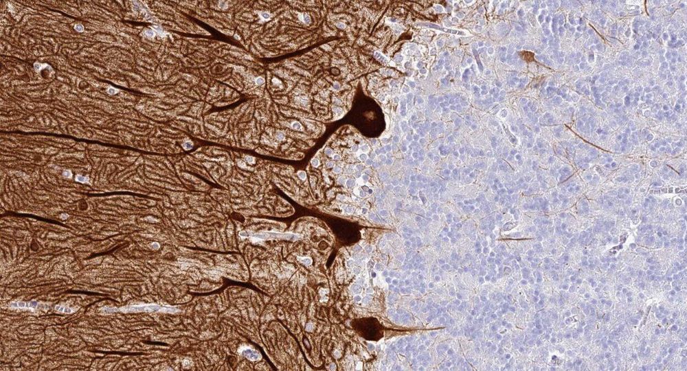
An international team of scientists led by researchers at Karolinska Institutet in Sweden has launched a comprehensive overview of all proteins expressed in the brain, published today in the journal Science. The open-access database offers medical researchers an unprecedented resource to deepen their understanding of neurobiology and develop new, more effective therapies and diagnostics targeting psychiatric and neurological diseases.
The brain is the most complex organ, both in structure and function. The new Brain Atlas resource is based on the analysis of nearly 1,900 brain samples covering 27 brain regions, combining data from the human brain with corresponding information from the brains of the pig and mouse. It is the latest database released by the Human Protein Atlas (HPA) program which is based at the Science for Life Laboratory (SciLifeLab) in Sweden, a joint research centre aligned with KTH Royal Institute of Technology, Karolinska Institutet, Stockholm University and Uppsala University. The project is a collaboration with the BGI research centre in Shenzhen and Qingdao in China and Aarhus University in Denmark.
“As expected, the blueprint for the brain is shared among mammals, but the new map also reveals interesting differences between human, pig and mouse brains,” says Mathias Uhlén, Professor at the Department of Protein Science at KTH Royal Institute of Technology, Visiting professor at the Department of Neuroscience at Karolinska Institutet and Director of the Human Protein Atlas effort.

A new joint study by Tel Aviv University (TAU) and Weizmann Institute of Science researchers has yielded an innovative method for bolstering memory processes in the brain during sleep.
The method relies on a memory-evoking scent administered to one nostril. It helps researchers understand how sleep aids memory, and in the future could possibly help to restore memory capabilities following brain injuries, or help treat people with post–traumatic stress disorder (PTSD) for whom memory often serves as a trigger.
The new study was led by Ella Bar, a Ph.D. student at TAU and the Weizmann Institute of Science. Other principal investigators include Prof. Yuval Nir of TAU’s Sackler Faculty of Medicine and Sagol School of Neuroscience, as well as Profs. Yadin Dudai, Noam Sobel and Rony Paz, all of Weizmann’s Department of Neurobiology. It was published in Current Biology on March 5.

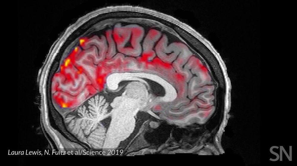
Getting plenty of deep, restful sleep is essential for our physical and mental health. Now comes word of yet another way that sleep is good for us: it triggers rhythmic waves of blood and cerebrospinal fluid (CSF) that appear to function much like a washing machine’s rinse cycle, which may help to clear the brain of toxic waste on a regular basis.
The video above uses functional magnetic resonance imaging (fMRI) to take you inside a person’s brain to see this newly discovered rinse cycle in action. First, you see a wave of blood flow (red, yellow) that’s closely tied to an underlying slow-wave of electrical activity (not visible). As the blood recedes, CSF (blue) increases and then drops back again. Then, the cycle—lasting about 20 seconds—starts over again.
The findings, published recently in the journal Science, are the first to suggest that the brain’s well-known ebb and flow of blood and electrical activity during sleep may also trigger cleansing waves of blood and CSF. While the experiments were conducted in healthy adults, further study of this phenomenon may help explain why poor sleep or loss of sleep has previously been associated with the spread of toxic proteins and worsening memory loss in people with Alzheimer’s disease.
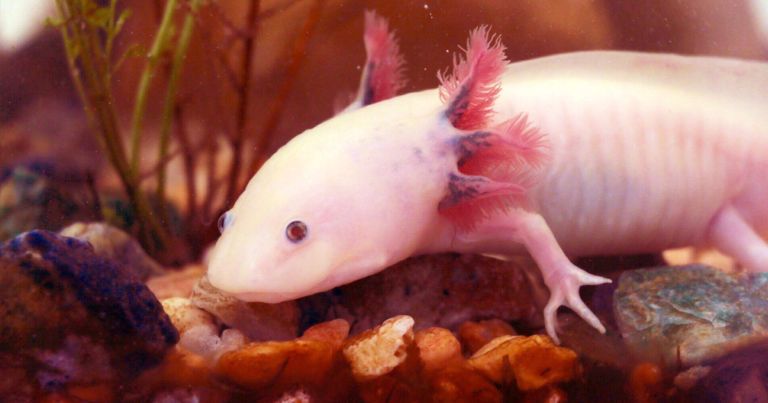
“It’s hard to find a body part they can’t regenerate: the limbs, the tail, the spinal cord, the eye, and in some species, the lens, even half of their brain has been shown to regenerate,” Kentucky researcher Randal Voss said in the release.
“Just a few years ago, no one thought it possible to assemble a 30+GB genome,” said Kentucky biologist Jeramiah Smith. “We have now shown it is possible using a cost effective and accessible method, which opens up the possibility of routinely sequencing other animals with large genomes.”
With that capability, the team hopes to begin probing the full DNA sequence for insights into the axolotl’s regenerative abilities.
“Now that we have access to genomic information, we can really start to probe axolotl gene functions and learn how they are able to regenerate body parts,” said Voss. “Hopefully someday we can translate this information to human therapy, with potential applications for spinal cord injury, stroke, joint repair… the sky’s the limit, really.”
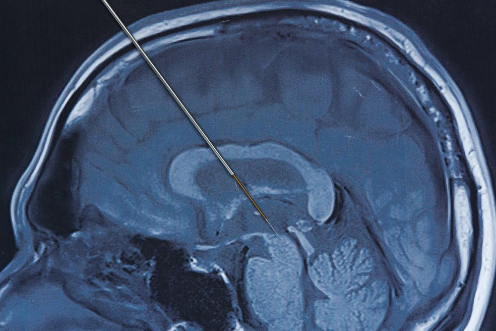
The patients always knew that when he stimulated their arm, it was him doing it, not them. And when they stimulated their arm, they were doing it, not him. So Penfield said, he couldn’t stimulate the will. He could never trick the patients into thinking it was them doing it. He said, the patients always retained a correct sense of agency. They always know if they did it or if he did it.
So he said the will was not something he could stimulate, meaning it was not material.
So he had three lines of evidence: His inability to stimulate intellectual thought, the inability of seizures to cause intellectual thought, and his inability to stimulate the will. … So he concluded that the intellect and the will are not from the brain. Which is precisely what Aristotle said.
