Toggle light / dark theme

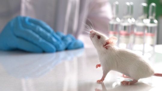
The team compared germ-free (sterile) mice and mice with normal microbes. They used a laboratory technique called mass spectrometry to characterize the non-living molecules in every mouse organ. They identified as many molecules as possible by comparing them to reference structures in the GNPS database, a crowdsourced mass spectrometry repository developed by Dorrestein and collaborators. They also determined which living microbes co-locate with these molecules by sequencing a specific genetic region that acts as a barcode for bacterial types.

In total, they analyzed 768 samples from 96 sites of 29 different organs from four germ-free mice and four mice with normal microbes. The result was a map of all of the molecules found throughout the body of a normal mouse with microbes, and a map of molecules throughout a mouse without microbes.

A comparison of the maps revealed that as much as 70 percent of a mouse’s gut chemistry is determined by its gut microbiome. Even in distant organs, such as the uterus or the brain, approximately 20 percent of molecules were different in the mice with gut microbes.
Drug overdoses can be life threatening, but for two women who accidentally took massive hits of LSD the experience was life changing — and in a good way.

A 46-year-old woman snorted a staggering 550 times the normal recreational dose of LSD and not only survived, but found that the foot pain she had suffered from since her 20s was dramatically reduced.

Separately, a 15-year-old girl with bipolar disorder overdosed on 10 times the normal dose of the drug, which she said resulted in a massive improvement in her mental health.

Researchers in the UK claim to have developed a microfluidic chip that can rapidly tell whether someone has suffered a traumatic brain injury from a finger-prick blood sample. The optofluidic device detects a biomarker linked to brain injury, based on the way that it scatters light (Nat. Biomed. Eng. 10.1038/s41551-019‑0510-4).


An optofluidic device uses Raman spectroscopy to detect a biomarker in blood associated with traumatic brain injury.

One of the most remarkable characteristics of the vertebrate eye is its retina. Surprisingly, the sensitive portions of the photoreceptor cells are found on the hind side of the retina, meaning that light needs to travel through living neural tissue before it can be detected. While the origin of the high optical quality of the retina remain largely uninvestigated, it has long been proposed that a peculiar DNA organization would serve to improve vision in nocturnal mammals. Researchers at the Max Planck Institute of Molecular Cell Biology and Genetics in Dresden now showed that the optical quality of the mouse retina increases in the first month after birth that imparts improved visual sensitivity under low light conditions. This improvement is caused by a compact organization of the genetic material in the cell nucleus of rod photoreceptor cells that responsible for dim light vision.

Our retina is an amazing feature of the eye of vertebrates. This -sensitive layer of is lining the back of the eye-ball and acts as a screen for images projected by the lens. The retina has a thickness of 130 to 500 micrometer and is composed of five layers of dense . Since the sensitive portions of the photoreceptor cells are found on the hind side of the retina, light needs to travel through this dense neural tissue to reach the photoreceptors. Researchers suggested that a certain compact arrangement of DNA in the cell nucleus of the rod photoreceptors could improve night vision in nocturnal animals but it remained unclear if and how would benefit from this organization of genetic material.

Scientists around the research group leader Moritz Kreysing at the Max Planck Institute of Molecular Cell Biology and Genetics together with colleagues from the TU Dresden and the Biozentrum at the Ludwig Maximilians Universität in Munich wanted to find out, if and why cells of retinal neural cells are optically special and what the implications for the transparency of the retina are. Transparency in this context means that each rod cell scatters less light, which causes it to be more transparent.

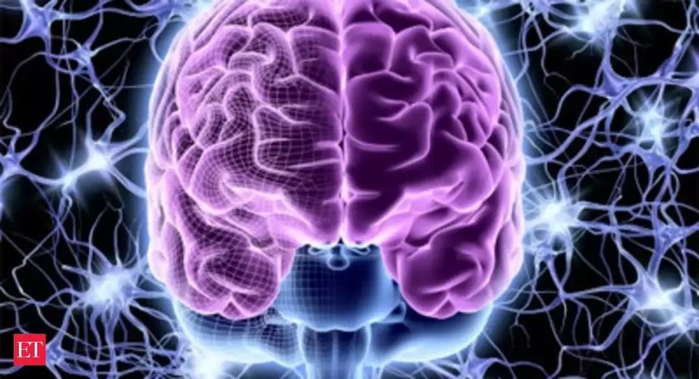
WASHINGTON: Scientists have found that people who have a variant of a longevity gene have improved brain skills such as thinking, learning and memory. Researchers found that increasing levels of the gene, called KLOTHO, in mice made them smarter, possibly by increasing the strength of connections between nerve cells in the brain.

“This could be a major step toward helping millions around the world who are suffering from Alzheimer’s disease and other dementias,” said Dena Dubal, an assistant professor of neurology, the David A Coulter Endowed Chair in Aging and Neurodegeneration at the University of California San Francisco (UCSF) and the lead author of the study published in Cell Reports. “If we could boost the brain’s ability to function, we may be able to counter dementias,” Dubal said.

People who have one copy of a variant, or form, of the KLOTHO gene, called KL-VS, tend to live longer and have lower chances of suffering a stroke whereas people who have two copies may live shorter lives and have a higher risk of stroke. In the study, researchers found that people who had one copy of the KL-VS variant performed better on a battery of cognitive tests than subjects who did not have it, regardless of age, sex or the presence of the apolipoprotein 4 gene, the main genetic risk factor for Alzheimer’s disease.

A bionic revolution is brewing, as recent advancements in bioengineering have brought about scientific breakthroughs in rehabilitation for people with disabilities. The most cutting edge research is happening inside the human brain, where implanted technology allows people to communicate directly with computers, using their thoughts.

VICE’s Wilbert L. Cooper travels to Zurich to see the first-ever bionic Olympics and discovers a host of technologies that are expanding what it means to be human.

Check out VICE News for more: http://vicenews.com
Follow VICE News here:
Facebook: https://www.facebook.com/vicenews
Twitter: https://twitter.com/vicenews
Tumblr: http://vicenews.tumblr.com/
Instagram: http://instagram.com/vicenews
More videos from the VICE network: https://www.fb.com/vicevideo
#VICEonHBO

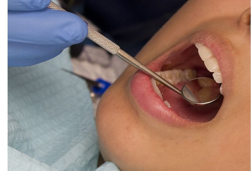
Atherosclerosis progresses is driven by chronic inflammation, the more sources of low-grade, smoldering inflammation, the faster atherosclerosis could develop. Gum disease is one such source of inflammation and is associated with a higher incidence of strokes.


Patients with gum disease were three times as likely to have a stroke involving blood vessels in the back of the brain, which controls vision, coordination and other vital bodily functions; and.

Built on decades of previous research, a team from the University of Wisconsin-Madison teased out a tiny chunk of brain tissue within the thalamus, a nub above the brain stem, as a critical part of NCC. As proof of concept, they gave it several bouts of electrical shocks, and restored awareness in unconscious monkeys under heavy anesthesia.

The crux? As soon as the electrical stimulation stopped, the monkeys’ awareness also slipped away.

Although the thalamus has long been thought of as somehow involved in supporting consciousness, the study is one of the first to pinpoint exact neural circuits—highways between the thalamus and parts of the cortex—as “switches” for consciousness that we can control using brain stimulation. And that’s wonderful news for comatose patients.