Toggle light / dark theme

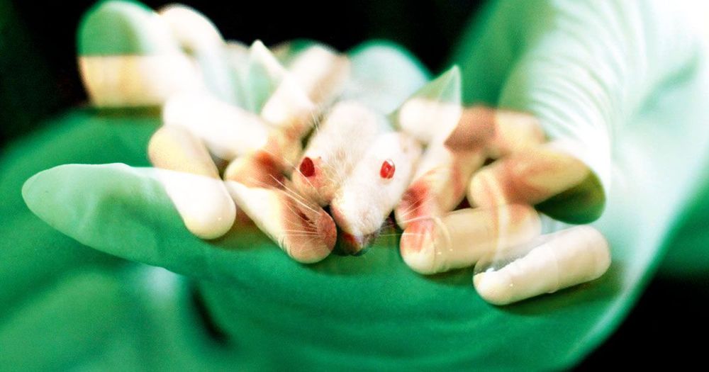
To treat the mice, the team gave them brain implants: a fiber optic that shined light onto a region called the paraventricular thalamus and blocked withdrawal symptoms. A day later, the mice no longer sought out morphine and relapse — or at least do the lab mouse version of relapsing — even after two weeks.

According to the new research, published Thursday in the journal Neuron, people relapse partially because they miss the high, but more so because the symptoms of withdrawal can often be overwhelming. By down those symptoms, the mice appear to be able to kick the habit more easily.

“Our success in preventing relapse in rodents may one day translate to an enduring treatment of opioid addiction in people,” CAS researcher Zhu Yingjie said in a press release.
We are highly sensitive to people around us. As infants, we observe our parents and teachers, and from them we learn how to walk, talk, read—and use smartphones. There seems to be no limit to the complexity of behavior we can acquire from observational learning.

But social influence goes deeper than that. We don’t just copy the behavior of people around us. We also copy their minds. As we grow older, we learn what other people think, feel, and want—and adapt to it. Our brains are really good at this—we copy computations inside the brains of others. But how does the brain distinguish between thoughts about your own mind and thoughts about the minds of others? Our new study, published in Nature Communications, brings us closer to an answer.

Our ability to copy the minds of others is hugely important. When this process goes wrong, it can contribute to various mental health problems. You might become unable to empathize with someone, or, at the other extreme, you might be so susceptible to other people’s thoughts that your own sense of “self” is volatile and fragile.

A team of researchers with interdisciplinary expertise in psychology, informatics (the application of information science to solve problems with data) and engineering along with the Vanderbilt Brain Institute (VBI) gained critical insights into one of the biggest mysteries in neuroscience, identifying the location and critical nature of these neurons.”


New research on cognitive flexibility points to a small class of brain cells that support switching attention strategies when old strategies fail.

There are 86 billion neurons, or cells, in the human brain. Of these, an infinitely small portion of them handle cognitive flexibility—our ability to adjust to new environments and concepts.

A team of researchers with interdisciplinary expertise in psychology, informatics (the application of information science to solve problems with data) and engineering along with the Vanderbilt Brain Institute (VBI) gained critical insights into one of the biggest mysteries in neuroscience, identifying the location and critical nature of these neurons.

The article was published in the journal Proceedings of the National Academy of Science (PNAS) on July 13. The discovery presents an opportunity to enhance researchers’ understanding and treatment of mental illnesses rooted in cognitive flexibility.

Glioblastoma is the most aggressive type of cancer that begins with the brain and develops from astrocytes, star-shaped brain cells that help protect the brain from diseases in the blood and provide the brain’s neurons with nutrients, with around 12,000 cases diagnosed in the United States each year. Glioblastoma cells have more genetic abnormalities than the cells of other types of astrocytoma brain cancer. Now researchers from the University of Virginia (UVA) School of Medicine report they have identified an oncogene responsible for this deadly cancer.

Their study, “A cytoskeleton regulator AVIL drives tumorigenesis in glioblastoma,” is published in Nature Communications and led by Hui Li, PhD, associate professor, pathology, at the University of Virginia School of Medicine and the UVA Cancer Center.

“Glioblastoma is a deadly cancer, with no effective therapies. Better understanding and identification of selective targets are urgently needed. We found that advillin (AVIL) is overexpressed in all the glioblastomas we tested including glioblastoma stem/initiating cells, but hardly detectable in non-neoplastic astrocytes, neural stem cells or normal brain,” the researchers wrote.

Animals have an innate preference for certain scents and tastes. Attractive scents are linked to things like good food. Less attractive scents—that of spoiled food, for example—instinctively give the animal a signal which says: “There could be danger here!” When it comes to taste, all animals have similar preferences: Sugars and fats are perceived positively, whereas a bitter taste is perceived rather negatively.

In order to be able to make such evaluations, we need signals in the that tell us “This is good” or “This is bad.” The in the brain, better known as the reward system, plays an important role in these evaluations.

Groups of neurons in the human brain produce patterns of activity that represent information about the stimuli that one is perceiving and then convey these patterns to different brain regions via nerve cell junctions known as synapses. So far, most neuroscience studies have focused on the two primary components of neuron information processing individually (i.e., the representation of stimuli in the form of neural activity and the transmission of this information in networks that model neural interactions), rather than exploring them together.

A team of researchers at the University of Pennsylvania recently reviewed literature investigating each of these two components, in order to develop a holistic framework that better describes how groups of neurons process information. Their paper, published in Nature Neuroscience, introduces a holistic theoretical perspective that could inform future neuroscience research focusing on neural information processing.

“In the past decade or so, neuroscientists have used more sophisticated tools to understand how the represents things that it sees or hears in its environment,” Harang Ju and Danielle Bassett, the two researchers who carried out the study, told Medical Xpress. “Some researchers studied brain representations as single patterns of brain activity, while others studied representations as changing patterns of activity. The aim of our paper was to explore how understanding the brain as a of neural units and their connections could frame the recent developments in a way that helps push the field towards a better understanding of the dynamic nature of neural representations.”

Queen’s University researchers uncover brain-based marker of new thoughts and discover we have more than 6,000 thoughts each day.

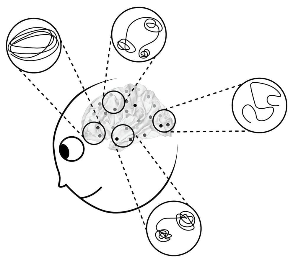
Researchers at Queen’s University have established a method that, for the first time, can detect indirectly when one thought ends and another begins. Dr. Jordan Poppenk (Psychology) and his master’s student, Julie Tseng, devised a way to isolate “thought worms,” consisting of consecutive moments when a person is focused on the same idea. This research was recently published in Nature Communications.

“What we call thought worms are adjacent points in a simplified representation of activity patterns in the brain. The brain occupies a different point in this ‘state space’ at every moment. When a person moves onto a new thought, they create a new thought worm that we can detect with our methods,” explains Dr. Poppenk, who is the Canada Research Chair in Cognitive Neuroscience. “We also noticed that thought worms emerge right as new events do when people are watching movies. Drilling into this helped us validate the idea that the appearance of a new thought worm corresponds to a thought transition.”

Scientists from Trinity College Dublin have discovered a new link between impaired brain energy metabolism and delirium—a disorienting and distressing disorder particularly common in the elderly and one that is currently occurring in a large proportion of patients hospitalized with COVID-19 [15th of July 2020].

While much of the research was conducted in mice, additional work suggests overlapping mechanisms are at play in humans because cerebrospinal fluid (CSF) collected from patients suffering from delirium also contained tell-tale markers of altered brain glucose .

Collectively, the research, which has just been published in the Journal of Neuroscience, suggests that therapies focusing on brain energy metabolism may offer new routes to mitigating delirium.