Toggle light / dark theme

New research, based on earlier results in mice, suggests that our brains are never at rest, even when we are not learning anything about the world around us.

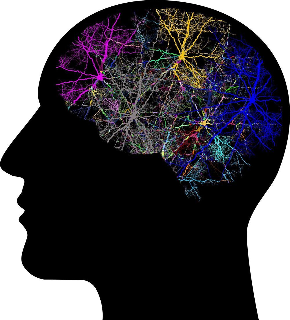
Our brains are often likened to computers, with learned skills and memories stored in the of billions of . However, new research shows that memories of specific events and experiences may never settle down. Instead, the activity patterns that store information can continually change, even when we are not learning anything new.

Why does this not cause the to forget what it has learned? The study, from the University of Cambridge, Harvard Medical School and Stanford University, reveals how the brain can reliably access stored information despite drastic changes in the brain signals that represent it.

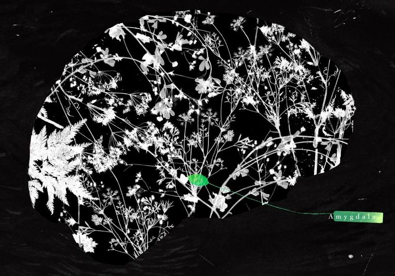
The amygdala is the brain’s surveillance hub: involved in recognizing when someone with an angry face and hostile body language gets closer, tamping down alarm when a honeybee buzzes past, and paying attention when your mother teaches you how to cross the street safely and points out which direction traffic will be coming from — in other words, things people should run away from, but also those they should look toward, attend to and remember.

In that sense, researchers say, this little knot of brain tissue shows just how tangled up emotion and social behavior are for humans. “Important events tend to be emotional in nature,” as do most aspects of social behavior, says John Herrington, assistant professor of psychiatry at the Children’s Hospital of Philadelphia in Pennsylvania.

As a result, the amygdala has long been a focus of autism research, but its exact role in the condition is still unclear.

Nelson Dellis is a four-time USA Memory Champion and Grandmaster of Memory. Some of his feats of recollection include memorizing 10,000 digits of pi, the order of more than nine shuffled decks of cards, and lists of hundreds of names after only hearing them once.

But with a little dedication, Dellis says that anyone can improve their memory. Here are five steps to follow that will get your filling your head with information.

1. Start With Strong Images

You can find our beautiful education posters in our shop: https://shop.kurzgesagt.org

This video was made possible by a grant from the Templeton World Charity Foundation.

Sources & further reading:
https://sites.google.com/view/sourcesintelligence

Humans are proud of a lot of things, from particle accelerators, to poetry to pokemon. All of them made possible because of something humans value extremely highly: intelligence.

OUR CHANNELS
▀▀▀▀▀▀▀▀▀▀▀▀▀▀▀▀▀▀▀▀▀▀▀▀▀▀
German Channel: https://kgs.link/youtubeDE
Spanish Channel: https://kgs.link/youtubeES

HOW CAN YOU SUPPORT US?
▀▀▀▀▀▀▀▀▀▀▀▀▀▀▀▀▀▀▀▀▀▀▀▀▀▀
This is how we make our living and it would be a pleasure if you support us!

Neurons, specialized cells that transmit nerve impulses, have long been known to be a vital element for the functioning of the human brain. Over the past century, however, neuroscience research has given rise to the false belief that neurons are the only cells that can process and learn information. This misconception or ‘neurocomputing dogma’ is far from true.

An is a different type of cell that has recently been found to do a lot more than merely fill up spaces between neurons, as researchers believed for over a century. Studies are finding that these cells also play key roles in brain functions, including learning and central pattern generation (CPG), which is the basis for critical rhythmic behaviors such as breathing and walking.

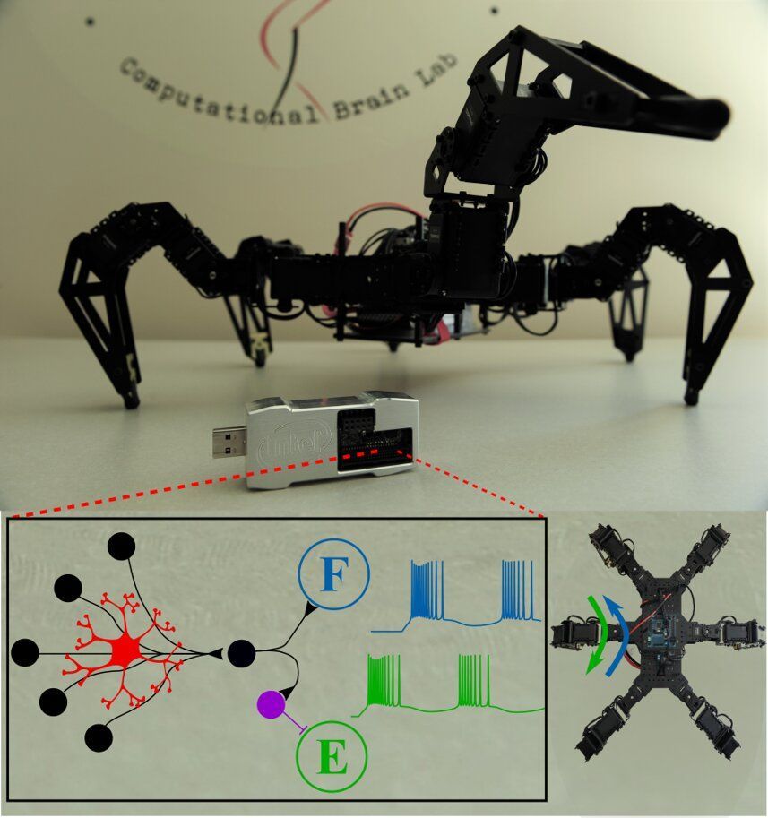
Although astrocytes are now known to underlie numerous brain functions, most existing inspired by the only target the structure and function of neurons. Aware of this gap in existing literature, researchers at Rutgers University are developing brain-inspired algorithms that also account for and replicate the functions of astrocytes. In a paper pre-published on arXiv and set to be presented at the ICONS 2020 Conference in July, they introduce a neuromorphic central pattern generator (CPG) modulated by artificial astrocytes that successfully entrained several rhythmic walking behaviors in their in-house robots.

Various diseases of the digestive tract, for example severe intestinal inflammation in humans, are closely linked to disturbances in the natural mobility of the intestine. What role the microbiome—i.e. the natural microbial community colonizing the digestive tract—plays in these rhythmic contractions of the intestine, also known as peristalsis, is currently the subject of intensive research. It is particularly unclear how the contractions are controlled and how the cells of the nervous system, that act as pacemakers, function together with the microorganisms.

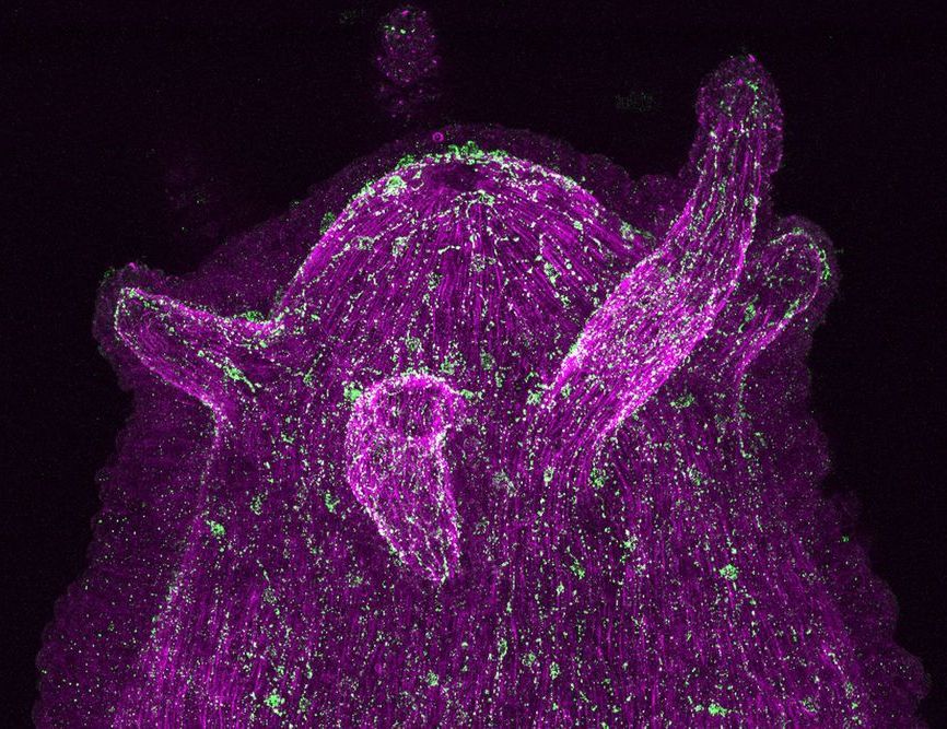
A research team from the Cell and Developmental Biology group at Kiel University has now succeeded in demonstrating for the first time, using the freshwater polyp Hydra as an example, that phylogenetically old neurons and bacteria actually communicate directly with each other. Surprisingly, they discovered that the are able to cross-talk with the microorganisms via immune receptors, i.e., to some extent with the mechanisms of the immune system.

On this basis, the scientists of the Collaborative Research Center (CRC) 1182 “Origin and Function of Metaorganisms” formulated the hypothesis that the has not only taken over sensory and motor functions from the onset of evolution, but is also responsible for communication with the microbes. The Kiel researchers around Professor Thomas Bosch published their results together with international colleagues today in the journal Proceedings of the National Academy of Sciences (PNAS).

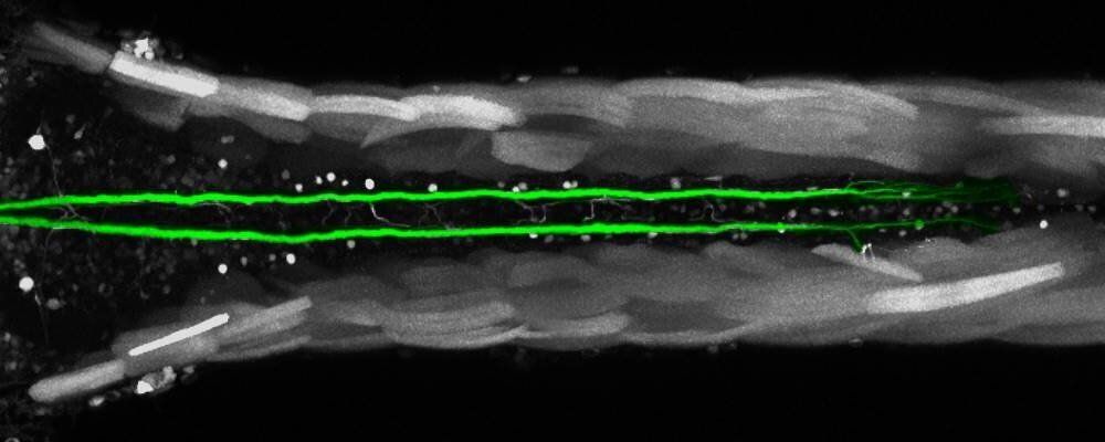
Biologists from the University of Bayreuth have discovered a uniquely rapid form of regeneration in injured neurons and their function in the central nervous system of zebrafish. They studies the Mauthner cells, which are solely responsible for the escape behavior of the fish, and previously regarded as incapable of regeneration. However, their ability to regenerate crucially depends on the location of the injury. In central nervous systems of other animal species, such a comprehensive regeneration of neurons has not yet been proven beyond doubt. The scientists report their findings in the journal Communications Biology.

Mauthner are the largest cells found in animal brains. They are part of the central nervous system of most fish and amphibian species and trigger life-saving escape responses when predators approach. The transmission of signals in Mauthner cells to their motoneurons is only guaranteed if a certain part of these cells, the axon, is intact. The axon is an elongated structure that borders the cell body with its at one of its two ends. If the injury of the axon occurs close to the cell body, the Mauthner cell dies. If the axon is damaged at its opposite end, lost functions are either not restored at all or only slowly and to a limited extent. However, the Mauthner cell reacts to an injury in the middle of the axon with rapid and complete regeneration. Indeed, within a week after the , the axon and its function are fully restored, and the fish is able to escape approaching predators again.

“Such a rapid regeneration of a neuron was never observed anywhere in the of other animal species until now. Here, regeneration processes usually extend over several weeks or months,” says Dr. Alexander Hecker, first author of the new study and member of the Department of Animal Physiology. This finding clearly disproves the widely accepted view in the that Mauthner cells are unable to regenerate.