Toggle light / dark theme

If given the chance, a Kenyan herder is likely to keep a mix of goats and camels. It seems like an irrational economic choice because goats reproduce faster and thus offer higher near-term herd growth. But by keeping both goats and camels, the herder lowers the variability in growth from year to year. All of this helps increase the odds of household survival, which is essentially a gamble that depends on a multiplicative process with no room for catastrophic failure. It turns out, the choice to keep camels also makes evolutionary sense: families that keep camels have a much higher probability of long-term persistence. Unlike businesses or governments, organisms can’t go into evolutionary debt—there is no borrowing one’s way back from extinction.

How biological survival relates to economic choice is the crux of a new paper published in Evolutionary Human Sciences, co-authored by Michael Price, an anthropologist and Applied Complexity Fellow at the Santa Fe Institute, and James Holland Jones, a biological anthropologist and associate professor at Stanford’s Earth System Science department.

“People have wanted to make this association between evolutionary ideas and economic ideas for a long time,” Price says, and “they’ve gone about it quite a lot of different ways.” One is to equate the economic idea of maximizing utility—the satisfaction received from consuming a good—with the evolutionary idea of maximizing fitness, which is long-term reproductive success. “That utility equals fitness was simply assumed in a lot of previous work,” Price says, but it’s “a bad assumption.” The human brain evolved to solve proximate problems in ways that avoid an outcome of zero. In the Kenyan example, mixed herding diversifies risk. But more importantly, the authors note, the growth of these herds, like any biological growth process, is multiplicative and the rate of increase is stochastic.
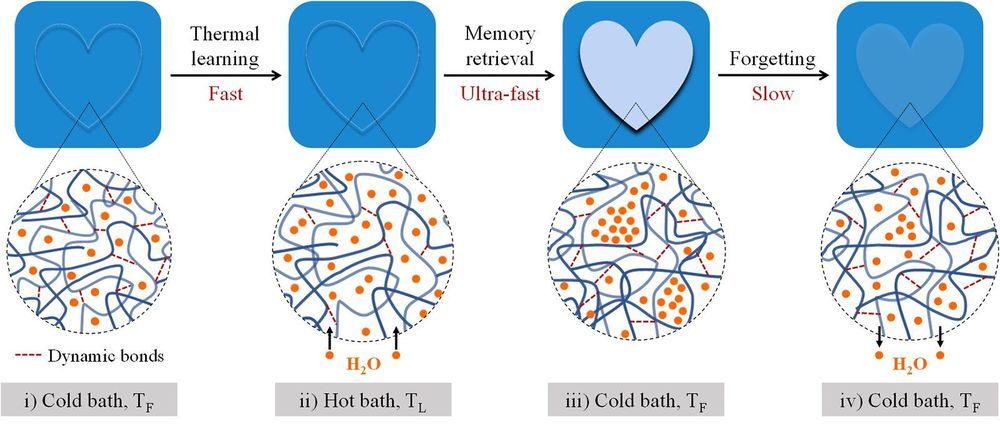
Hokkaido University researchers have found a soft and wet material that can memorize, retrieve, and forget information, much like the human brain. They report their findings in the journal Proceedings of the National Academy of Sciences (PNAS).

The learns things, but tends to forget them when the is no longer important. Recreating this dynamic process in manmade materials has been a challenge. Hokkaido University researchers now report a hydrogel that mimics the dynamic memory function of the brain: encoding information that fades with time depending on the memory intensity.

Hydrogels are flexible materials composed of a large percentage of water—in this case about 45%—along with other chemicals that provide a scaffold-like structure to contain the water. Professor Jian Ping Gong, Assistant Professor Kunpeng Cui and their students and colleagues in Hokkaido University’s Institute for Chemical Reaction Design and Discovery (WPI-ICReDD) are seeking to develop hydrogels that can serve biological functions.

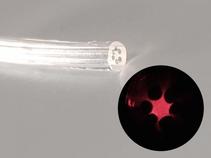
An optical fiber made of agar has been produced at the University of Campinas (UNICAMP) in the state of São Paulo, Brazil. This device is edible, biocompatible and biodegradable. It can be used in vivo for body structure imaging, localized light delivery in phototherapy or optogenetics (e.g., stimulating neurons with light to study neural circuits in a living brain), and localized drug delivery.

Another possible application is the detection of microorganisms in specific organs, in which case the probe would be completely absorbed by the body after performing its function.

The research project, which was supported by São Paulo Research Foundation—FAPESP, was led by Eric Fujiwara, a professor in UNICAMP’s School of Mechanical Engineering, and Cristiano Cordeiro, a professor in UNICAMP’s Gleb Wataghin Institute of Physics, in collaboration with Hiromasa Oku, a professor at Gunma University in Japan.

Scientists discover that oxytocin could be used to treat cognitive disorder like Alzheimer’s disease.


Alzheimer’s disease is a progressive disorder in which the nerve cells (neurons) in a person’s brain and the connections among them degenerate slowly, causing severe memory loss, intellectual deficiencies, and deterioration in motor skills and communication. One of the main causes of Alzheimer’s is the accumulation of a protein called amyloid β (Aβ) in clusters around neurons in the brain, which hampers their activity and triggers their degeneration.

Studies in animal models have found that increasing the aggregation of Aβ in the hippocampus—the brain’s main learning and memory center—causes a decline in the signal transmission potential of the neurons therein. This degeneration affects a specific trait of the neurons, called ‘,’ which is the ability of synapses (the site of signal exchange between neurons) to adapt to an increase or decrease in signaling activity over time. Synaptic plasticity is crucial to the development of learning and cognitive functions in the hippocampus. Thus, Aβ and its role in causing cognitive memory and deficits have been the focus of most research aimed at finding treatments for Alzheimer’s.

Now, advancing this research effort, a team of scientists from Japan, led by Professor Akiyoshi Saitoh from the Tokyo University of Science, has looked at oxytocin, a hormone conventionally known for its role in the female reproductive system and in inducing the feelings of love and well-being. “Oxytocin was recently found to be involved in regulating learning and memory performance, but so far, no previous study deals with the effect of oxytocin on Aβ-induced ,” Prof Saitoh says. Realizing this, Prof Saitoh’s group set out to connect the dots. Their findings are published in Biochemical and Biophysical Research Communication.

Scientists uncover a potential mechanism behind sleep-induced memory changes.

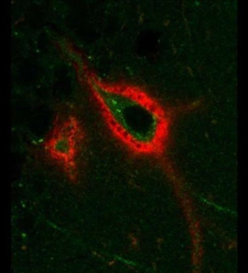
The morphing structure of the brain’s “cartilage cells” may regulate how memories change while you snooze, according to new research in eNeuro.

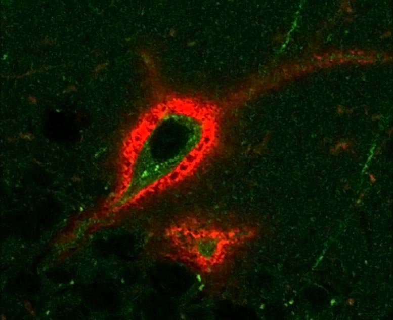
Sleep lets the body rest, but not the brain. During sleep, the brain accounts for a day of learning by making strong memories stronger and weak memories weaker, a process known as memory consolidation. But changing memories requires changing synapses, the connections between neurons. Sleep-induced changes need to overcome perineuronal nets, cartilage-like sheaths that not only surround and protect neurons, but also prevent changes in synapses.

Two groups of nerve cells may serve as “on-off switches” for male mating and aggression, suggests a new study in rodents. These neurons appear to send signals between two parts of the brain—the back tip, or posterior, of the amygdala and the hypothalamus—that together regulate emotions including fear, anxiety, and aggression.

Led by researchers at NYU Grossman School of Medicine, the study showed that male mice struggled to have sex in experiments that blocked signals from one cell group that communicates with the hypothalamus (MPN-signaling ). When the same signals were instead bolstered, the animals were not only able to mate but would repeatedly court unreceptive females, something they would not do normally.

Similarly, when the action of a second cell group in the amygdala that also communicates with the hypothalamus (VMHvl-signaling cells) was blocked, the rodents attacked unfamiliar males half as often. When these same neurons were triggered, the mice became unusually aggressive, even attacking their female mates and familiar males.

“‘Aurora will enable us to explore new frontiers in artificial intelligence and machine learning,’ said Narayanan ‘Bobby’ Kasthuri, assistant professor of neurobiology at the University of Chicago and researcher at Argonne. ‘This will be the first time scientists have had a machine powerful enough to match the kind of computations the brain can do.’”

Super computer Aurora will help map the human brain at “quintillion—or one billion billion—calculations per second, 50 times quicker than today’s most powerful supercomputers.”

Note: the article discusses implications beyond neuroscience.


Argonne, DOE and Intel announce exascale computer built for next-generation AI and machine learning.

Click to read the paper published in Frontiers in Immunology: https://fro.ntiers.in/tp1U

Summary: Cortical thickness and regional brain connectivity pay an equally important role in linking brain and behavior.

Source: Penn State

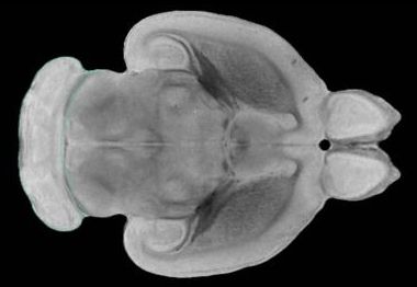
Most people think of the brain as divided into regions that are each responsible for different functions, such as language and fine motor skills. A new study by Penn State researchers suggests that there’s more to the story: The thickness of the brain’s tissue and a brain region’s connectivity may play an equally important role in linking brain and behavior.