Toggle light / dark theme

Crucially, plasma treatment of the old rats reduced the epigenetic ages of blood, liver and heart by a very large and significant margin, to levels that are comparable with the young rats. According to the six epigenetic clocks, the plasma fraction treatment rejuvenated liver by 73.4%, blood by 52%, heart by 52%, and hypothalamus by 11%. The rejuvenation effects are even more pronounced if we use the final versions of our epigenetic clocks: liver 75%, blood 66%, heart 57%, hypothalamus 19%. According to the final version of the epigenetic clocks, the average rejuvenation across four tissues was 54.2%.


Researchers have demonstrated that epigenetic age can be halved in rats by using signals commonly found in the blood.

Epigenetic changes

One of the proposed reasons we age are the changes to gene expression that our cells experience as we get older; these are commonly called epigenetic alterations. These alterations harm the fundamental functions of our cells and can increase the risk of cancer and other age-related diseases.
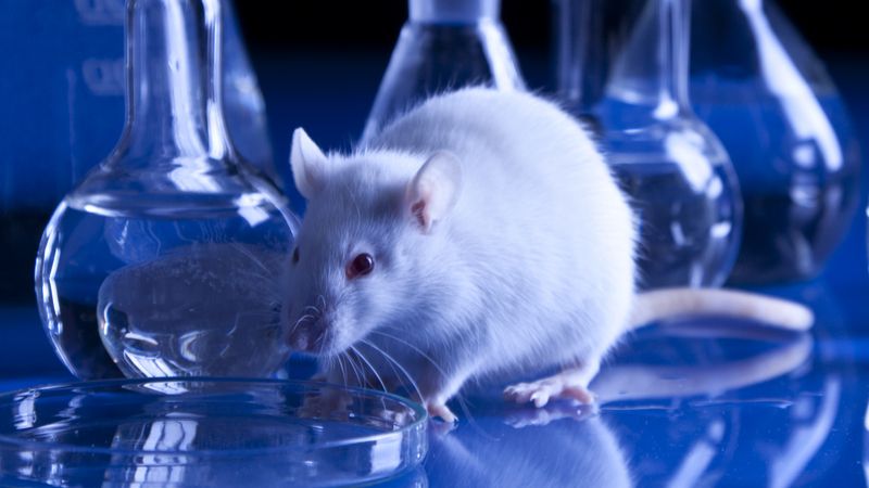
Young blood plasma is known to confer beneficial effects on various organs in mice. However, it was not known whether young plasma rejuvenates cells and tissues at the epigenetic level; whether it alters the epigenetic clock, which is a highly-accurate molecular biomarker of aging. To address this question, we developed and validated six different epigenetic clocks for rat tissues that are based on DNA methylation values derived from n=593 tissue samples. As indicated by their respective names, the rat pan-tissue clock can be applied to DNA methylation profiles from all rat tissues, while the rat brain-, liver-, and blood clocks apply to the corresponding tissue types. We also developed two epigenetic clocks that apply to both human and rat tissues by adding n=850 human tissue samples to the training data. We employed these six clocks to investigate the rejuvenation effects of a plasma fraction treatment in different rat tissues. The treatment more than halved the epigenetic ages of blood, heart, and liver tissue. A less pronounced, but statistically significant, rejuvenation effect could be observed in the hypothalamus. The treatment was accompanied by progressive improvement in the function of these organs as ascertained through numerous biochemical/physiological biomarkers and behavioral responses to assess cognitive functions. Cellular senescence, which is not associated with epigenetic aging, was also considerably reduced in vital organs. Overall, this study demonstrates that a plasma-derived treatment markedly reverses aging according to epigenetic clocks and benchmark biomarkers of aging.

Several authors are founders, owners, employees (Harold Katcher and Akshay Sanghavi) or consultants of Nugenics Research (Steve Horvath and Agnivesh Shrivastava) which plans to commercialize the “Elixir” treatment. Other authors (Kavita Singh, Shraddha Khairnar) received financial support from Nugenics Research. The other authors do not have conflict of interest.

Your cells teem with small machinery and devices of all kinds, including teeny tiny batteries called mitochondria. Just like real batteries, they can malfunction, but replacing them isn’t that easy. In this episode, Veera explains what happens to our microbatteries with aging and what we can do about it.

Circa 2012


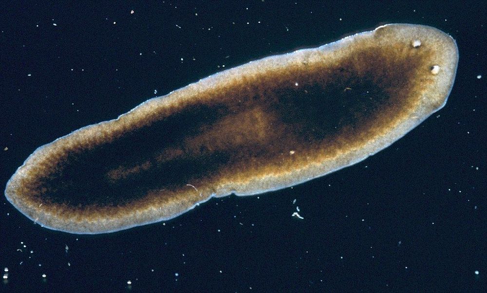
Forget expensive lotions and potions – the key to becoming immortal could be found in flatworms, scientists say.

The worms, which live in lakes and ponds, hold the remarkable ability to regenerate time and time again – effectively living forever.

If one is cut in half, the head portion grows a tail and the tail portion grows a head. Cut it into 20 pieces and 20 new worms, each an exact copy of the first, are created.

:3 could heal body parts in humans.


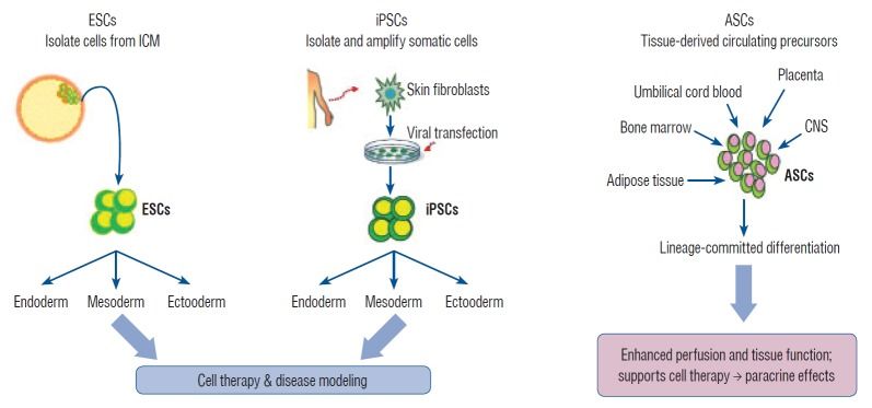
The generation of human induced pluripotent stem cells (iPSCs) from somatic cells using gene transfer opens new areas for precision medicine with personalized cell therapy and encourages the discovery of essential platforms for targeted drug development. iPSCs retain the genome of the donor, may regenerate indefinitely, and undergo differentiation into virtually any cell type of interest using a range of published protocols. There has been enormous interest among researchers regarding the application of iPSC technology to regenerative medicine and human disease modeling, in particular, modeling of neurologic diseases using patient-specific iPSCs. For instance, Parkinson’s disease, Alzheimer’s disease, and spinal cord injuries may be treated with iPSC therapy or replacement tissues obtained from iPSCs. In this review, we discuss the work so far on generation and characterization of iPSCs and focus on recent advances in the use of human iPSCs in clinical setting.

Stem cells exhibit the capacity of self-renewal and may undergo differentiation into various tissue types. These are divided into pluripotent stem cells (PSCs; embryonic stem cells [ESCs] and induced pluripotent stem cells [iPSCs]) and multipotent stem cells (adult stem cells [ASCs]) based on their differentiation capacity [45]. PSCs, including ESCs derived from embryos and iPSCs derived by gene transfer, may undergo indefinite proliferation and differentiate into different types of tissues depending on the treatment conditions [86]. Multipotent stem cells, however, may be obtained from tissue-derived precursors (umbilical cord blood, bone marrow, adipose tissue, placenta, or blood), which are already grown tissues.

A study just released by Columbia University Mailman School of Public Health is reporting a blood-DNA-methylation measure that is sensitive to variation in the pace of biological aging among individuals born the same year. The tool—DunedinPoAm—offers a unique measurement for intervention trials and natural experiment studies investigating how the rate of aging may be changed by behavioral or drug therapy, or by changes to the environment. The study findings are published online in the journal eLife.

“The goal of our study was to distill a measurement of the rate of biological aging based on 12-years of follow-up on 18 different clinical tests into a blood test that can be administered at a single time point.” said lead author Daniel Belsky, Ph.D., assistant professor of epidemiology at Columbia Mailman School and a researcher at the Columbia Aging Center.

Midlife adults measured to be aging faster according to the new measurement showed faster declines in physical and cognitive functioning and looked older in facial photographs. Older adults measured to be aging faster by the tool were at increased risk for chronic disease and mortality. In other analyses, the researchers showed that DunedinPoAm captured new information not measured by proposed measures of biological aging known as epigenetic clocks, that 18-year-olds with histories of childhood poverty and victimization showed faster aging as measured by DunedinPoAm, and that DunedinPoAm predictions were disrupted by a caloric restriction intervention in a randomized trial.

World-first research led by neuroimaging expert Dr. Jiyang Jiang at UNSW’s Centre for Healthy Brain Ageing (CHeBA) has found that those aged 95 and over demonstrated more activation between the left and ride side of their brain than their younger counterparts.

Given the prevalence of dementia increases with age, near-centenarians and centenarians without dementia are generally considered as models of successful aging and resistance against .

“We wanted to see if there was something particularly special about the ’s functional connectivity of those aged 95 and older that helps them preserve brain function into the 11th decade of their life,” says Dr. Jiang.