Toggle light / dark theme

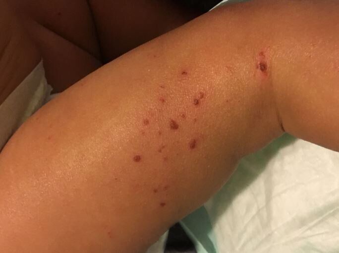
New research supported by the National Institutes of Health delineates how two relatively common variations in a gene called KIF3A are responsible for an impaired skin barrier that allows increased water loss from the skin, promoting the development of atopic dermatitis, commonly known as eczema. This finding could lead to genetic tests that empower parents and physicians to take steps to potentially protect vulnerable infants from developing atopic dermatitis and additional allergic diseases.

Atopic dermatitis is an that affects up to 20% of children in developed countries. This chronic is characterized by dry, thickened and intensely itchy skin, particularly in skin folds. People with eczema are more susceptible to bacterial, viral and fungal skin infections and frequently develop additional allergic diseases such as asthma.

KIF3A is a gene that codes for a protein involved in generating signals from the outside to the inside of a cell, part of a complex sensory apparatus. Previously, scientists had identified an association between two genetic variations in KIF3A and asthma in children who also had eczema. In the new study, the researchers found that these variations, or (SNPs), changed parts of the KIF3A gene to a form that can regulate, through a process called methylation, the rate at which a gene is transcribed into the blueprint for protein production. The investigators confirmed that skin and nasal-lining cells from people with the KIF3A SNP variants had more methylation and contained fewer blueprints for the KIF3A protein than cells in which KIF3A lacked the SNPs. In addition, the researchers demonstrated that people with the SNP-created regulating sites had higher levels of from the skin.

The US Food and Drug Administration (FDA) has approved viltolarsen (Viltepso; NS Pharma) for the treatment of patients with Duchenne muscular dystrophy amenable to exon 53 skipping, making it only the second FDA-approved therapy for this specific DMD gene mutation.

The agent from NS Pharma, delivered via weekly intravenous infusion, was granted accelerated approval via its priority review, fast track, orphan drug, and rare disease designations after its new drug application was accepted earlier this year. In March, NS Pharma launched an expanded access program for qualified patients.

The approval was granted based on findings from a phase 2 clinical trial (NCT02740972) and long-term extension study, details of which were recently published in JAMA Neurology. Among 16 participants age 4 to 9, significant drug-induced dystrophin production was observed in both viltolarsen dose cohorts (40 mg/kg per week: mean, 5.7% [range, 3.2–10.3] of normal; 80 mg/kg per week: mean, 5.9% [range, 1.1–14.4] of normal), with 15 (94%) patients achieving dystrophin levels greater than 2% of normal and 14 of 16 (88%) achieving levels greater than 3% of normal.

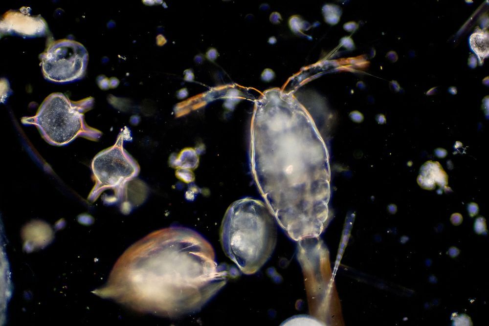
While satellite imaging lets researchers observe the outer life of plankton populations, the complex genetics in microscopic marine life have made looking inward more challenging. According to a new study published in Nature Methods, researchers from the University of East Anglia were able to deliver and express foreign DNA in 13 species that have never before transformed. They were also able to evaluate the potential cause of non-transformation in 17 other species; in turn, laying the foundation for an expanded understanding of genomes discovered in plankton.

The sheer variety of plankton potential — from antibacterial compounds to antiviral and antifungal solutions — makes this a worthwhile endeavor. If scientists can create reliable methods to modify phytoplankton, it should be possible to reduce their toxic impact, better control their bloom cycle and even increase the photosynthetic output — all critical in the fight to keep our oceans blue and our terra firma green.

As noted by Science Magainze, the international research team used a variety of methods to modify plankton DNA. For some species, shooting tiny gold or tungsten particles covered with DNA through cell walls produced the best result. For others, jolts of electricity made cell walls “leaky” and allowed new DNA to seep through. Specific protist successes included modification of a fish-killing toxic plankton species, and one that infects both mollusks and amphibians. While these discoveries don’t present a complete understanding of the genetics in microscopic marine life, they provide a key testing protocol: By modifying genetic structure and then observing how plankton react, teams could uncover ways to boost antibiotic resistance or lower infectious impact. According to lead UK study author Thomas Mock, “These insights will improve our understanding about their role in the oceans, and they are invaluable for biotechnological applications such as building factories for biofuel or the production of bioactive compounds.”

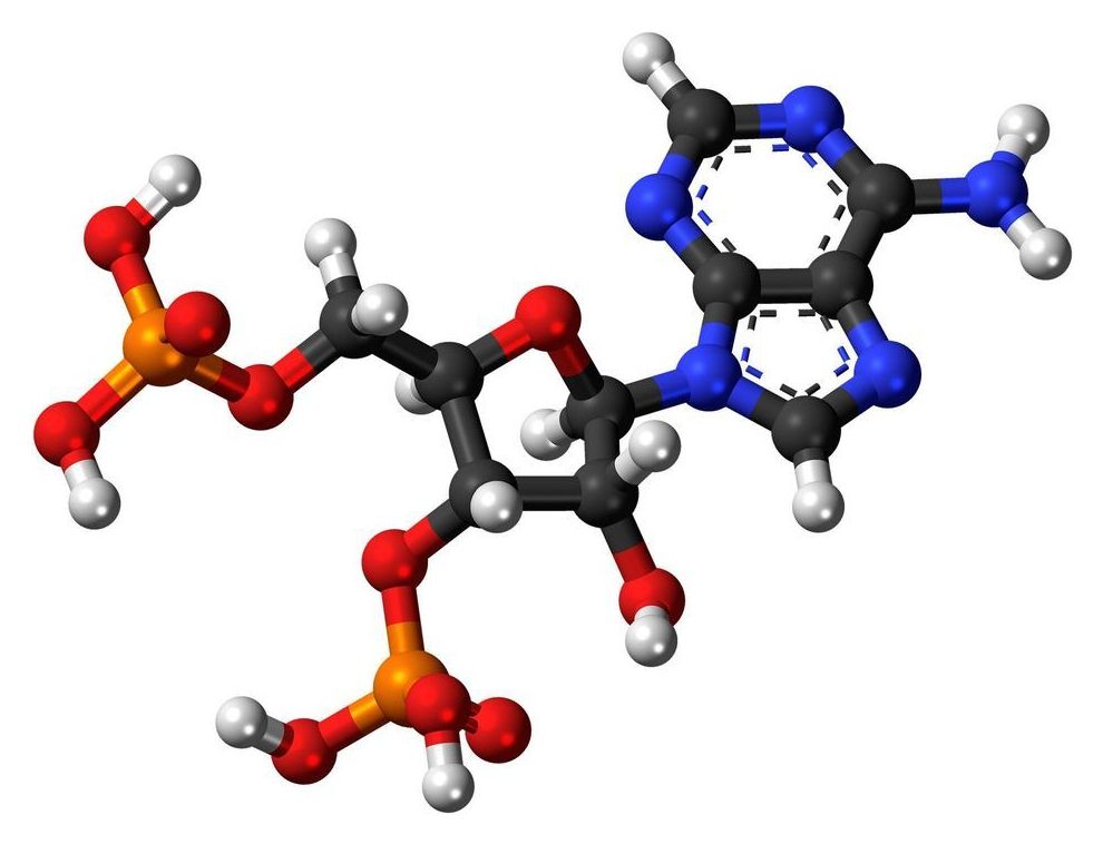
Before the first oncogene mutations were discovered in human cancer in the early 1980s, the 1970s provided the first data suggesting alterations in the genetic material of tumors. In this context, the prestigious journal Nature published in 1975 the existence of a specific alteration in the transformed cell: an RNA responsible for carrying an amino acid to build proteins (transfer RNA) was missing a piece, the enigmatic nucleotide ‘Y.’

After that outstanding observation, virtually no developments were made for forty-five years on the causes and consequences of not having the correct base in RNA.

In an article published in Proceedings of the National Academy of Sciences (PNAS) by the group of Dr. Manel Esteller, Director of the Josep Carreras Leukaemia Research Institute, ICREA Research Professor and Professor of Genetics at the University of Barcelona has solved this mystery by observing that in the protein that generates the Y is epigenetically inactivated, causing small but highly aggressive tumors.

Last year information was released concerning rejuvenation of the thymus which resulted in a reversal of the epigenetic clock an average of 2.5 years in a small trial of 9 people costing $10,000 per person. You can get this done too. A company has formed called Intervene Immune which will take on volunteers for the process. It is not funded so you would have to pay out pf pocket though eventually the cost may come down and they can provide financing. You do not have to travel to California to get this done. Cost prohibits me, and I may or may not be eligible as I have IBS though that is not on the exclusion list. I emailed them concerning all this which is how I got the information.

http://interveneimmune.com/

https://www.surveymonkey.com/r/TRIIMX


The TRIIM-X trial is an expanded pilot clinical study that will evaluate a personalized combination treatment regimen for thymus regeneration. The thymus is a part of the immune system that declines markedly with age, and regenerating it may prevent or reverse key aspects of immunosenescence (immune system aging) and potentially prevent or reverse key parts of the aging process more generally. The study will evaluate biomarkers for epigenetic aging and immunosenescence, as well as evaluate established clinical measures and risk factors for prevention of physical frailty, cancer, cardiovascular disease, diabetes, dementia, and also infectious diseases, including flu and COVID-19.

The study uses multiple agents in combination with personalized doses of recombinant human growth hormone (somatropin), metformin, and DHEA, in a similar manner to how the combination treatment was applied in the earlier TRIIM trial at Stanford, which demonstrated strong statistical significance for the primary efficacy endpoints that will be evaluated in TRIIM-X. Somatropin is approved by the FDA for adult growth hormone deficiency and its use in the study is guided by prior safety data established for that use and also based on safety data available on its prior use in the TRIIM trial and in clinical practice in healthy elderly individuals. There will also be control groups that enable testing of biomarker variability and the contribution of individual medications within the combination treatment.

The objective of the study is to obtain information needed for designing an effective personalized and adaptive treatment regimen for a larger and more diverse study population, and to obtain additional proof of principle for the new use of the medications and biomarkers for preventive medicine. The duration of treatment in the TRIIM-X trial will be 12 months.

Hereditary hearing loss is one of the most common disabilities among newborns, affecting approximately 1 in 1000 live-born babies. Most forms of hereditary hearing loss are nonsyndromic; 80% of affected newborns have hearing loss that is inherited in an autosomal recessive pattern, and in the remaining 20%, inheritance shows a dominant pattern.

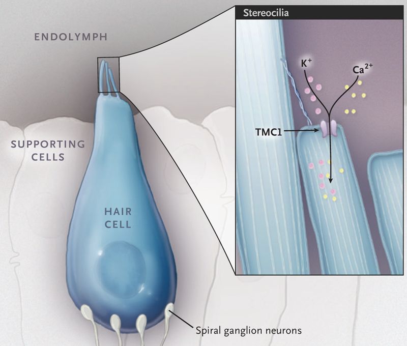
Many forms of hereditary hearing loss are caused by mutations in genes that affect the formation and function of cochlear hair cells — highly specialized sensory cells that play an important role in the detection and processing of sound. The hair cell has bundles of hair-like projections, called stereocilia, on its apical surface ( Fig. 1 ). The deflection of these bundles by sound results in the opening of mechanotransduction ion channels, which are located at the tips of the stereocilia, and consequently, in the depolarization of the hair-cell membrane. Mutations that affect the protein transmembrane channel-like 1 (TMC1), an integral component of the mechanotransduction complex, cause autosomal dominant and autosomal recessive forms of hearing loss. Correction of the dominant form of hearing loss in a mouse model of Tmc1 (termed “Beethoven”) was recently reported by Gao and colleagues.

An external file that holds a picture, illustration, etc. Object name is nihms-1017746-f0001.jpg

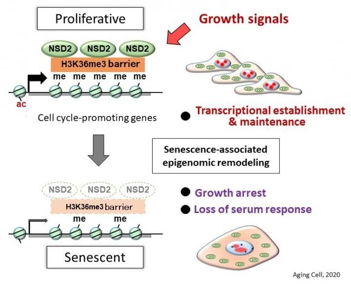
NSD2 is the fourth protective factor of cellular senescence that our team has identified,” said Professor Mitsuyoshi Nakao. “With the discovery that NSD2 protects against cellular senescence, this study clarifies a basic mechanism of aging.


Researchers from Kumamoto University in Japan have used comprehensive genetic analysis to find that the enzyme NSD2, which is known to regulate the actions of many genes, also works to block cell aging. Their experiments revealed 1) inhibition of NSD2 function in normal cells leads to rapid senescence and 2) that there is a marked decrease in the amount of NSD2 in senescent cells. The researchers believe their findings will help clarify the mechanisms of aging, the development of control methods for maintaining NSD2 functionality, and age-related pathophysiology.

As the cells of the body continue to divide (cell reproduction), their function eventually declines and they stop growing. This cellular senescence is an important factor in health and longevity. Cell aging can also be stimulated when genomic DNA is damaged by physical stress, such as radiation or ultraviolet rays, or by chemical stress that occurs with certain drugs. However, the detailed mechanisms of aging are still unknown. Cell aging can be beneficial when a cell becomes cancerous; it prevents malignant changes by causing cellular senescence. On the other hand, it makes many diseases more likely with age. It is therefore important that cell aging is properly controlled.

Although senescent cells lose their proliferative ability, it has recently become clear that senescent cells secrete various proteins that act on surrounding cells to promote chronic inflammation and cancer development. Since senescent cells are more active than expected, cellular aging is thought to be responsible for whole body aging. This idea has been supported by reports of systemic aging suppression in aged mice after removal of accumulated senescent cells. In other words, if you can control cell aging, you may be able to control the progression of aging throughout the body.