Toggle light / dark theme

Within a mere eight years, CRISPR-Cas9 has become the go-to genome editor for both basic research and gene therapy. But CRISPR-Cas9 also has spawned other potentially powerful DNA manipulation tools that could help fix genetic mutations responsible for hereditary diseases.

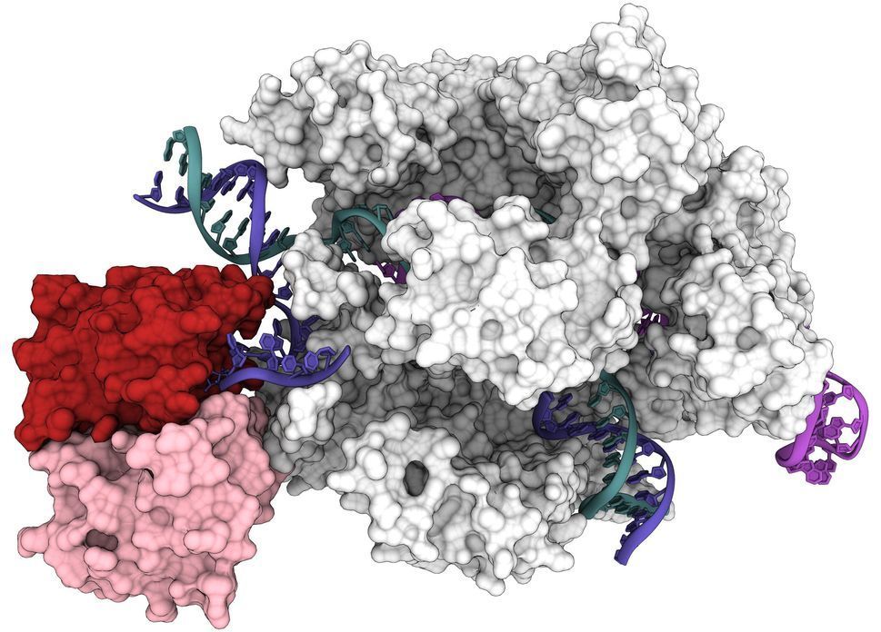
Researchers at the University of California, Berkeley, have now obtained the first 3D structure of one of the most promising of these tools: base , which bind to DNA and, instead of cutting, precisely replace one nucleotide with another.

First created four years ago, base editors are already being used in attempts to correct single-nucleotide mutations in the human genome. Base editors now available could address about 60% of all known genetic diseases—potentially more than 15,000 inherited disorders—caused by a mutation in only one nucleotide.
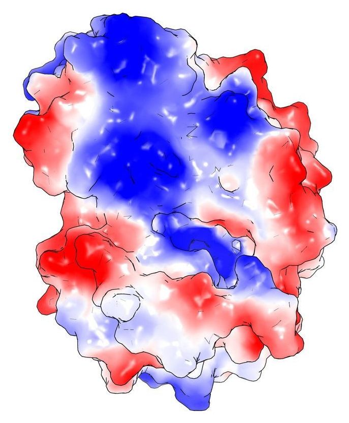
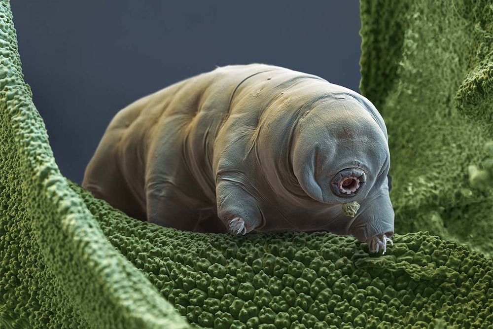
The toxin can block the use of important amino acids required by the bacteria to produce essential proteins needed for survival.

An international team of researchers, led by Durham University, UK, and the Laboratory of Molecular Microbiology and Genetics/Centre Integrative Biology in Toulouse, France, are aiming to exploit this to develop new anti-TB drugs.

Their findings are published in the journal Science Advances.

Summary: Researchers successfully applied a gene therapy platform to completely correct brain defects in a large animal model of a human genetic disease.

Source: University of Pennsylvania

A lone genetic mutation can cause a life-changing disorder with effects on multiple body systems. Lysosomal storage diseases, for example, of which there are dozens, arise due to single mutations that affect production of critical enzymes required to metabolize large molecules in cells. These disorders affect multiple organs including, notably, the brain, causing intellectual disability of varying degrees.

TORONTO — In a lab at the Hospital for Sick Children in Toronto, scientists went on a hunt through the DNA of some 10,000 families — many whom have children with autism.

Through this research, they identified something they call “genetic wrinkles” in DNA itself, a breakthrough they believe could explain why some individuals find themselves on the autistic spectrum.

The hope is that this could be an important new clue into how to diagnose autism spectrum disorder (ASD) early, or even treat it.

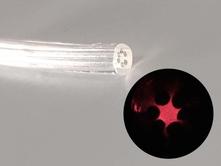
An optical fiber made of agar has been produced at the University of Campinas (UNICAMP) in the state of São Paulo, Brazil. This device is edible, biocompatible and biodegradable. It can be used in vivo for body structure imaging, localized light delivery in phototherapy or optogenetics (e.g., stimulating neurons with light to study neural circuits in a living brain), and localized drug delivery.

Another possible application is the detection of microorganisms in specific organs, in which case the probe would be completely absorbed by the body after performing its function.

The research project, which was supported by São Paulo Research Foundation—FAPESP, was led by Eric Fujiwara, a professor in UNICAMP’s School of Mechanical Engineering, and Cristiano Cordeiro, a professor in UNICAMP’s Gleb Wataghin Institute of Physics, in collaboration with Hiromasa Oku, a professor at Gunma University in Japan.

When our neurons—the principle cells of the brain—die, so do we.

Most neurons are created during and have no “backup” after birth. Researchers have generally believed that their survival is determined nearly extrinsically, or by outside forces, such as the tissues and that neurons supply with .

A research team led by Sika Zheng, a biomedical scientist at the University of California, Riverside, has challenged this notion and reports the continuous survival of neurons is also intrinsically programmed during development.

The thalamus is a “Grand Central Station” for sensory information coming to our brains. Almost every sight, sound, taste and touch we perceive travels to our brain’s cortex via the thalamus. It is theorized that the thalamus plays a major role in consciousness itself. Not only does sensory information pass through the thalamus, it is also processed and transformed by the thalamus so our cortex can better understand and interpret these signals from the world around us.

One powerful type of transformation comes from interactions between excitatory neurons that carry data to the neocortex and inhibitory neurons of the thalamic reticular nucleus, or TRN, that regulate flow of that data. Although the TRN has long been recognized as important, much less has been known about what kinds of cells are in the TRN, how they are organized and how they function.

Now a paper published in the journal Nature addresses those questions. Researchers led by corresponding author Scott Cruikshank, Ph.D., and co-authors Rosa I. Martinez-Garcia, Ph.D., Bettina Voelcker, Ph.D., and Barry Connors, Ph.D., show that the somatosensory part of the TRN is divided into two functionally distinct sub-circuits. Each has its own types of genetically defined neurons that are topographically segregated, are physiologically distinct and connect reciprocally with independent thalamocortical nuclei via dynamically divergent synapses.

Scientists have discovered extinct strains of smallpox in the teeth of Viking skeletons — proving for the first time that the killer disease plagued humanity for at least 1400 years.

Smallpox spread from person to person via infectious droplets, killed around a third of sufferers and left another third permanently scarred or blind. Around 300 million people died from it in the 20th century alone before it was officially eradicated in 1980 through a global vaccination effort — the first human disease to be wiped out.

Now an international team of scientists have sequenced the genomes of newly discovered strains of the virus after it was extracted from the teeth of Viking skeletons from sites across northern Europe. The findings have been published in Science today (July 23, 2020).