Toggle light / dark theme

MSU’s expertise in fish biology, genetics helping researchers rewrite evolutionary history and shape future health studies.

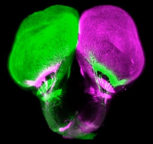
The network of nerves connecting our eyes to our brains is sophisticated and researchers have now shown that it evolved much earlier than previously thought, thanks to an unexpected source: the gar fish.

Michigan State University’s Ingo Braasch has helped an international research team show that this connection scheme was already present in ancient fish at least 450 million years ago. That makes it about 100 million years older than previously believed.

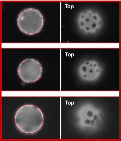
A team of polymer science and engineering researchers at the University of Massachusetts Amherst has demonstrated for the first time that the positions of tiny, flat, solid objects integrated in nanometrically thin membranes—resembling those of biological cells—can be controlled by mechanically varying the elastic forces in the membrane itself. This research milestone is a significant step toward the goal of creating ultrathin flexible materials that self-organize and respond immediately to mechanical force.

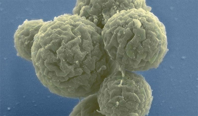
The team has discovered that rigid solid plates in biomimetic fluid membranes experience interactions that are qualitatively different from those of biological components in cell membranes. In cell membranes, fluid domains or adherent viruses experience either attractions or repulsions, but not both, says Weiyue Xin, lead author of the paper detailing the research, which recently appeared in Science Advances. But in order to precisely position solid objects in a membrane, both attractive and repulsive forces must be available, adds Maria Santore, a professor of polymer science and engineering at UMass. In the Santore Lab at UMass, Xin used giant unilamellar vesicles, or GUVs, which are cell-like membrane sacks, to probe the interactions between solid objects in a thin, sheet-like material. Like biological cells, GUVs have fluid membranes and form a nearly spherical shape. Xin modified the GUVs so that the membranes included tiny, solid, stiff plate-like masses.
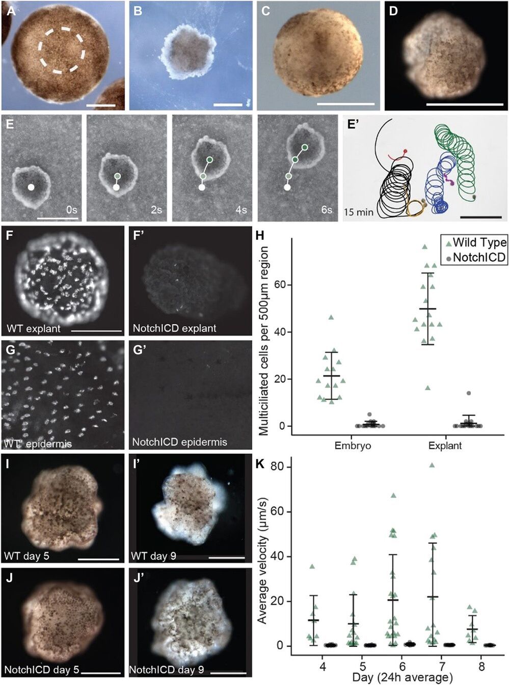
Robot swarms have, to date, been constructed from artificial materials. Motile biological constructs have been created from muscle cells grown on precisely shaped scaffolds. However, the exploitation of emergent self-organization and functional plasticity into a self-directed living machine has remained a major challenge. We report here a method for generation of in vitro biological robots from frog (Xenopus laevis) cells. These xenobots exhibit coordinated locomotion via cilia present on their surface. These cilia arise through normal tissue patterning and do not require complicated construction methods or genomic editing, making production amenable to high-throughput projects.

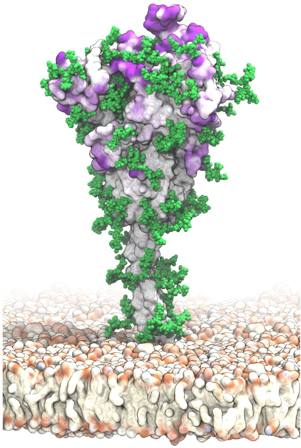
A new, detailed model of the surface of the SARS-CoV-2 spike protein reveals previously unknown vulnerabilities that could inform development of vaccines. Mateusz Sikora of the Max Planck Institute of Biophysics in Frankfurt, Germany, and colleagues present these findings in the open-access journal PLOS Computational Biology.

SARS-CoV-2 is the virus responsible for the COVID-19 pandemic. A key feature of SARS-CoV-2 is its spike , which extends from its and enables it to target and infect human cells. Extensive research has resulted in detailed static models of the spike protein, but these models do not capture the flexibility of the spike protein itself nor the movements of protective glycans—chains of sugar molecules—that coat it.

To support vaccine development, Sikora and colleagues aimed to identify novel potential target sites on the surface of the spike protein. To do so, they developed that capture the complete structure of the spike protein and its motions in a realistic environment.

Bacteria have been found exploiting quantum physics to survive.


Oxygen is life to animals like us. But for many species of microbe, the smallest whiff of the highly reactive element puts their delicate chemical machinery at risk of rusting up.

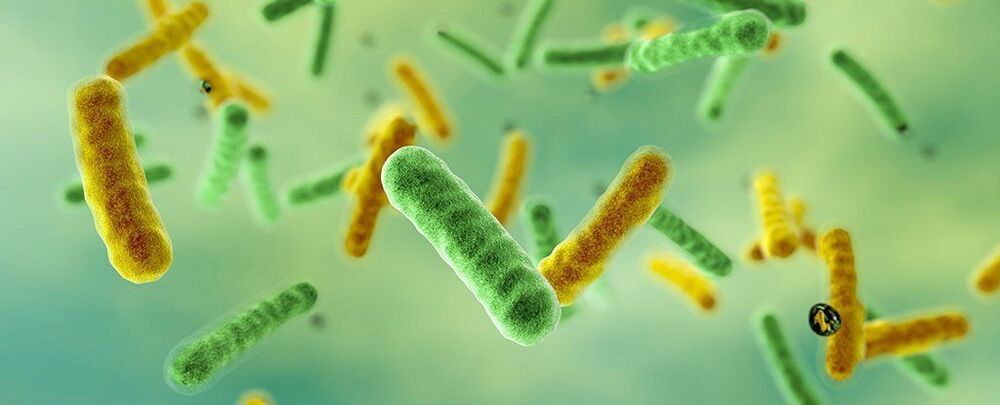
The photosynthesizing bacterium Chlorobium tepidum has evolved a clever way to shield its light-harvesting processes from oxygen’s poisonous effects, using a quantum effect to shift its energy production line into low gear.

A study conducted by scientists from the University of Chicago and Washington University in St. Louis has shown how the bacterium throws a spanner into its quantum resonance to ‘tune’ its system so that it loses energy in the presence of oxygen, preventing it from wrecking its photosynthetic apparatus.

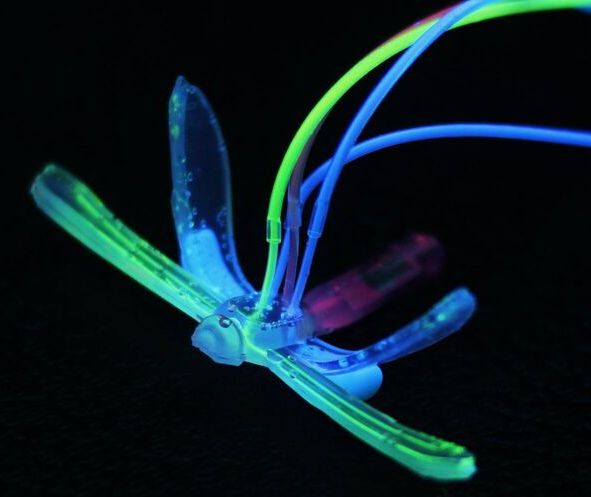
Engineers at Duke University have developed an electronics-free, entirely soft robot shaped like a dragonfly that can skim across water and react to environmental conditions such as pH, temperature or the presence of oil. The proof-of-principle demonstration could be the precursor to more advanced, autonomous, long-range environmental sentinels for monitoring a wide range of potential telltale signs of problems.

The soft robot is described online March 25 in the journal Advanced Intelligent Systems.

Soft robots are a growing trend in the industry due to their versatility. Soft parts can handle delicate objects such as biological tissues that metal or ceramic components would damage. Soft bodies can help robots float or squeeze into tight spaces where rigid frames would get stuck.

Here’s my latest video!


Papers referenced in the video:

The Hallmarks of Aging: https://www.ncbi.nlm.nih.gov/pmc/articles/PMC3836174/

Telomere Length as a Marker of Biological Age: State-of-the-Art, Open Issues, and Future Perspectives: https://www.ncbi.nlm.nih.gov/pmc/articles/PMC7859450/

Telomeres and the natural lifespan limit in humans: https://www.ncbi.nlm.nih.gov/pmc/articles/PMC5425118/

Telomere Length and All-Cause Mortality: A Meta-analysis: https://pubmed.ncbi.nlm.nih.gov/30254001/

Comparability of biological aging measures in the National Health and Nutrition Examination Study, 1999-2002: https://pubmed.ncbi.nlm.nih.gov/30999227/

Longitudinal trajectories, correlations and mortality associations of nine biological ages across 20-years follow-up: https://elifesciences.org/articles/51507

(From left) Expedition 65 crew members Pyotr Dubrov, Oleg Novitskiy and Mark Vande Hei, pose for a photo during Soyuz qualification exams in Moscow.


The Expedition 64 crew continued researching how microgravity affects biology aboard the International Space Station today. The orbital residents also conducted vein and eye checks and prepared for three new crew members due in early April.

NASA Flight Engineer Shannon Walker joined Russian cosmonauts Sergey Ryzhikov and Sergey Kud-Sverchkov for vein and eye scans on Thursday. Japan Aerospace Exploration Agency astronaut Soichi Noguchi led the effort scanning veins in the trio’s neck, clavicle and shoulder areas using the Ultrasound 2 device in the morning. In the afternoon, Noguchi examined Walker’s eyes using the orbiting lab’s optical coherence tomography gear.

Walker also assisted fellow Flight Engineer Kate Rubins of NASA setting up samples of tiny worms for viewing in a microscope. Rubins captured video of the microscopic worms wriggling around to learn how microgravity affects genetic expression and muscle function. Insights from the Micro-16 study may benefit human health on and off the Earth.