Toggle light / dark theme

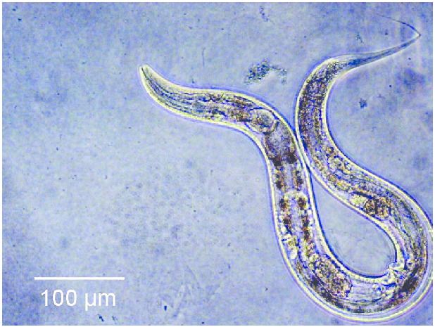
To answer this question, an internal team of scientists, consisting of researchers affiliated with the Buck Institute for Research on Ageing, and researchers from Nanjing University decided to modify both the Insulin and the rapamycin pathways of a group of C.elegans worms, expecting to see a cumulative result of a 130% increase in lifespan. However, instead of seeing a cumulative effect in lifespan, the worms lived five times longer than they normally would.

“The synergistic extension is really wild. The effect isn’t one plus one equals two, it’s one plus one equals five. Our findings demonstrate that nothing in nature exists in a vacuum; in order to develop the most effective anti-aging treatments we have to look at longevity networks rather than individual pathways.” – Jarad Rollins of Nanjing University.

What could this mean for human regenerative medicine? Humans are not worms, however on a cellular level they do possess very similar biology. Both the insulin pathway and the rapamycin pathway are what is known as ‘conserved’ between humans and C.elegans, meaning that these pathways have been maintained in both organisms. In the distant past, both humans and C.elegans had a common ancestor, in exactly the same way as humans and Chimpanzees have a common ancestor. Evolution has changed our bodies significantly over the millions of years that humans and C.elegans have diverged from one another, but a lot of our fundamental biological functions remain largely unchanged.
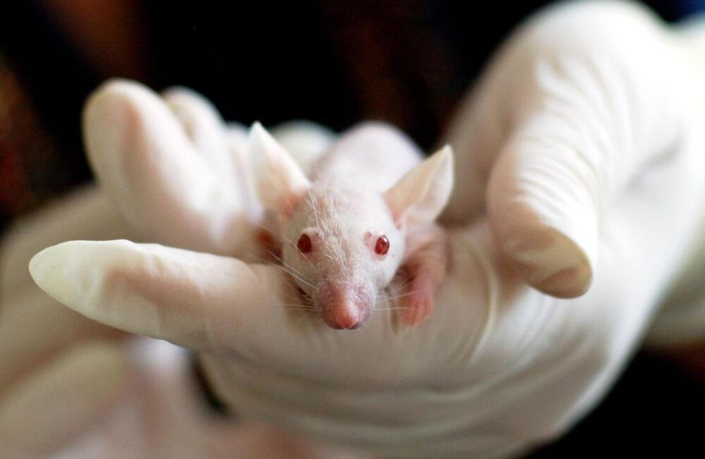
To begin with, why do we use mice in medical and biological research? The answer to this question is fairly straight forward. Mice are cheap, they grow quickly, and the public rarely object to experimentations involving mice. However, mice offer something that is far more important than simple pragmatism, as despite being significantly smaller and externally dissimilar to humans, our two species share an awful lot of similarities. Almost every gene found within mice share functions with genes found within humans, with many genes being essentially identical (with the obvious exception of genetic variation found within all species). This means that anatomically mice are remarkably similar to humans.

Now, this is where for the sake of clarity it would be best to break down biomedical research into two categories. Physiological research and pharmaceutical research, as the success of the mouse model should probably be judges separately depending upon the research that is being carried out. Separating the question of the usefulness of the mouse model down into these two categories also solves the function of more accurately focusing the ire of its critics.

The usefulness of the mouse model in the field of physiological research is largely unquestioned at this point. We have quite literally filled entire textbooks with the information we have gained from studying mice, especially in the field of genetics and pathology. The similarities between humans and mice are so prevalent that it is in fact possible to create functioning human/mouse hybrids, known as ‘genetically engineered mouse models’ or ‘GEMMs’. Essentially, GEMMs are mice that have had the mouse version of a particular gene replaced with its human equivalent. This is an exceptionally powerful tool for medical research, and has led to numerous medical breakthroughs, including most notably our current treatment of acute promyelocytic leukaemia (APL), which was created using GEMMs.

There’s some really interesting CRISPR news out today, and it’s likely to be a forerunner of much more news to come. A research team has demonstrated what looks like robust, long-lasting effects in a primate model after one injection of the CRISPR enzymatic machinery. There have been plenty of rodent reports on various forms of CRISPR, and there are some human trials underway, but these is the first primate numbers that I’m aware of.

The gene they chose to inactivate is PCSK9, which has been a hot topic in drug discovery for some years now. It’s a target validated by several converging lines of evidence from the human population (see the “History” section of that first link). People with overactive PCSK9 have high LDL lipoproteins and cholesterol, and people with mutations that make it inactive have extremely low LDL and seem to be protected from a lot of cardiovascular disease. There are several drugs and drug candidates out there targeting the protein, as well there might be.

It’s a good proof-of-concept, then, because we know exactly what the effects of turning down the expression of active PCSK9 should look like. It’s also got the major advantage of being mostly a liver target – as I’ve mentioned several times on the blog already, many therapies aimed at gene editing or RNA manipulation have a pharmacokinetic complication. The formulations used to get such agents intact into the body (and in a form that they can penetrate cells) tend to get combed out pretty thoroughly by the liver – which after all, is (among other things) in the business of policing the bloodstream for weird, unrecognized stuff that is then targeted for demolition by hepatocytes. Your entire bloodstream goes sluicing through the liver constantly; you’re not going to able to dodge it if your therapy is out there in the circulation. It happens to our small-molecule drugs all the time: hepatic “first pass” metabolism is almost always a factor to reckon with.

“Gene editing offers unique opportunities to make food production more sustainable and to further improve the quality, but also the safety, of food. With the help of these new molecular tools, more robust plants can be developed that deliver high yields for high-quality nutrition, even with less fertiliser,” says co-author Stephan Clemens, Professor of Plant Physiology at the University of Bayreuth and founding Dean of the new Faculty of Life Sciences: Food, Nutrition & Health on the Kulmbach campus.


For more sustainability on a global level, EU legislation should be changed to allow the use of gene editing in organic farming. This is what an international research team involving the Universities of Bayreuth and Göttingen demands in a paper published in the journal “Trends in Plant Science”.

In May 2020, the EU Commission presented its “Farm-to-Fork” strategy, which is part of the “European Green Deal”. The aim is to make European agriculture and its food system more sustainable. In particular, the proportion of organic farming in the EU’s total agricultural land is to be increased to 25 percent by 2030. However, if current EU legislation remains in place, this increase will by no means guarantee more sustainability, as the current study by scientists from Bayreuth, Göttingen, Düsseldorf, Heidelberg, Wageningen, Alnarp, and Berkeley shows.

Great new episode with renowned geneticist Christopher Mason who talks about his book on how we will need to bioengineer our own species in order to expand beyond our solar system.


Geneticist Christopher Mason chats about his new book, “The Next 500 Years: Engineering Life to Reach New Worlds” from MIT Press. We discuss both the nuts and bolts and the philosophy driving our expansion offworld. Mason’s goal is to preserve our species by expanding to an Earth 2.0 in order to avoid our star’s own Red Giant endgame.

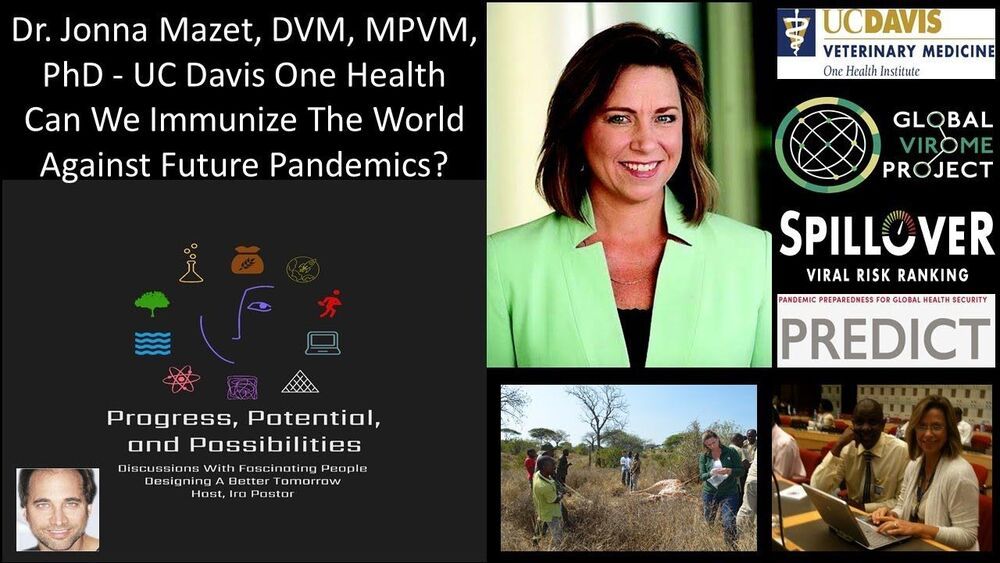
Can We Immunize The World Against Future Pandemics? Dr Jonna Mazet, DVM, MPVM, PhD, UC Davis School of Veterinary Medicine — Global Virome Project.


Dr. Jonna Mazet, DVM, MPVM, PhD, is a Professor of Epidemiology and Disease Ecology at the UC Davis School of Veterinary Medicine, Founding Executive Director of the UC Davis One Health Institute, and Vice Provost For Grand Challenges At UC Davis.

Additionally, Dr. Mazet in on the Steering Committee of the Global Virome Project, Principal Investigator of the PREDICT project, Chair, National Academies’ One Health Action Collaborative, and Co-Vice Chair, UC Global Health Institute Board of Directors.

Dr. Mazet’s work focuses on global health problem solving for emerging infectious diseases and conservation challenges. She is active in international One Health education, service, and research programs, most notably in relation to pathogen emergence; disease transmission among wildlife, domestic animals, and people; and the ecological drivers of novel disease dynamics.

Currently, Dr. Mazet is the Co-Director of the US Agency for International Development’s One Health Workforce – Next Generation, an $85 million educational strengthening project to empower professionals in Central/East Africa and Southeast Asia to address complex and emerging health threats, including antimicrobial resistance and zoonotic diseases.

Dr. Mazet is the Principal Investigator of, and served as the Global Director of, the PREDICT Project for 10 years, a greater than $200 million viral emergence early warning project under USAID’s Emerging Pandemic Threats Division, which served as an early-warning system-strengthening effort aimed at finding emerging viruses before they spread to humans.

Dr. Mazet was elected to the US National Academy of Medicine in 2013 in recognition of her successful and innovative approach to emerging environmental and global health threats, and serves on the National Academies of Science, Engineering, and Medicine’s Forum on Microbial Threats and chairs the Academies’ One Health Action Collaborative. She was appointed to the National Academies Standing Committee on Emerging Infectious Diseases and 21st Century Health Threats, which was created to assist the federal government with critical science and policy issues related to the COVID-19 crisis and other emerging health threats.

First discovered in 1984, retrons are floating ribbons of DNA in some bacteria cells that can be converted into a specific type of DNA—a single chain of DNA bases dubbed ssDNAs (yup, it’s weird). But that’s fantastic news for gene editing, because our cells’ double-stranded DNA sequences become impressionable single chains when they divide. Perfect timing for a retron bait-and-switch.

Normally, our DNA exists in double helices that are tightly wrapped into 23 bundles, called chromosomes. Each chromosome bundle comes in two copies, and when a cell divides, the copies separate to duplicate themselves. During this time, the two copies sometimes swap genes in a process called recombination. This is when retrons can sneak in, inserting their ssDNA progeny into the dividing cell instead. If they carry new tricks—say, allowing a bacteria cell to become resistant against drugs—and successfully insert themselves, then the cell’s progeny will inherit that trait.

Because of the cell’s natural machinery, retrons can infiltrate a genome without cutting it. And they can do it in millions of dividing cells at the same time.

Widely used to monitor and map biological signals, to support and enhance physiological functions, and to treat diseases, implantable medical devices are transforming healthcare and improving the quality of life for millions of people. Researchers are increasingly interested in designing wireless, miniaturized implantable medical devices for in vivo and in situ physiological monitoring. These devices could be used to monitor physiological conditions, such as temperature, blood pressure, glucose, and respiration for both diagnostic and therapeutic procedures.

To date, conventional implanted electronics have been highly volume-inefficient—they generally require multiple chips, packaging, wires, and external transducers, and batteries are often needed for . A constant trend in electronics has been tighter integration of electronic components, often moving more and more functions onto the integrated circuit itself.

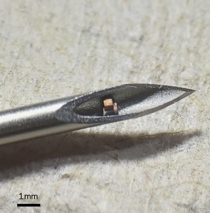
Researchers at Columbia Engineering report that they have built what they say is the world’s smallest single– system, consuming a total volume of less than 0.1 mm3. The system is as small as a dust mite and visible only under a microscope. In order to achieve this, the team used ultrasound to both power and communicate with the device wirelessly. The study was published online May 7 in Science Advances.

7:01 they talk about Church’s comments of ending aging by 2030. Also this appears to be a part one.


In this video Professor Church talks about his theory of aging and touches on his ideas on the future of aging.

George Church is the Robert Winthrop Professor of Genetics at Harvard Medical School, a Professor of Health Sciences and Technology at Harvard and MIT. Professor Church helped initiate the Human Genome Project in 1984 and the Personal Genome Project in 2005. He is widely recognized for his innovative contributions to genomic science and his many pioneering contributions to chemistry and biomedicine. He has co-authored 580 paper, 143 patent publications & the book “Regenesis”.

George Church Links.
Professor Church’s Lab at Harvard.
https://arep.med.harvard.edu/

Professor Church’s Book on Amazon.
Regenesis: How Synthetic Biology Will Reinvent Nature and Ourselves.
https://amzn.to/3vTAVKo.

Twitter Link.
https://twitter.com/geochurch.
***********************************************************************************
If you would like to support our channel, we’d love a coffee…thank you! https://www.buymeacoffee.com/mhealthspan.

#TheoryOfAging #GeorgeChurch #Harvard

I still don’t get how there seems to be No organized effort anywhere to achieve the ability to 3D print a perfect genetic match of all organs by 2025 — 2030. You would think some government somewhere would want to work round the clock on this.


NIBIB-funded engineers at the University of Buffalo have fine-tuned the use of stereolithography for 3D printing of organ models that contain live cells. The new technique is capable of printing the models 10–50 times faster than the industry standard-;in minutes instead of hours-; a major step in the quest to create 3D-printed replacement organs.

Conventional 3D printing involves the meticulous addition of material to the 3D model with a small needle that produces fine detail but is extremely slow —taking six or seven hours to print a model of a human part, such as a hand, for instance. The lengthy process causes cellular stress and injury inhibiting the ability to seed the tissues with live, functioning cells.

The method developed by the SUNY Buffalo group, led by Rougang Zhao, PhD, Associate Professor of Biomedical Engineering in the Jacobs School of Medicine & Biomedical Sciences, takes a different approach that minimizes damage to live cells. The rapid, cell friendly technique is a significant step towards creating printed tissues infused with large numbers of living cells.