Toggle light / dark theme

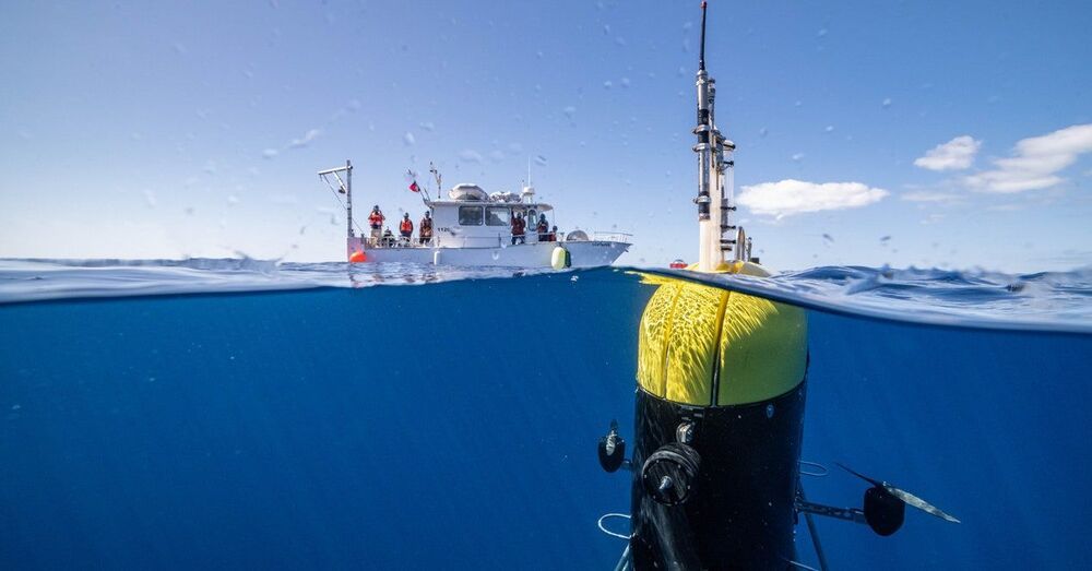
But now a spy swims among them: Mesobot. Today in the journal Science Robotics, a team of engineers and oceanographers describes how they got a new autonomous underwater vehicle to lock onto movements of organisms and follow them around the ocean’s “twilight zone,” a chronically understudied band between 650 feet and 3200 feet deep, which scientists also refer to as mid-water. Thanks to some clever engineering, the researchers did so without flustering these highly sensitive animals, making Mesobot a groundbreaking new tool for oceanographers.

“It’s super cool from an engineering standpoint,” says Northeastern University roboticist Hanumant Singh, who develops ocean robots but wasn’t involved in this research. “It’s really an amazing piece of work, in terms of looking at an area that’s unexplored in the ocean.”

Mesobot looks like a giant yellow-and-black AirPods case, only it’s rather more waterproof and weighs 550 pounds. It can operate with a fiber-optic tether attached to a research vessel at the surface, or it can swim around freely.
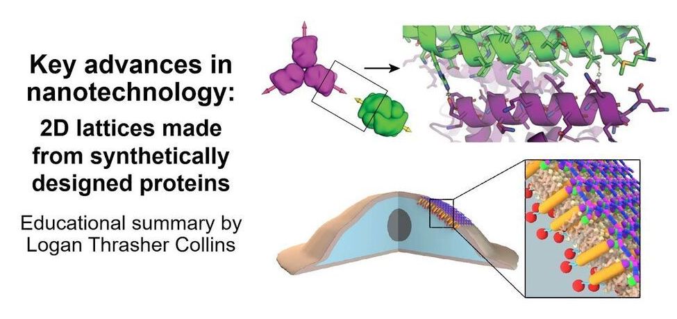
Check out my short video in which I explain some super exciting research in the area of nanotechnology: de novo protein lattices! I specifically discuss a journal article by Ben-Sasson et al. titled “Design of biologically active binary protein 2D materials”.


Here, I explain an exciting nanotechnology paper “Design of biologically active binary protein 2D materials” (https://doi.org/10.1038/s41586-020-03120-8).

Though I am not involved in this particular research myself, I have worked in adjacent areas such as de novo engineering of aggregating antimicrobial peptides, synthetic biology, nanotechnology-based tools for neuroscience, and gene therapy. I am endlessly fascinated by this kind of computationally driven de novo protein design and would love to incorporate it in my own research at some point in the future.

I am a PhD candidate at Washington University in St. Louis and the CTO of the startup company Conduit Computing. I am also a published science fiction writer and a futurist. To learn more about me, check out my website: https://logancollinsblog.com/.

Circa 2020


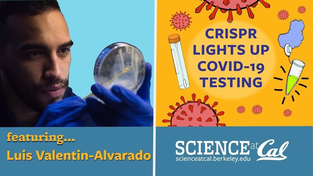
Researchers at UC Berkeley have developed a rapid test for SARS-CoV-2 that uses an enzyme to cleave viral RNA, initiating a fluorescent signal that can be detected using a smartphone camera, and which can provide a quantitative measurement of the level of viral particles in the sample. The test produce a result in as little as 30 minutes and does not require bulky or expensive laboratory equipment.

Rapid testing is key to measuring and stopping the spread of COVID-19, but current tests, such as PCR, are time consuming and require expensive laboratory equipment, creating a bottleneck in obtaining results. Researchers have been developing alternatives, and this latest technology was rapidly repurposed when the pandemic began. Originally intended to detect HIV in blood samples, the Berkeley researchers have pivoted to allow the device to detect SARS-CoV-2 in nasal swab samples.

The test relies on CRISPR-Cas, originally developed as a gene editing technology. When a pre-programmed Cas13 enzyme is added to the sample, it can cleave RNA sequences from the SARS-CoV-2 virus. This results in other nearby sequences being cleaved also, including a probe that releases fluorescent light when cleaved. The device uses a laser to excite this fluorescence and a smartphone camera can then detect the light, providing a quantitative measurement of the viral particles present in the sample.

✅ Instagram: https://www.instagram.com/pro_robots.

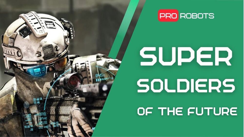
You are on the Pro Robot channel and today we are going to talk about the soldiers of the future. Exoskeletons, ballistic helmets, military suits, chips and more are already being introduced into the armaments of different countries. In this issue we will find out what the super-soldier of the future will be like and what developments are being conducted in the military industry. Watch the video to the end and write your opinion in the comments: will robots replace humans in military service?

0:00 In this video.
0:30 Combat glasses.
2:26 Devtac Ronin Kevlar ballistic helmet.
3:00 STILE smart fabric.
3:42 Stealth Cloak.
4:10 Future Soldier System Full Suit.
5:15 Sotnik Suit.
5:55 Exoskeleton Military.
6:32 PowerWalk current generator exoskeletons.
7:00 Human Universal Load Carrier exoskeleton with hydraulic drive.
7:24 A Flying Suit for Military.
7:48 Jetpack.
8:09 Invasive chips and genetic engineering.
9:02 Man-Made Lightning.

More interesting and useful content:

✅ Elon Musk Innovation https://www.youtube.com/playlist?list=PLcyYMmVvkTuQ-8LO6CwGWbSCpWI2jJqCQ
✅Future Technologies Reviews https://www.youtube.com/playlist?list=PLcyYMmVvkTuTgL98RdT8-z-9a2CGeoBQF
✅ Technology news.

#prorobots #technology #roboticsnews.

Circa 2019


The evolution of micro and nanofabrication approaches significantly spurred the advancements of cardiac tissue engineering over the last decades. Engineering in the micro and nanoscale allows for the rebuilding of heart tissues using cardiomyocytes. The breakthrough of human induced pluripotent stem cells expanded this field rendering the development of human tissues from adult cells possible, thus avoiding the ethical issues of the usage of embryonic stem cells but also creating patient-specific human engineered tissues. In the case of the heart, the combination of cardiomyocytes derived from human induced pluripotent stem cells and micro/nano engineering devices gave rise to new therapeutic approaches of cardiac diseases. In this review, we survey the micro and nanofabrication methods used for cardiac tissue engineering, ranging from clean room-based patterning (such as photolithography and plasma etching) to electrospinning and additive manufacturing. Subsequently, we report on the main approaches of microfluidics for cardiac culture systems, the so-called “Heart on a Chip”, and we assess their efficacy for future development of cardiac disease modeling and drug screening platforms.

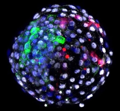
The way the team made the human–monkey embryo is similar to previous attempts at half-human chimeras.

Here’s how it goes. They used de-programmed, or “reverted,” human stem cells, called induced pluripotent stem cells (iPSCs). These cells often start from skin cells, and are chemically treated to revert to the stem cell stage, gaining back the superpower to grow into almost any type of cell: heart, lung, brain…you get the idea. The next step is preparing the monkey component, a fertilized and healthy monkey egg that develops for six days in a Petri dish. By this point, the embryo is ready for implantation into the uterus, which kicks off the whole development process.

This is where the chimera jab comes in. Using a tiny needle, the team injected each embryo with 25 human cells, and babied them for another day. “Until recently the experiment would have ended there,” wrote Drs. Hank Greely and Nita Farahany, two prominent bioethicists who wrote an accompanying expert take, but were not involved in the study.

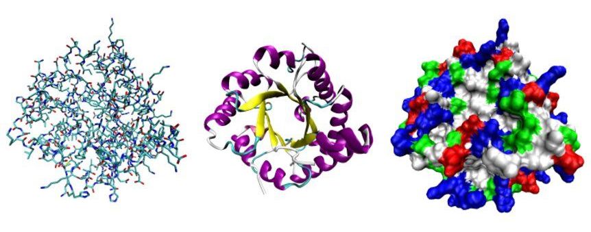
While DNA provides the genetic recipe book for biological form and function, it is the job of the body’s proteins to carry out the complex commands dictated by DNA’s genetic code.

Stuart Lindsay, a researcher at the Biodesign Institute at ASU, has been at the forefront of efforts to improve rapid DNA sequencing and has more recently applied his talents to explore the much thornier problem of sequencing molecules, one molecule at a time.

In a new overview article, Lindsay’s efforts are described along with those of international colleagues, who are applying a variety of innovative strategies for protein sequencing at the single-cell, and even single-molecule level.

A research team from the University of Massachusetts Amherst has created an electronic microsystem that can intelligently respond to information inputs without any external energy input, much like a self-autonomous living organism. The microsystem is constructed from a novel type of electronics that can process ultralow electronic signals and incorporates a device that can generate electricity “out of thin air” from the ambient environment.

The groundbreaking research was published June 7 in the journal Nature Communications.

Jun Yao, an assistant professor in the electrical and computer engineering (ECE) and an adjunct professor in biomedical engineering, led the research with his longtime collaborator, Derek R. Lovley, a Distinguished Professor in microbiology.

Imagine you’re a fisherman living by a lake with a rowboat. Every day, you row out on the calm waters and life is good. But then your family grows, and you need more fish, so you go to the nearby river. Then, you realize you go farther and faster on the river. You can’t take your little rowboat out there—it’s not built for those currents. So, you learn everything you can about how rivers work and build a better boat. Life is good again…until you realize you need to go farther still, out on the ocean. But ocean rules are nothing like river rules. Now you have to learn how ocean currents work, and then design something even more advanced that can handle that new space.

Communication frequencies are just like those water currents. And the boats are just like the tools we build to communicate. The challenge is twofold: learning enough about the nature of each frequency and then engineering novel devices that will work within them. In a recent paper published in Proceedings of the IEEE, the flagship publication of the largest engineering society in the world, one USC Viterbi School of Engineering researcher has done just that for the next generation of cellular networks—6G.

Andy Molisch, professor of electrical and computer engineering at USC Viterbi and the holder of the Solomon Golomb—Andrew and Erna Viterbi Chair, together with colleagues from Lund University in Sweden, New Zealand Telecom, and King’s College London, explained that we have more options for communications at 6G frequency than previously thought. Think of it as something like early explorers suddenly discovering the gulf stream.

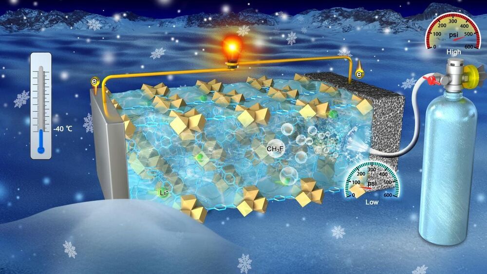
A new technology could dramatically improve the safety of lithium-ion batteries that operate with gas electrolytes at ultra-low temperatures. Nanoengineers at the University of California San Diego developed a separator—the part of the battery that serves as a barrier between the anode and cathode—that keeps the gas-based electrolytes in these batteries from vaporizing. This new separator could, in turn, help prevent the buildup of pressure inside the battery that leads to swelling and explosions.

“By trapping , this can function as a stabilizer for volatile electrolytes,” said Zheng Chen, a professor of nanoengineering at the UC San Diego Jacobs School of Engineering who led the study.

The new separator also boosted performance at ultra–. Battery cells built with the new separator operated with a high capacity of 500 milliamp-hours per gram at-40 C, whereas those built with a commercial separator exhibited almost no capacity. The battery cells still exhibited high capacity even after sitting unused for two months—a promising sign that the new separator could also prolong shelf life, the researchers said.