Toggle light / dark theme

Post-mortem changes may shed light on important brain studies.

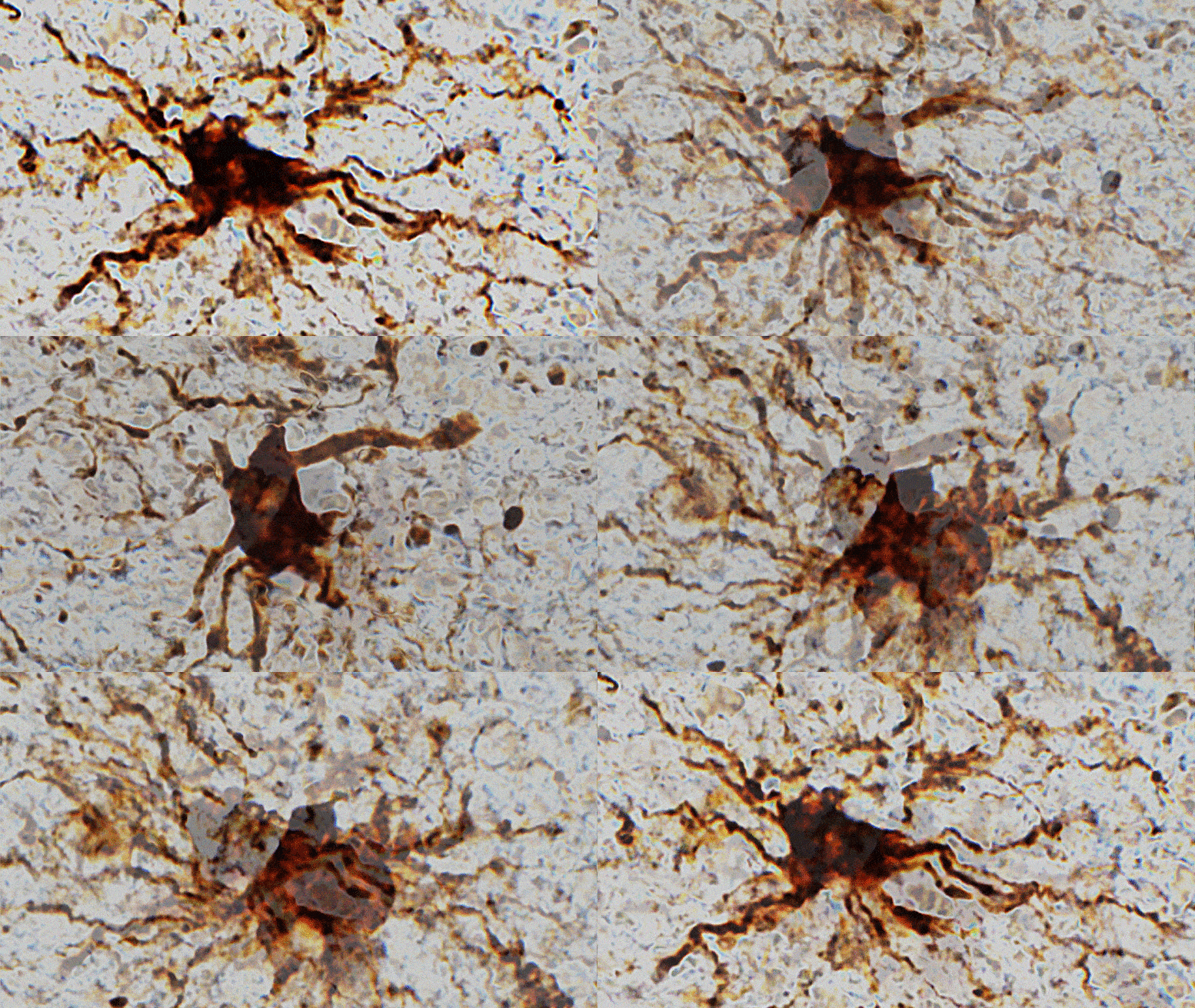
In the hours after we die, certain cells in the human brain are still active. Some cells even increase their activity and grow to gargantuan proportions, according to new research from the University of Illinois Chicago.

In a newly published study in the journal Scientific Reports, the UIC researchers analyzed gene expression in fresh brain tissue — which was collected during routine brain surgery — at multiple times after removal to simulate the post-mortem interval and death. They found that gene expression in some cells actually increased after death.

Using a new class of nanoparticles that are two thousand times thinner than a human hair, Sakhrat Khizroev, a professor of electrical and computer engineering at the University’s College of Engineering, hopes to unlock the secrets of the brain.

The neurosurgeon who examined Sakhrat Khizroev after he lost his eyesight in a horrible accident told the young scientist that his vision would come back slowly. Then, after months of living in darkness, it finally started to return.

At first, the images were blurry and fragmented, as if someone were looking through a narrow window and seeing only part of a picture. But with each passing day, everything Khizroev looked at appeared clearer, sharper.

LOS ANGELES — UCLA neuroscientists reported Monday that they have transferred a memory from one animal to another via injections of RNA, a startling result that challenges the widely held view of where and how memories are stored in the brain.

The finding from the lab of David Glanzman hints at the potential for new RNA-based treatments to one day restore lost memories and, if correct, could shake up the field of memory and learning.


“It’s pretty shocking,” said Dr. Todd Sacktor, a neurologist and memory researcher at SUNY Downstate Medical Center in Brooklyn, N.Y. “The big picture is we’re working out the basic alphabet of how memories are stored for the first time.” He was not involved in the research, which was published in eNeuro, the online journal of the Society for Neuroscience.

Many scientists are expected to view the research more cautiously. The work is in snails, animals that have proven a powerful model organism for neuroscience but whose simple brains work far differently than those of humans. The experiments will need to be replicated, including in animals with more complex brains. And the results fly in the face of a massive amount of evidence supporting the deeply entrenched idea that memories are stored through changes in the strength of connections, or synapses, between neurons.

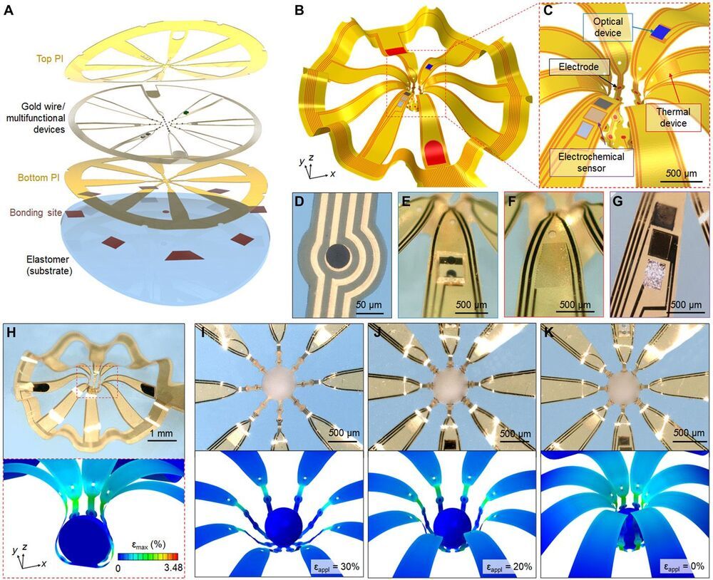
Three-dimensional (3D), submillimeter-scale constructs of neural cells, known as cortical spheroids, are of rapidly growing importance in biological research because these systems reproduce complex features of the brain in vitro. Despite their great potential for studies of neurodevelopment and neurological disease modeling, 3D living objects cannot be studied easily using conventional approaches to neuromodulation, sensing, and manipulation. Here, we introduce classes of microfabricated 3D frameworks as compliant, multifunctional neural interfaces to spheroids and to assembloids. Electrical, optical, chemical, and thermal interfaces to cortical spheroids demonstrate some of the capabilities. Complex architectures and high-resolution features highlight the design versatility. Detailed studies of the spreading of coordinated bursting events across the surface of an isolated cortical spheroid and of the cascade of processes associated with formation and regrowth of bridging tissues across a pair of such spheroids represent two of the many opportunities in basic neuroscience research enabled by these platforms.

Progress in elucidating the development of the human brain increasingly relies on the use of biosystems produced by three-dimensional (3D) neural cultures, in the form of cortical spheroids, organoids, and assembloids (1–3). Precisely monitoring the physiological properties of these and other types of 3D biosystems, especially their electrophysiological behaviors, promises to enhance our understanding of the interactions associated with development of the nervous system, as well as the evolution and origins of aberrant behaviors and disease states (4–8). Conventional multielectrode array (MEA) technologies exist only in rigid, planar, and 2D formats, thereby limiting their functional interfaces to small areas of 3D cultures, typically confined to regions near the bottom contacting surfaces.

Summary: Using brain organoid models, researchers have identified how the brain grows much larger and has three times as many neurons, as the brains of chimpanzees and gorillas.

Source: UK Research and Innovation.

A new study is the first to identify how human brains grow much larger, with three times as many neurons, compared with chimpanzee and gorilla brains. The study, led by researchers at the Medical Research Council (MRC) Laboratory of Molecular Biology in Cambridge, UK, identified a key molecular switch that can make ape brain organoids grow more like human organoids, and vice versa.

Should interest those into links on aging/longevity and neuroscience.


The mammalian center for learning and memory, hippocampus, has a remarkable capacity to generate new neurons throughout life. Newborn neurons are produced by neural stem cells (NSCs) and they are crucial for forming neural circuits required for learning and memory, and mood control. During aging, the number of NSCs declines, leading to decreased neurogenesis and age-associated cognitive decline, anxiety, and depression. Thus, identifying the core molecular machinery responsible for NSC preservation is of fundamental importance if we are to use neurogenesis to halt or reverse hippocampal age-related pathology.

While there are increasing number of tools available to study NSCs and neurogenesis in mouse models, one of the major hurdles in exploring this fundamental biological process in the human brain is the lack of specific NSCs markers amenable for advanced imaging and in vivo analysis. A team of researchers led by Dr. Mirjana Maletić-Savatić, associate professor at Baylor College of Medicine and investigator at the Jan and Dan Duncan Neurological Research Institute at Texas Children’s Hospital, and Dr. Louis Manganas, associate professor at the Stony Brook University, decided to tackle this problem in a rather unusual way. They reasoned that if they could find proteins that are present on the surface of NSCs, then they could eventually make agents to “see” NSCs in the .

“The ultimate goal of our research is to maintain neurogenesis throughout life at the same level as it is in the young brains, to prevent the decline in our cognitive capabilities and reduce the tendency towards mood disorders such as depression, as we age. To do that, however, we first need to better understand this elusive, yet fundamental process in humans. However, we do not have the tools to study this process in live humans and all the knowledge we have gathered so far comes from analyses of the postmortem brains. And we cannot develop tools to detect this process in people because existing NSC markers are present within cells and unreachable for in vivo visualization,” Maletić-Savatić said. “So, in collaboration with our colleagues from New York and Spain, we undertook this study to find surface markers and then develop tools such as ligands for positron emission tomography (PET) to visualize them using advanced real-time in vivo brain imaging.”
Circa 2013


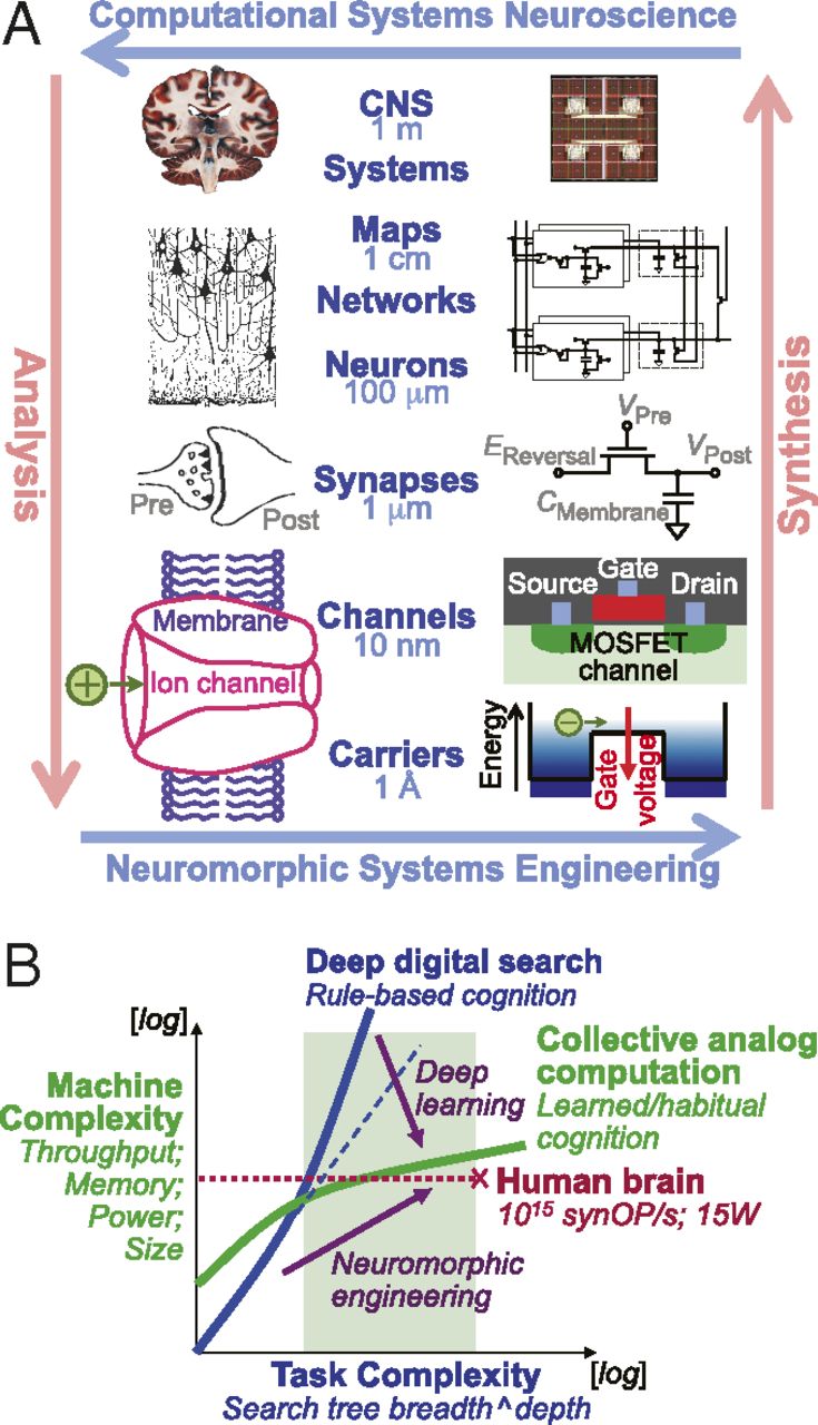
One of the greatest aspirations of the human mind has been to realize machines that surpass its cognitive intelligence. The rapid expansion in computing power, about to exceed the equivalent of the human brain, has yet to produce such a machine. The article by Neftci et al. in PNAS (1) offers a refreshing and humbling reminder that the brain’s cognition does not arise from exacting digital precision in high-performance computing, but rather emerges from an extremely efficient and resilient collective form of computation extending over very large ensembles of sluggish, imprecise, and unreliable analog components. This observation, first made by John von Neumann in his final opus (2), continues to challenge scientists and engineers several decades later in figuring and reproducing the mechanisms underlying brain-like forms of cognitive computing.

Related developments are currently unfolding in collaborative initiatives engaging scientists and engineers, on a grander scale, in advancing neuroscience toward understanding the brain. In parallel with the Human Brain Project in Europe, the Brain Research through Advancing Innovative Neurotechnologies Initiative promises groundbreaking advances in enabling tools for revolutionizing neuroscience by developing nanotechnology to probe brain function at greatly increased spatial and temporal detail. Engineers are poised to contribute even further in revolutionizing such developments in neuroscience. In this regard it is helpful to relate the inquisitive nature of science—analysis—to the constructive power of engineering, synthesis.

The research team, led by Todd Lencz, PhD, with Itsik Pe’er, PhD, Tom Maniatis, PhD, and Erin Flaherty, PhD, of Columbia University, carried out a genetic study identifying a single letter change in the DNA code in the PCDHA3 gene that is associated with schizophrenia. The affected gene makes a type of protein called a protocadherin, which generates a cell surface “barcode” required for neurons to recognize, and communicate with, other neurons. They found that the PCDHA3 variant blocks this normal protocadherin function.

The discovery was made possible by the special genetic characteristics of the samples studied by Lencz’s team—patients with schizophrenia and healthy volunteers drawn from the Ashkenazi Jewish population. The Ashkenazi Jewish population represents an important population for study based on its unique history. Just a few hundred individuals who migrated to Eastern Europe less than 1000 years ago are the ancestors of nearly 10 million Ashkenazi Jews today. This lineage, combined with a tradition of marriage within the community, has resulted in a more uniform genetic background in which to identify disease-related variants.

“In addition to our primary findings regarding PCDHA3 and related genes, we were able— due to the unique characteristics of the Ashkenazi population—to replicate several prior findings in schizophrenia despite relatively small sample sizes,” said Lencz, professor in the Institute of Behavioral Science at the Feinstein Institutes. “In our study, we demonstrated this population represents a smart, cost-effective strategy for identifying disease-related genes. Our findings allow us to zero in on a novel aspect of brain development and function in our quest to develop new treatments for schizophrenia.”