Toggle light / dark theme

Studies of hibernating animals suggest that the molecular and synaptic integrity of neurons in the cerebral cortex that underlie self and consciousness is maintained in many cases when from the outside the brain appears dead.


A striking feature of medicine over the past few centuries has been our growing ability to bring people back from the “dead.” For most of human history, patients who were unconscious and not breathing were treated as though they had died. But the concept of resuscitation emerged as doctors grew to understand the basic function of the lungs and airways. That led to new techniques and tools capable of restoring both breathing and heartbeat — and the realization that cardiac arrest was not always a death sentence. That, in turn, gave rise to a distinction between what’s now called clinical death versus brain death.

Today that brain focus continues, but with a growing glimmer of hope that even brain death might be reversible in some instances. These dreams are fueled by research showing that the disappearance of brain function is not the same as deletion of computer files. Rather, it represents a deterioration of the pathways that normally enable different parts of the brain to communicate. This idea was bolstered recently with the 2017 success in France, where a patient was partially revived from a 15-year vegetative state. It also dovetails with insights from the study of hibernating animals.

Medical magic: Expansion of resuscitation of capability over the centuries

Resuscitation entered the field of medicine beginning in the 1500s, not surprisingly with practices that may have helped victims occasionally, but with a low success rate. Such practices included flagellation, and were based on experience rather than an understanding of the underlying physiological processes. This started to change with the use of air bellows, based on an understanding that air needed to flow in and out of the lungs. But the 1740s, mouth-to-mouth resuscitation was standard practice in France for resuscitating drowning victims.

Brain implants are neural implants that are used to stimulate the parts & structures of the nervous system. These implants are technical systems that communicate with the nervous system and help to enhance senses, physical movement, and memory after a stroke or other head injuries. Deep brain stimulation and spinal cord stimulation are used to treat depression, obsessive-compulsive disorder and epilepsy, among other neural disorders.

Technology has touched all aspects of our lives in this 21st century world that we live in and has, in fact, become an integral part of our lives. So much so that we start feeling incomplete as soon as we manage to get away from it.

No doubt, it has enhanced our lives in many different ways and today we can do things that we couldn’t have even imagined a few decades ago. I mean, sending a text to someone half way around the world in an instant? Almost feels like magic, doesn’t it?

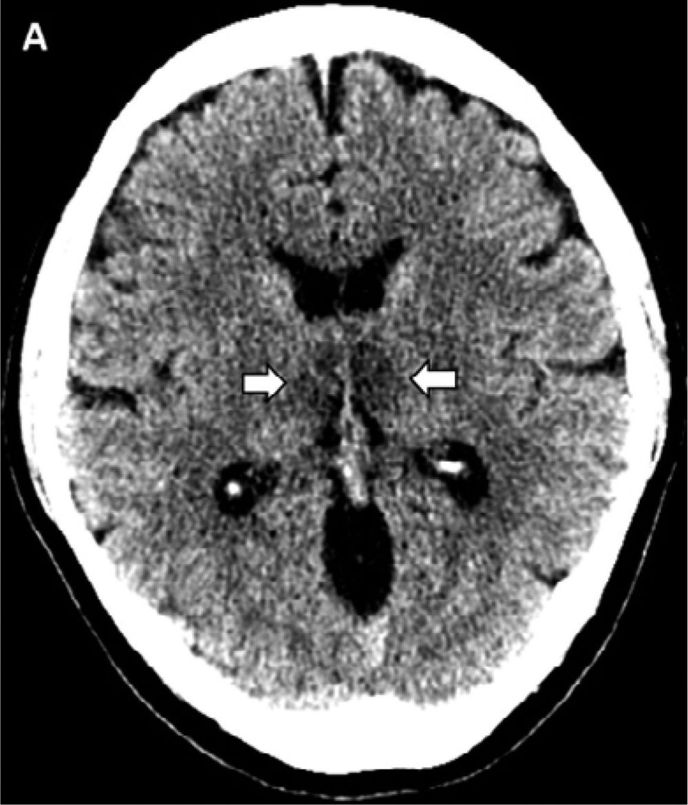
This is the first reported case of COVID-19–associated acute necrotizing hemorrhagic encephalopathy. As the number of patients with COVID-19 increases worldwide, clinicians and radiologists should be watching for this presentation among patients presenting with COVID-19 and altered mental status.


Home Radiology Recently Published PreviousNext Reviews and CommentaryFree AccessImages in Radiology COVID-19–associated Acute Hemorrhagic Necrotizing Encephalopathy: CT and MRI FeaturesNeo Poyiadji, Gassan Shahin, Daniel Noujaim, Michael Stone, Suresh Patel, Brent Griffith Neo Poyiadji, Gassan S…

Dr. Susan White and her genetics team treated two triplets from a family who had an undiagnosed neurodegenerative disorder in 2014. After one year of age, the children’s developmental skills declined. They lost visual coordination. Feeding and swallowing food became impossible. The children developed intractable seizures.

Exactly what led to their neurodegeneration was a mystery.

“As you can imagine, that was just a horrendous experience for their family and we suspected a genetic condition because of that pattern of problems occurring in both children,” White, an associate professor at Murdoch Children’s Research Institute (MCRI) and Victorian Clinical Genetics Services (VCGS), said in an interview with Being Patient.

Given the rapid development of virtual reality technology, we may very well be moving toward a time when we’re able to manage the brain’s memories.


Could we develop a similar capability? That may depend heavily upon a handful of ambitious attempts at brain-computer interfacing. But science is moving in baby steps with other tactics in both laboratory animals and humans.

Thus far, there have been some notable achievements in rodent experiments, that haven’t done so well with humans. We don’t have a beam that can go into your mind and give you 60 years worth of new experiences. Nevertheless, the emerging picture is that the physical basis of memory is understandable to the point that we should be able to intervene — both in producing and eliminating specific memories.

At MIT’s Center for Neural Circuit Genetics, for example, scientists have modified memories in mice using an optogenetic interface. This technology involves genetic modification of tissues, in this case within the brain, to express proteins that respond to light. Triggered by implants that deliver laser beams, brain cells can be triggered to be more or less active. In research that has been published in the prestigious journal Nature, the MIT team used the approach in specific brain circuits important to memory consolidation. The researchers were able to enhance the development of negative memories — for instance a shock given to an animal’s leg — and also to convert those negative memories into positive memories. The latter was achieved by letting male mice enjoy some time with females, while nerve cells that usually deliver the negative impulses associated with the former shock were stimulated through the optogenetic interface.

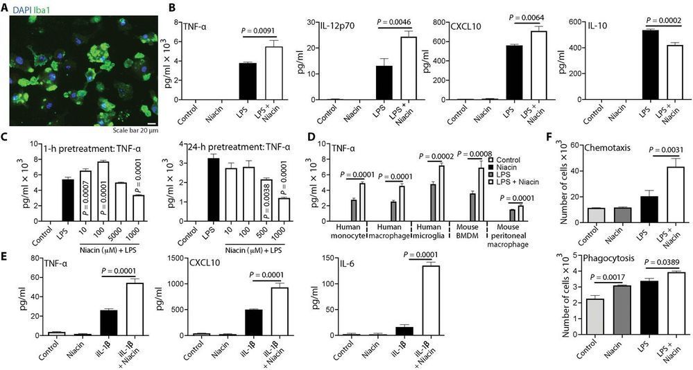
Although innate immune cells are typically present inside tumors, they often have an inactive phenotype such that they are ineffective at killing the cancer cells or even promote tumor growth. Sarkar et al. discovered that it may be possible to reprogram these cells to a more active type using niacin (vitamin B3). The authors showed that niacin-exposed monocytes can inhibit the growth of brain tumor–initiating cells. Moreover, niacin treatment of intracranial mouse models of glioblastoma increased monocyte and macrophage infiltration into the tumors, stimulated antitumor immune responses, and extended the animals’ survival, especially when combined with the chemotherapeutic drug temozolomide.

Glioblastomas are generally incurable partly because monocytes, macrophages, and microglia in afflicted patients do not function in an antitumor capacity. Medications that reactivate these macrophages/microglia, as well as circulating monocytes that become macrophages, could thus be useful to treat glioblastoma. We have discovered that niacin (vitamin B3) is a potential stimulator of these inefficient myeloid cells. Niacin-exposed monocytes attenuated the growth of brain tumor–initiating cells (BTICs) derived from glioblastoma patients by producing anti-proliferative interferon-α14. Niacin treatment of mice bearing intracranial BTICs increased macrophage/microglia representation within the tumor, reduced tumor size, and prolonged survival. These therapeutic outcomes were negated in mice depleted of circulating monocytes or harboring interferon-α receptor–deleted BTICs. Combination treatment with temozolomide enhanced niacin-promoted survival.

As research on Alzheimer’s disease (AD) advances, a desperate need remains for an easy blood test to help diagnose the condition as early as possible. Ideally, such a test could also distinguish AD from other forms of dementia that produce similar symptoms. As published recently in Nature Medicine, an NIH-funded research team has designed a simple blood test that is on course to meet these criteria [1].

The latest work builds on a large body of work showing that one secret to predicting a person’s cognitive decline and treatment response in AD lies in a protein called tau. Using the powerful, but expensive, approach of PET scan imaging, we know that tau builds up in the brain as Alzheimer’s disease progresses. We also know that some tau spills from the brain into the bloodstream.

The trouble is that the circulating tau protein breaks down far too quickly for a blood test to offer a reliable measure of what’s happening in a person’s brain. A few years ago, researchers discovered a possible solution: test for blood levels of a slightly different and more stable version of the protein called pTau181 [2]. (The “p” in its name comes from the addition of phosphorus in a particular part of the protein’s structure.)

Scientists at Flinders University have, for the first time, identified a specific type of sensory nerve ending in the gut and how these may ‘talk’ to the spinal cord, communicating pain or discomfort to the brain.

This discovery is set to inform the development of new medications to treat problems associated with gut-to-brain communication, paving the way for targeted treatments to mitigate related dysfunction.

While our understanding of the gut’s neurosensory abilities has grown rapidly in recent years, two of the great mysteries have been where and how the different types of sensory nerve endings in the gut lie, and how they are activated.