Toggle light / dark theme

This is a story about math educator Mark Saul, and his Math on The Border program for migrant children. Mark and his team are trying to work with these children, and to encourage them. Mark is not only one of the best math educators in the world, he is also an amazing human being.


Having an opportunity to use one’s brain is a basic human need, says Saul. Back at the Templeton Foundation, he studied under-exploited human capital and the boundless human potential. Despite their difficult past and uncertain future, migrant children are eager to build their math skills. Resourceful and resilient in the face of failure, they reshuffle the pieces and try again. They work in groups and make new friends along the way. Many of them are highly gifted – Saul can attest to that. It doesn’t take him long to see what these children, abandoned by life, are capable of with just a little encouragement. And he can tell from the looks on their faces how delighted they are at having their abilities recognized and valued.

Important that people read this given how much this spread.


If you have been following the mainstream media recently, you have probably seen a story about hyperbaric oxygen treatment and claims that it can reverse aging. Unfortunately, the media hype surrounding the results is nothing like the reality of the actual research paper, and this is another example of how shoddy journalism harms our field.

Welcome to the media circus

Back in July, we talked about how hyperbaric oxygen therapy may reduce age-related cognitive decline in older people, which was based on the results of another study. A new publication from the same team of Israeli scientists led by Prof. Shai Efrati has further explored these original findings, and while the results are interesting, the media hype and marketing surrounding those results is frankly ridiculous and entirely unwarranted.

Tiny Dancer

Chakrabartty’s tunneling barrier was built in just such a way that “you can control the flow of electrons. You can make it reasonably slow, down to one electron every minute and still keep it reliable.”

The team is hoping the technology could one day power glucose or even brain activity monitors without the need for batteries.

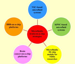
Neurodegenerative diseases (NDDs) include more than 600 types of nervous system disorders in humans that impact tens of millions of people worldwide. Estimates by the World Health Organization (WHO) suggest NDDs will increase by nearly 50% by 2030. Hence, development of advanced models for research on NDDs is needed to explore new therapeutic strategies and explore the pathogenesis of these disorders. Different approaches have been deployed in order to investigate nervous system disorders, including two-and three-dimensional (2D and 3D) cell cultures and animal models. However, these models have limitations, such as lacking cellular tension, fluid shear stress, and compression analysis; thus, studying the biochemical effects of therapeutic molecules on the biophysiological interactions of cells, tissues, and organs is problematic. The microfluidic “organ-on-a-chip” is an inexpensive and rapid analytical technology to create an effective tool for manipulation, monitoring, and assessment of cells, and investigating drug discovery, which enables the culture of various cells in a small amount of fluid (10−9 to 10−18 L). Thus, these chips have the ability to overcome the mentioned restrictions of 2D and 3D cell cultures, as well as animal models. Stem cells (SCs), particularly neural stem cells (NSCs), induced pluripotent stem cells (iPSCs), and embryonic stem cells (ESCs) have the capability to give rise to various neural system cells. Hence, microfluidic organ-on-a-chip and SCs can be used as potential research tools to study the treatment of central nervous system (CNS) and peripheral nervous system (PNS) disorders. Accordingly, in the present review, we discuss the latest progress in microfluidic brain-on-a-chip as a powerful and advanced technology that can be used in basic studies to investigate normal and abnormal functions of the nervous system.

Circa 2018

Livescience.com | By LIVESCIENCE


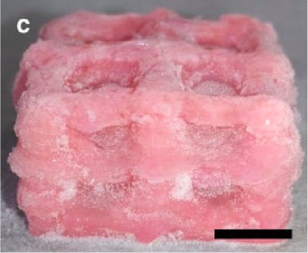
A new 3D-printing technique can create tissues as soft as a human’s squishy brain or spongy lungs — something that has not been possible before.

“Additive manufacturing,” or 3D printing, promises to allow doctors to produce tailored organs for patients using the patients’ own cells, which could help bring down the severe shortage of organs available for people who need transplants.

The replacement of lost neurons is a holy grail for neuroscience. A new promising approach is the conversion of glial cells into new neurons. Improving the efficiency of this conversion or reprogramming after brain injury is an important step towards developing reliable regenerative medicine therapies. Researchers at Helmholtz Zentrum München and Ludwig Maximilians University Munich (LMU) have identified a hurdle towards an efficient conversion: the cell metabolism. By expressing neuron-enriched mitochondrial proteins at an early stage of the direct reprogramming process, the researchers achieved a four times higher conversion rate and simultaneously increased the speed of reprogramming.

Neurons (nerve cells) have very important functions in the brain such as information processing. Many brain diseases, injuries and neurodegenerative processes, are characterized by the loss of neurons that are not replaced. Approaches in regenerative medicine therefore aim to reconstitute the neurons by transplantation, stem cell differentiation or direct conversion of endogenous non-neuronal cell types into functional neurons.

Researchers at Helmholtz Zentrum München and LMU are pioneering the field of direct conversion of glial cells into neurons which they have originally discovered. Glia are the most abundant cell type in the brain and can proliferate upon injury. Currently, researchers are able to convert glia cells into neurons — but during the process many cells die. This means that only few glial cells convert into functional nerve cells, making the process inefficient.

Here are just a few ways exercise changes the structure of our brain.

Memory

Many studies suggest that exercise can help protect our memory as we age. This is because exercise has been shown to prevent the loss of total brain volume (which can lead to lower cognitive function), as well as preventing shrinkage in specific brain regions associated with memory. For example, one magnetic resonance imaging (MRI) scan study revealed that in older adults, six months of exercise training increases brain volume.