Toggle light / dark theme

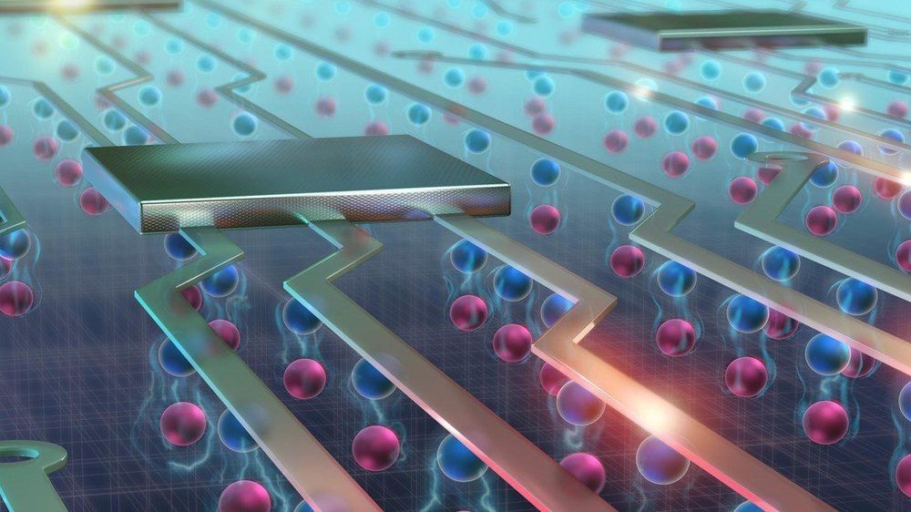
After developing a method to control exciton flows at room temperature, EPFL scientists have discovered new properties of these quasiparticles that can lead to more energy-efficient electronic devices.

They were the first to control flows at . And now, the team of scientists from EPFL’s Laboratory of Nanoscale Electronics and Structures (LANES) has taken their technology one step further. They have found a way to control some of the properties of excitons and change the polarization of the light they generate. This can lead to a new generation of electronic devices with transistors that undergo less energy loss and heat dissipation. The scientists’ discovery forms part of a new field of research called valleytronics and has just been published in Nature Photonics.

Excitons are created when an electron absorbs light and moves into a higher energy level, or “energy band” as they are called in solid quantum physics. This excited electron leaves behind an “electron hole” in its previous band. And because the electron has a and the hole a positive charge, the two are bound together by an electrostatic force called a Coulomb force. It’s this electron-electron hole pair that is referred to as an exciton.
Read more

A quick and easy test devised by scientists from the University of Queensland could transform cancer diagnosis as we know it.

Cancer is a difficult disease to diagnose because different types are characterised by different signatures. Until now, scientists have been unable to find a unique signature common to all forms of cancer that would set it apart from healthy cells.

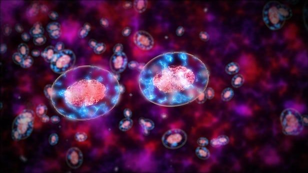
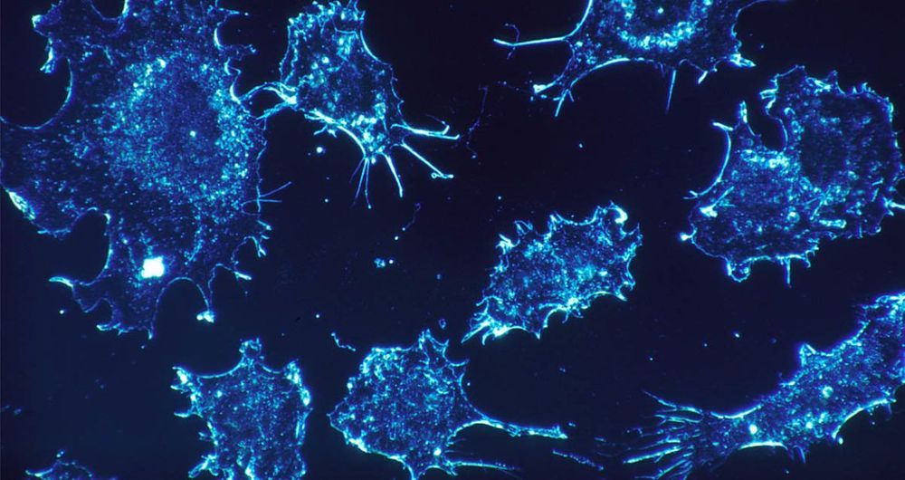
That’s what University of Queensland researchers Dr Laura Carrascosa, Dr Abu Sina and Professor Matt Trau have addressed. They have discovered a unique DNA nanostructure that seems to be common to all types of cancer and is visible when cancer cells are placed in water.

Read more

Histology is used to identify structural details of tissue at the microscale in the pathology lab, but analyses remain two-dimensional (2D) as they are limited to the same plane. Nondestructive 3D technologies including X-ray micro and nano-computed tomography (nanoCT) have proven validity to understand anatomical structures, since they allow arbitrary viewing angles and 3D structural detail. However, low attenuation of soft tissue has hampered their application in the field of 3D virtual histology. In a recent study, now published on Scientific Reports, Mark Müller and colleagues at the Department of Physics and Bioengineering have developed a hematein-based X-ray staining method to specifically target cell nuclei, followed by demonstrations on a whole liver lobule of a mouse.


Scientists have created a rubbery, shape-shifting material that morphs from one sophisticated form to another on demand.

The shapes programmed into a polymer appear in ambient conditions and melt away when under heat. The process also works in reverse.

The smooth operation belies a battle at the nanoscale, where liquid crystals and the elastomer in which they’re embedded fight for control. When cool, the shape programmed into the liquid crystals dominates, but when heated, the crystals relax within the rubber band-like elastomer, like ice melting into water.

Read more

Coming up with potent anti-cancer drugs is one thing, delivering them to the site of a tumor inside the body is very much another. With a complicated organism guarded by a highly evolved immune system to navigate, getting these particles to there target in one piece is a challenging task, and one that scientists are continuing to tackle from all angles. A promising new approach developed at Virginia Tech leans on the penetrative properties of a salmonella infection, which they’ve found can be used as a vehicle to smuggle cancer-fighting nanoparticles into a tumor in a huge abundance.

Read more

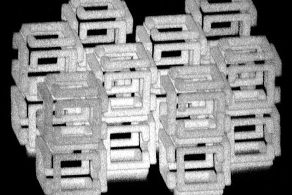
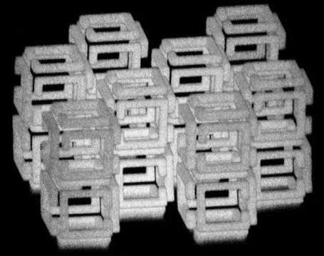
MIT researchers invented a method of shrinking objects to the nanoscale.

The team can generate structures one-thousandth the volume of the original using a variety of materials, including metals, quantum dots, and DNA.

Existing techniques—like etching patterns onto a surface with light—work for 2D nanostructures, but not 3D. And while it’s possible to make 3D nanostructures, the process is slow, challenging, and restrictive.

Read more