Toggle light / dark theme

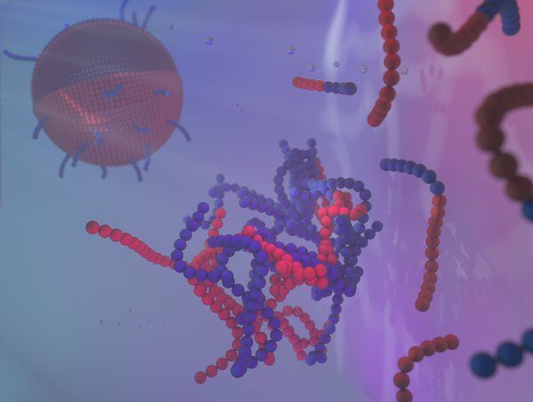
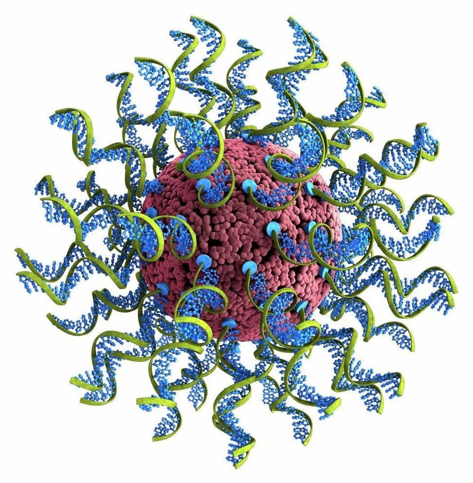
  • Spherical nucleic acids are a class of personalized medicines for treating cancer and other diseases
  • SNAs are challenging to optimize because their structures can vary in many ways
  • Northwestern University team developed a library approach and machine learning to rapidly synthesize, analyze and select for potent SNA medicines

EVANSTON, Ill.— With their ability to treat a wide a variety of diseases, (SNAs) are poised to revolutionize medicine. But before these digitally designed nanostructures can reach their full potential, researchers need to optimize their various components.

A Northwestern University team led by nanotechnology pioneer Chad A. Mirkin has developed a direct route to optimize these challenging particles, bringing them one step closer to becoming a viable treatment option for many forms of cancer, , neurological disorders and more.

Read more

Biological membranes, and man-made variants, consist of amphiphilic molecules, of which soap is an example. These molecules have a head that bonds with water, but a tail that turns away from water. You can imagine that a group of such molecules in water, preferably puts the tails together, and sticks the heads out, towards the water. Similar processes also dominate the creation of membranes. Often they are spherical, like liposomes, so you can, for example, put a medicine in it. And also the ultimate , the cell wall, is constructed in a similar way.

How nano-droplets self-assemble

Until now, the formation of ‘micelles’ was considered to be the first step in membrane formation. A micelle is an extremely small spherical structure (about 100 nanometers) of amphiphilic molecules—all with the tails inwards and the heads outwards. However, researchers at Eindhoven University of Technology discovered a different beginning: the formation of nano-droplets in with a higher concentration of amphiphilic molecules. At the interface of that drop, the amphiphilic molecules, as it were, take each others’ hands: first they form spheres, which then change into cylinders or plates, and then a closed membrane is created that encloses the nano-droplet. With this so-called ‘self-assembly’ process, the droplet has become a liposome.

Read more

stretchy nanotubesLighter than air! Stronger than steel! More flexible than rubber! No, it’s not an upcoming superhero flick: It’s the latest marvelous formulation of carbon nanotubes–at least as reported by the creators of the new super-material. Researchers working on artificial muscles say they’ve created nanotech ribbons that make our human muscles look puny by comparison. The ribbons, which are made of long, entangled 11-nanometer-thick nanotubes, can stretch to more than three times their normal width but are stiffer and stronger than steel… They can expand and contract thousands of times and withstand temperatures ranging from −190 to over 1,600 °C. What’s more, they are almost as light as air, and are transparent, conductive, and flexible [Technology Review].

The material is made from bundles of vertically aligned nanotubes that respond directly to electricity. Lengthwise, the muscle can expand and contract with tremendous speed; from side-to-side, it’s super-stiff. Its possibilities may only be limited by the imaginations of engineers. [The material’s composition] “is akin to having diamond-like behavior in one direction, and rubber-like behavior in the others” [Wired], says material scientist John Madden, who wasn’t involved in the research.

When voltage is applied to the material, the nanotubes become charged and push each other away, causing the material to expand at a rate of 37,000 percent per second, tripling its width. That’s 10 times as far and 1000 times as fast as natural muscle can move, and the material does so while generating 30 times as much force as a natural muscle [IEEE Spectrum]. The researchers put together a video to illustrate the concept, and will publish their work in Science tomorrow.

Read more

Developed in the 1940s, AA7075 is an aluminum alloy that’s almost as strong as steel, yet it weighs just one third as much. Unfortunately its use has been limited, due to the fact that pieces of it couldn’t be securely welded together. That’s recently changed, however, thanks to the use of titanium carbide nanoparticles.

Read more

Circa 2018


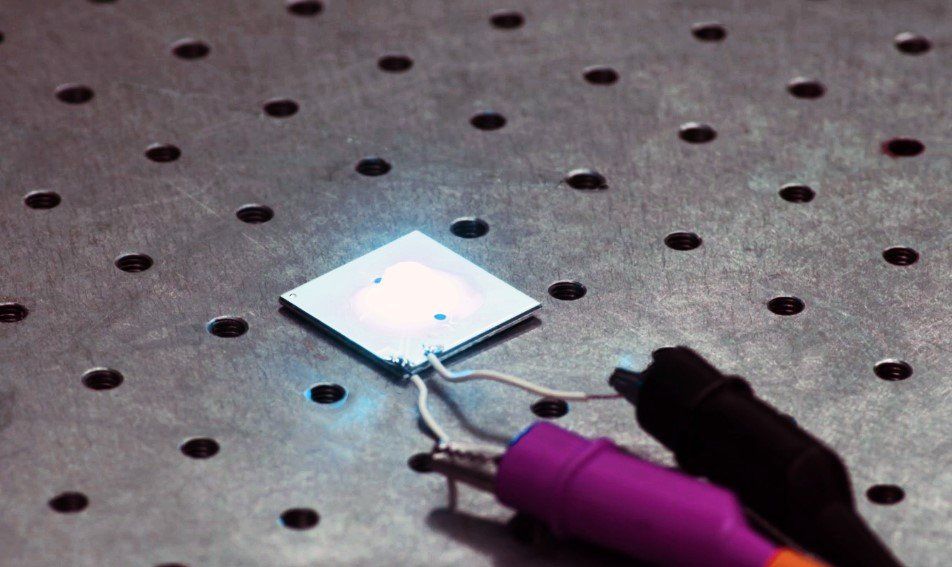
Researchers have demonstrated nanomaterial-based white-light-emitting diodes (LEDs) that exhibit a record luminous efficiency of 105 lumens per watt. Luminous efficiency is a measure of how well a light source uses power to generate light. With further development, the new LEDs could reach efficiencies over 200 lumens per watt, making them a promising energy-efficient lighting source for homes, offices and televisions.

“Efficient LEDs have strong potential for saving energy and protecting the environment,” said research leader Sedat Nizamoglu, Koç University, Turkey. “Replacing conventional lighting sources with LEDs with an of 200 lumens per watt would decrease the global electricity consumed for lighting by more than half. That reduction is equal to the electricity created by 230 typical 500-megawatt coal plants and would reduce greenhouse gas emissions by 200 million tons.”

The researchers describe how they created the high-efficiency white LEDs in Optica, The Optica l Society’s journal for high impact research. The new LEDs use commercially available blue LEDs combined with flexible lenses filled with a solution of nano-sized semiconductor particles called . Light from the blue LED causes the dots to emit green and red, which combines with the blue emission to create .

Read more

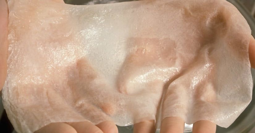
Researchers have developed a new kind of sensor designed to let artificial skin sense pressure, vibrations, and even magnetic fields. Developed by engineers, chemists, and biologists at the University of Connecticut and University of Toronto, the technology could help burn victims and amputees “feel” again through their prosthetic skin.

“The type of artificial skin we developed can be called an electronic skin or e-skin,” Islam Mosa, a postdoctoral fellow at UConn, told Digital Trends. “It is a new group of smart wearable electronics that are flexible, stretchable, shapable, and possess unique sensing capabilities that mimic human skin.”

To create the sensor for the artificial skin, Mosa and his team wrapped a silicone tube with a copper wire and filled the tube with an iron oxide nanoparticle fluid. As the nanoparticles move around the tube, they create an electrical current, which is picked up by the copper wire. When the tube experiences pressure, the current changes.

Read more

Due to their large size, charged surfaces, and environmental sensitivity, proteins do not naturally cross cell-membranes in intact form and, therefore, are difficult to deliver for both diagnostic and therapeutic purposes. Based upon the observation that clustered oligonucleotides can naturally engage scavenger receptors that facilitate cellular transfection, nucleic acid–metal organic framework nanoparticle (MOF NP) conjugates have been designed and synthesized from NU-1000 and PCN-222/MOF-545, respectively, and phosphate-terminated oligonucleotides. They have been characterized structurally and with respect to their ability to enter mammalian cells. The MOFs act as protein hosts, and their densely functionalized, oligonucleotide-rich surfaces make them colloidally stable and ensure facile cellular entry. With insulin as a model protein, high loading and a 10-fold enhancement of cellular uptake (as compared to that of the native protein) were achieved. Importantly, this approach can be generalized to facilitate the delivery of a variety of proteins as biological probes or potential therapeutics.

Proteins play key roles in living systems, and the ability to deliver active proteins to cells is attractive for both diagnostic and therapeutic purposes. Potential uses involve the evaluation of metabolic pathways, regulation of cellular processes, and treatment of disease involving protein deficiencies. (4−6) During the past decade, a series of techniques have been developed to facilitate protein internalization by live cells, including the use of complementary transfection agents, nanocarriers, (7−9) and protein surface modifications. (10−13) Although each strategy has its own merit, none are perfect solutions; they can cause cytotoxicity, reduce protein activity, and suffer from low delivery payloads. (14) For example, we have made the observation that one can take almost any protein and functionalize its surface with DNA to create entities that will naturally engage the cell-surface receptors involved in spherical nucleic acid (SNA) uptake. (13,15−17) While this method is extremely useful in certain situations, it requires direct modification of the protein and large amounts of nucleic acid, on a per-protein basis, to effect transfection. Ideally, one would like to deliver intact, functional proteins without the need to chemically modify them, and to do so in a nucleic-acid efficient manner.

Metal organic frameworks (MOFs) have emerged as a class of promising materials for the immobilization and storage of functional proteins. (18) Their mesoporous structures allow for exceptionally high protein loadings, and their framework architectures can significantly improve the thermal and chemical stabilities of the encapsulated proteins. (19−24) However, although MOF NPs have been recognized as potentially important intracellular delivery vehicles for proteins, (25−27) their poor colloidal stability and positively charged surfaces, (28,29) inhibit their cellular uptake and have led to unfavorable bioavailabilities. (30−33) Therefore, the development of general approaches for reducing MOF NP aggregation, minimizing positive charge (which can cause cytotoxicity), and facilitating cellular uptake is desirable. (34,35)

Read more
A prototype quantum radar that has the potential to detect objects which are invisible to conventional systems has been developed by an international research team led by a quantum information scientist at the University of York.

The new breed of radar is a hybrid system that uses correlation between microwave and optical beams to detect objects of low reflectivity such as cancer cells or aircraft with a stealth capability. Because the quantum radar operates at much lower energies than conventional systems, it has the long-term potential for a range of applications in biomedicine including non-invasive NMR scans.

The research team led by Dr Stefano Pirandola, of the University’s Department of Computer Science and the York Centre for Quantum Technologies, found that a special converter — a double-cavity device that couples the microwave beam to an optical beam using a nano-mechanical oscillator — was the key to the new system.

Read more