Toggle light / dark theme

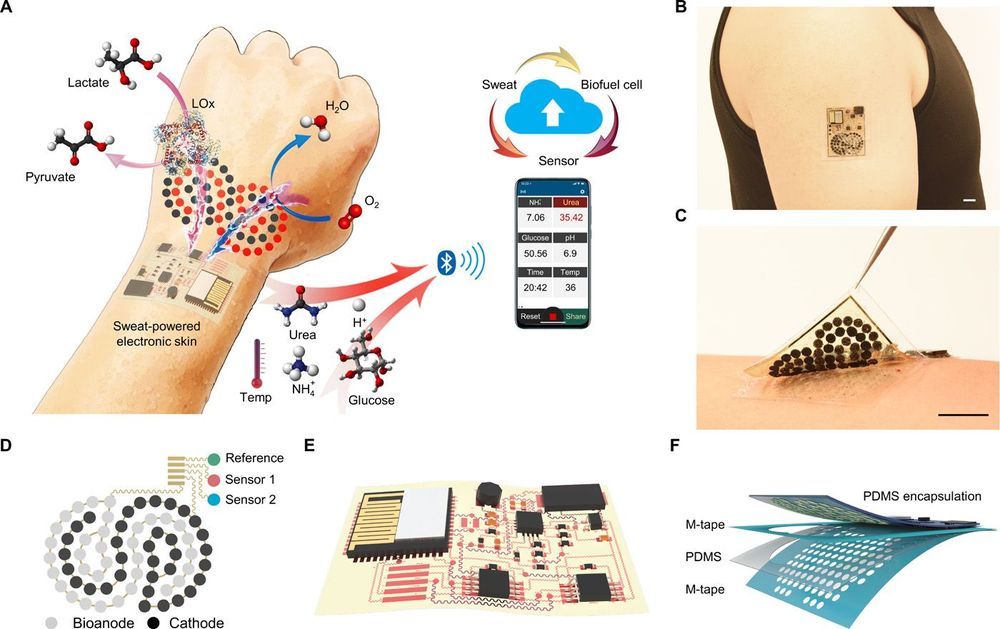
Existing electronic skin (e-skin) sensing platforms are equipped to monitor physical parameters using power from batteries or near-field communication. For e-skins to be applied in the next generation of robotics and medical devices, they must operate wirelessly and be self-powered. However, despite recent efforts to harvest energy from the human body, self-powered e-skin with the ability to perform biosensing with Bluetooth communication are limited because of the lack of a continuous energy source and limited power efficiency. Here, we report a flexible and fully perspiration-powered integrated electronic skin (PPES) for multiplexed metabolic sensing in situ. The battery-free e-skin contains multimodal sensors and highly efficient lactate biofuel cells that use a unique integration of zero- to three-dimensional nanomaterials to achieve high power intensity and long-term stability. The PPES delivered a record-breaking power density of 3.5 milliwatt·centimeter−2 for biofuel cells in untreated human body fluids (human sweat) and displayed a very stable performance during a 60-hour continuous operation. It selectively monitored key metabolic analytes (e.g., urea, NH4+, glucose, and pH) and the skin temperature during prolonged physical activities and wirelessly transmitted the data to the user interface using Bluetooth. The PPES was also able to monitor muscle contraction and work as a human-machine interface for human-prosthesis walking.

Recent advances in robotics have enabled soft electronic devices at different scales with excellent biocompatibility and mechanical properties; these advances have rendered novel robotic functionalities suitable for various medical applications, such as diagnosis and drug delivery, soft surgery tools, human-machine interaction (HMI), wearable computing, health monitoring, assistive robotics, and prosthesis (1–6). Electronic skin (e-skin) can have similar characteristics to human skin, such as mechanical durability and stretchability and the ability to measure various sensations such as temperature and pressure (7–11). Moreover, e-skin can be augmented with capabilities beyond those of the normal human skin by incorporating advanced bioelectronics materials and devices.
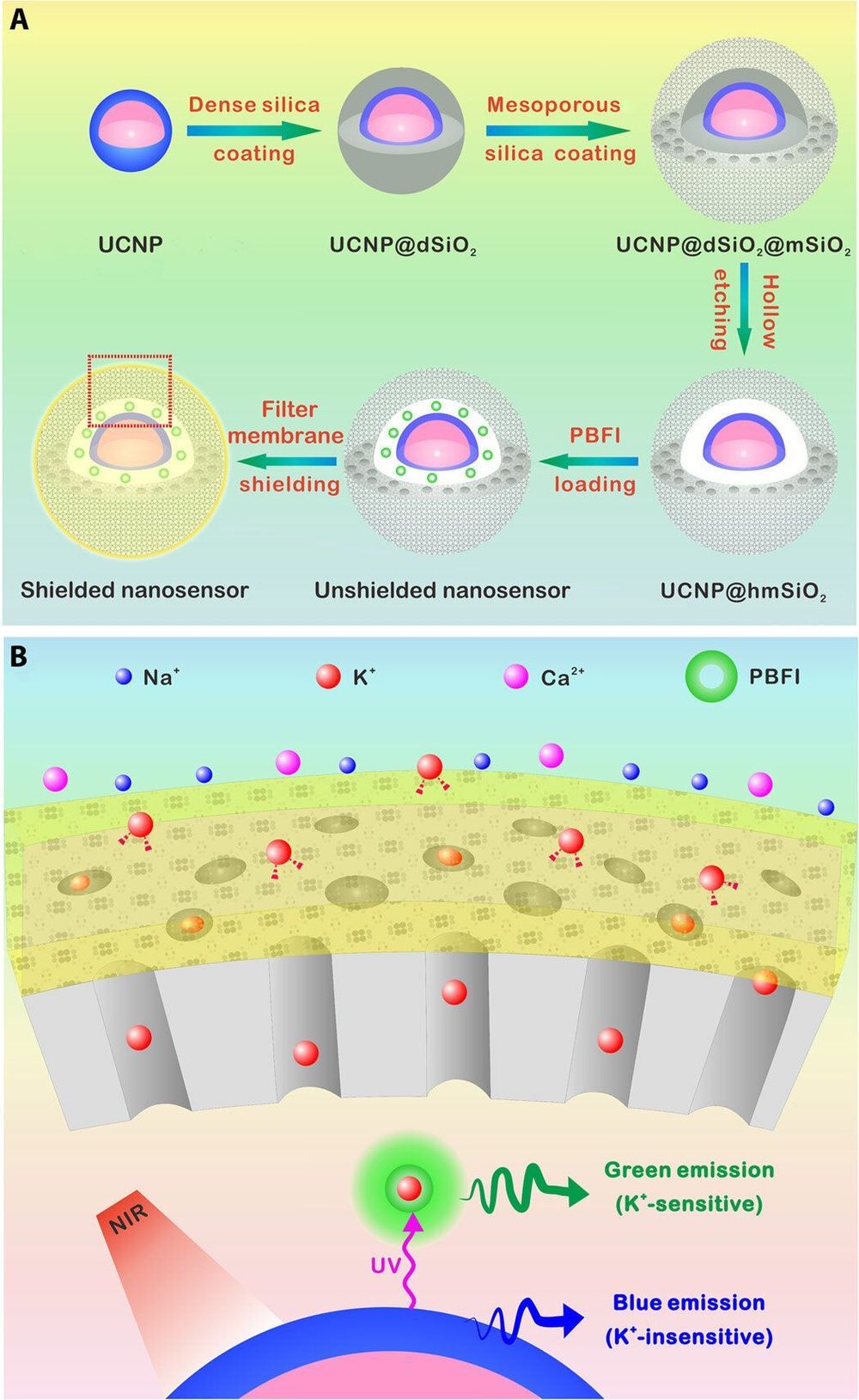
Researchers have developed a number of potassium ion (K+) probes to detect fluctuating K+ concentrations during a variety of biological processes. However, such probes are not sensitive enough to detect physiological fluctuations in living animals and it is not easy to monitor deep tissues with short-wavelength excitations that are in use so far. In a new report, Jianan Liu and a team of researchers in neuroscience, chemistry, and molecular engineering in China, describe a highly sensitive and selective nanosensor for near infrared (NIR) K+ ion imaging in living cells and animals. The team constructed the nanosensor by encapsulating upconversion nanoparticles (UCNPs) and a commercial potassium ion indicator in the hollow cavity of mesoporous silica nanoparticles and coated them with a K+ selective filter membrane. The membrane adsorbed K+ from the medium and filtered away any interfering cations. In its mechanism of action, UCNPs converted NIR to ultraviolet (UV) light to excite the potassium ion indicator and detect fluctuating potassium ion concentrations in cultured cells and in animal models of disease including mice and zebrafish larvae. The results are now published on Science Advances.

The most abundant intracellular cation potassium (K+) is extremely crucial in a variety of biological processes including neural transmission, heartbeat, muscle contraction and kidney function. Variations in the intracellular or extracellular K+ concentration (referred herein as [K+]) suggest abnormal physiological functions including heart dysfunction, cancer, and diabetes. As a result, researchers are keen to develop effective strategies to monitor the dynamics of [K+] fluctuations, specifically with direct optical imaging.

Most existing probes are not sensitive to K+ detection under physiological conditions and cannot differentiate fluctuations between [K+] and the accompanying sodium ion ([Na+]) during transmembrane transport in the Na+/K+ pumps. While fluorescence lifetime imaging can distinguish K+ and Na+ in water solution, the method requires specialized instruments. Most K+ sensors are also activated with short wavelength light including ultraviolet (UV) or visible light—leading to significant scattering and limited penetration depth when examining living tissues. In contrast, the proposed near-infrared (NIR) imaging technique will offer unique advantages during deep tissue imaging as a plausible alternative.

Researchers investigating cribellate spiders have discovered a unique comb structure that could help inform future equipment used to manipulate nanofibers. Nanofibers have been hard to handle in a lab setting as they can stick to the equipment attempting to manipulate them, but a new study published in the journal ACS Applied Nanomaterials reveals how spiders can help us to create non-stick tools for such scenarios.

Cribellate spiders are so named because of their unique web-spinning anatomy. Most spiders have a long single spinneret that they use to produce a single thread, whereas cribellate spiders have a silk-spinning organ. This organ acts like a plate with lots of small, ever so slightly raised protrusions, each of which produces a very fine silk just a few nanometers thick. The spiders then comb these thin fibers out using a calamistrum structure on their legs, producing silk with a woolly texture. This woolly-textured silk entraps the spider’s prey, but somehow, they are able to handle it without getting caught up in their own webs.

Nanofibers are a hot area of research right now but one of the difficulties in their handling is that they commonly stick to the equipment trying to manipulate them. Lead author Anna-Christin Joel, from RWTH Aachen University, and her colleagues wondered if the solution to this frustrating problem could be found within the silk-immune spiders’ anatomy.

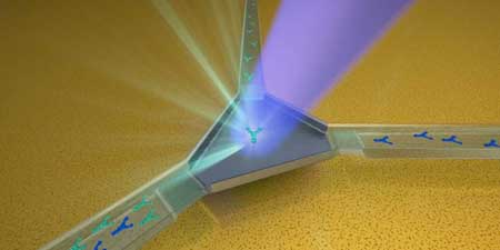
Engineering researchers developed a next-generation miniature lab device that uses magnetic nano-beads to isolate minute bacterial particles that cause diseases. Using this new technology improves how clinicians isolate drug-resistant strains of bacterial infections and difficult-to-detect micro-particles such as those making up Ebola and coronaviruses.

Ke Du and Blanca Lapizco-Encinas, both faculty-researchers in Rochester Institute of Technology’s Kate Gleason College of Engineering, worked with an international team to collaborate on the design of the new system — a microfluidic device, essentially a lab-on-a-chip.

Drug-resistant bacterial infections are causing hundreds of thousands of deaths around the world every year, and this number is continuously increasing. Based on a report from the United Nations, the deaths caused by antibiotics resistance could reach to 10 million annually by 2050, Du explained.

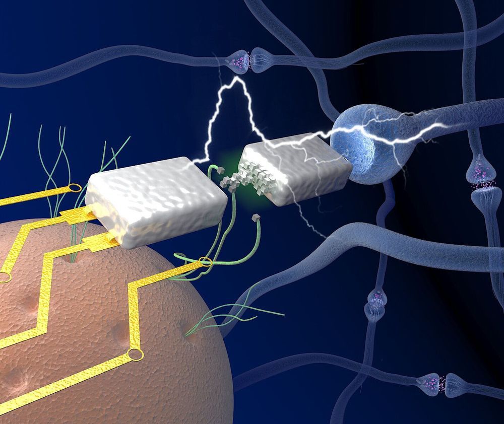
In a paper published on Nature Communications in 20 April 2020 by (read the original paper), Tianda Fu et al. from the University of Massachusetts Amherst proposed a new kind of diffusive memristor based on the protein nanowires sourced from the bacterium named Geobacter sulfurreducens that can potentially resolve the problem. The artificial neurons built on such memristors can function on the level of biological voltages, and they express “temporary integration feature that is similar to real neurons in our brain” according to the authors.

In a study published in Nature, a UCLA-led team of researchers describe how the nanomachine recognizes and kills bacteria, and report that they have imaged it at atomic resolution. The scientists also engineered their own versions of the nanomachine, which enabled them to produce variations that behaved differently from the naturally occurring version.

Their efforts could eventually lead to the development of new types of antibiotics that are capable of homing in on specific species of microbes. Drugs tailored to kill only a certain species or strain of bacteria could offer numerous advantages over conventional antibiotics, including lowering the likelihood that bacteria will develop resistance. In addition, the tailored drugs could destroy harmful cells without wiping out beneficial bugs in the gut microbiome, and they could eventually offer the possibilities of being deployed to prevent bacterial infections, to kill pathogens in food and to engineer human microbiomes so that favorable bacteria thrive.

The particle in the study, an R-type pyocin, is a protein complex released by the bacterium Pseudomonas aeruginosa as a way of sabotaging microbes that compete with it for resources. When a pyocin identifies a rival bacterium, it kills the bacterium by punching a hole in the cell’s membrane. P. aeruginosa, frequently a cause of hospital-acquired illness, is found in soil, in water and on fresh produce. The germ is commonly studied and its biology is well understood.

Only 10 years ago, scientists working on what they hoped would open a new frontier of neuromorphic computing could only dream of a device using miniature tools called memristors that would function/operate like real brain synapses.

But now a team at the University of Massachusetts Amherst has discovered, while on their way to better understanding protein , how to use these biological, electricity conducting filaments to make a neuromorphic memristor, or “memory transistor,” device. It runs extremely efficiently on very low power, as brains do, to carry signals between neurons. Details are in Nature Communications.

As first author Tianda Fu, a Ph.D. candidate in electrical and , explains, one of the biggest hurdles to neuromorphic computing, and one that made it seem unreachable, is that most conventional computers operate at over 1 volt, while the brain sends signals called action potentials between neurons at around 80 millivolts—many times lower. Today, a decade after early experiments, memristor voltage has been achieved in the range similar to conventional computer, but getting below that seemed improbable, he adds.

O,.o circa 2007.


Theoretical physicists at the University of St. Andrews have created ‘incredible levitation effects’ by engineering the force of nature which normally causes objects to stick together by quantum force. By reversing this phenomenon, known as ‘Casimir force’, the scientists hope to solve the problem of tiny objects sticking together in existing novel nanomachines.

Professor Ulf Leonhardt and Dr Thomas Philbin of the University’s School of Physics & Astronomy believe that they can engineer the Casimir force of quantum physics to cause an object to repel rather than attract another in a vacuum.

Casimir force (discovered in 1948 and first measured in 1997) can be demonstrated in a gecko’s ability to stick to a surface with just one toe. However, it can cause practical problems in nanotechnology, and ways of preventing tiny objects from sticking to each other is the source of much interest.

Many researchers from all around the world have joined forces to fight the ongoing coronavirus outbreak that very recently originated in Wuhan, China. As always, nanotechnology does have a solution for almost every problem we face, including now-struggling global health: a nanoparticle-based coronavirus vaccine was successfully developed.