Toggle light / dark theme

Spinels are oxides with chemical formulas of the type AB2O4, where A is a divalent metal cation (positive ion), B is a trivalent metal cation, and O is oxygen. Spinels are valued for their beauty, which derives from the molecules’ spatial configurations, but spinels in which the trivalent cation B consists of the element chrome (Cr) are interesting for a reason that has nothing to do with aesthetics: They have magnetic properties with an abundance of potential technological applications, including gas sensors, drug carriers, data storage media, and components of telecommunications systems.

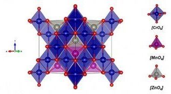
A study by Brazilian and Indian researchers investigated a peculiar kind of spinel: zinc-doped manganese chromite. Nanoparticles of this material, described by the formula Mn0.5 Zn0.5 Cr2O4 [where manganese (Mn) and zinc (Zn) compose the A-site divalent cation], were synthesized in the laboratory and characterized by calculations based on density functional theory (DFT), a method derived from that is used in solid-state physics and chemistry to resolve complex crystal structures.

The material’s structural, electronic, vibrational and were determined by X-ray diffraction, neutron diffraction, X-ray photoelectron spectroscopy and Raman spectroscopy. A report of the study has been published in the Journal of Magnetism and Magnetic Materials with the title “Structural, electronic, vibrational and magnetic properties of Zn2+ substituted MnCr2O4 nanoparticles.”
Since Robert Hooke’s first description of a cell in Micrographia 350 years ago, microscopy has played an important role in understanding the rules of life.

However, the smallest resolvable feature, the resolution, is restricted by the wave nature of light. This century-old barrier has restricted understanding of cellular functions, interactions and dynamics, particularly at the sub-micron to nanometer scale.

Super-resolution fluorescence microscopy overcomes this fundamental limit, offering up to tenfold improvement in resolution, and allows scientists to visualize the inner workings of cells and biomolecules at unprecedented spatial resolution.

Molecules are some of life’s most basic building blocks. When they work together in the right way, they become molecular machines that can solve the most amazing tasks. They are essential for all organisms by, for example, maintaining a wide range of cellular functions and mechanisms.

What if you could create and control an artificial molecular machine? And make it perform tasks that serve us humans?

Many researchers are looking for ways to create and control such , and research is going on in labs all over the world.

We discuss the state of the art and innovative micro- and nanoscale technologies that are finding niches and opening up new opportunities in medicine, particularly in diagnostic and therapeutic applications. We take the design of point-of-care applications and the capture of circulating tumor cells as illustrative examples of the integration of micro- and nanotechnologies into solutions of diagnostic challenges. We describe several novel nanotechnologies that enable imaging cellular structures and molecular events. In therapeutics, we describe the utilization of micro- and nanotechnologies in applications including drug delivery, tissue engineering, and pharmaceutical development/testing. In addition, we discuss relevant challenges that micro- and nanotechnologies face in achieving cost-effective and widespread clinical implementation as well as forecasted applications of micro- and nanotechnologies in medicine.

Here’s a new chapter in the story of the miniaturisation of machines: researchers in a laboratory in Singapore have shown that a single atom can function as either an engine or a fridge. Such a device could be engineered into future computers and fuel cells to control energy flows.

“Think about how your computer or laptop has a lot of things inside it that heat up. Today you cool that with a fan that blows air. In nanomachines or quantum computers, small devices that do cooling could be something useful,” says Dario Poletti from the Singapore University of Technology and Design (SUTD).

This work gives new insight into the mechanics of such devices. The work is a collaboration involving researchers at the Centre for Quantum Technologies (CQT) and Department of Physics at the National University of Singapore (NUS), SUTD and at the University of Augsburg in Germany. The results were published in the peer-reviewed journal npj Quantum Information on 1 May.

HOUSTON, May 1, 2020 — Rice University researchers plan to reconfigure their wastewater-treatment technology to capture and deactivate the virus that causes COVID-19. Their chemical-free nanotechnology, introduced earlier this year as a way to kill bacterial “superbugs” and degrade their antibiotic resistance genes in wastewater, will use graphitic carbon nitride to selectively adsorb viruses and then disable them by activating nearby catalysts with light. The team believes that this photocatalytic approach to disinfection — what it calls the “trap-and-zap” treatment approach — could be used to recognize coronaviruses that cause not only COVID-19 but also MERS and SARS.

North­eastern Uni­ver­sity and Wen­zhou Med­ical Uni­ver­sity in China have part­nered to com­bine their exper­tise in nan­otech­nology and med­i­cine, respec­tively, to col­lab­o­rate on research focusing on reducing eye infections caused by contact lenses and intraocular devices.

Through this part­ner­ship, the researchers will explore the emerging field of picotech­nology, which involves the manip­u­la­tion of matter on an even smaller scale than is used in nanotechnology.


Nanotechnology researchers at Northeastern are joining forces with Wenzhou Medical University in China to collaborate in the new field of picotechnology—and combat eye infections.

Not long ago nanotechnology was a fringe topic; now it’s a flourishing engineering field, and fairly mainstream. For example, while writing this article, I happened to receive an email advertisement for the “Second World Conference on Nanomedicine and Drug Delivery,” in Kerala, India. It wasn’t so long ago that nanomedicine seemed merely a flicker in the eyes of Robert Freitas and a few other visionaries!
But nano is not as small as the world goes. A nanometer is 10−9 meters – the scale of atoms and molecules. A water molecule is a bit less than one nanometer long, and a germ is around a thousand nanometers across. On the other hand, a proton has a diameter of a couple femtometers – where a femtometer, at 10−15 meters, makes a nanometer seem positively gargantuan. Now that the viability of nanotech is widely accepted (in spite of some ongoing heated debates about the details), it’s time to ask: what about femtotech? Picotech or other technologies at the scales between nano and femto seem relatively uninteresting, because we don’t know any basic constituents of matter that exist at those scales. But femtotech, based on engineering structures from subatomic particles, makes perfect conceptual sense, though it’s certainly difficult given current technology.

The nanotech field was arguably launched by Richard Feynman’s 1959 talk “There’s Plenty of Room at the Bottom.” As Feynman wrote there.

Alzheimer’s disease is the sixth leading cause of death in the United States, affecting one in 10 people over the age of 65. Scientists are engineering nanodevices to disrupt processes in the brain that lead to the disease.

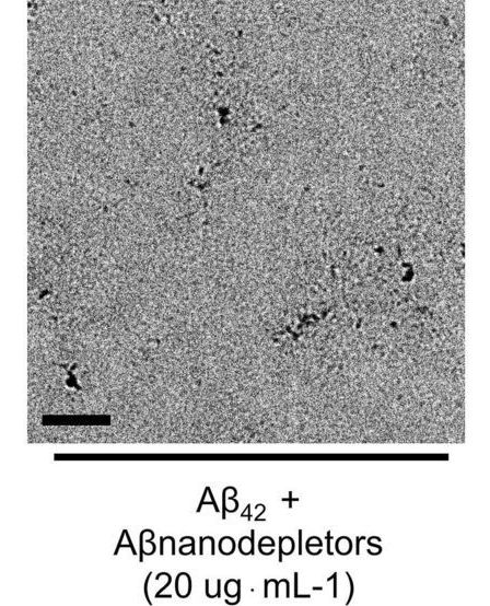
People who are affected by Alzheimer’s disease have a specific type of plaque, made of self-assembled molecules called β-amyloid (Aβ) , that build up in the brain over time. This buildup is thought to contribute to loss of neural connectivity and . Researchers are studying ways to prevent the peptides from forming these dangerous plaques in order to halt development of Alzheimer’s disease in the brain.

In a multidisciplinary study, scientists at the U.S. Department of Energy’s (DOE) Argonne National Laboratory, along with collaborators from the Korean Institute of Science and Technology (KIST) and the Korea Advanced Institute of Science and Technology (KAIST), have developed an approach to prevent plaque formation by engineering a nano-sized device that captures the dangerous peptides before they can self-assemble.

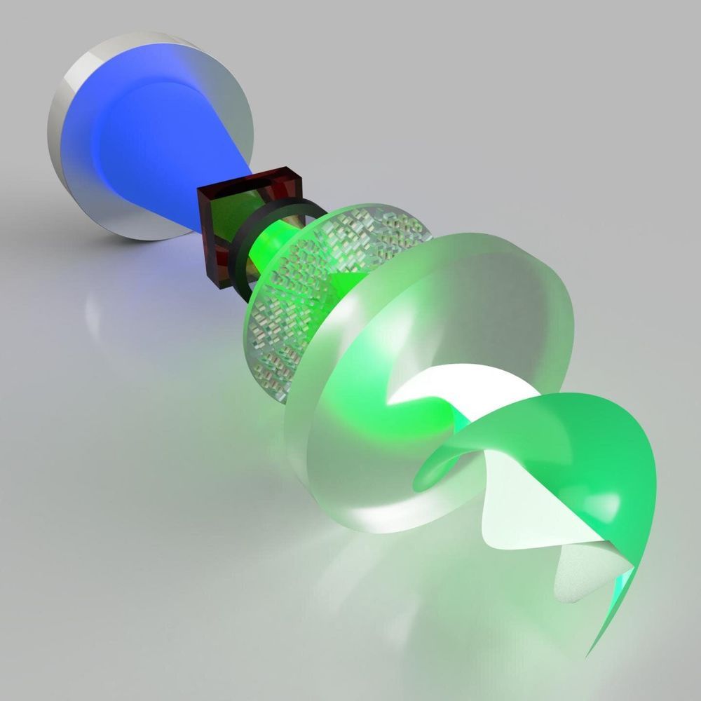
Researchers have demonstrated the world’s first metasurface laser that produces “super-chiral light”: light with ultra-high angular momentum. The light from this laser can be used as a type of “optical spanner” to or for encoding information in optical communications.

“Because can carry angular , it means that this can be transferred to matter. The more angular momentum light carries, the more it can transfer. So you can think of light as an ‘optical spanner’,” Professor Andrew Forbes from the School of Physics at the University of the Witwatersrand (Wits) in Johannesburg, South Africa, who led the research. “Instead of using a physical spanner to twist things (like screwing nuts), you can now shine light on the nut and it will tighten itself.”

The new produces a new high purity “twisted light” not observed from lasers before, including the highest angular momentum reported from a laser. Simultaneously the researchers developed a nano-structured that has the largest phase gradient ever produced and allows for high power operation in a compact design. The implication is a world-first laser for producing exotic states of twisted structured light, on demand.