Toggle light / dark theme

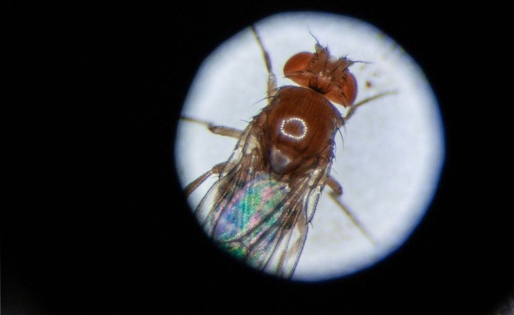
Ribonucleic acid, or RNA, is part of our genetic code and present in every cell of our body. The best known form of RNA is a single linear strand, of which the function is well known and characterized. But there is also another type of RNA, so-called “circular RNA,” or circRNA, which forms a continuous loop that makes it more stable and less vulnerable to degradation. CircRNAs accumulate in the brain with age. Still, the biological functions of most circRNAs are not known and are a riddle for the scientific community. Now scientists from the Max Planck Institute for Biology of Aging have come one step closer to answer the question what these mysterious circRNAs do: one of them contributes to the aging process in fruit flies.

Carina Weigelt and other researchers in the group led by Linda Partridge, Director at the Max Planck Institute for Biology of Aging, used to investigate the role of the circRNAs in the aging process. “This is unique, because it is not very well understood what circRNAs do, especially not in an aging perspective. Nobody has looked at circRNAs in a longevity context before,” says Carina Weigelt who conducted the main part of the study. She continues: “Now we have identified a circRNA that can extend lifespan of fruit flies when we increase it, and it is regulated by signaling.”

Learn how Databricks and the Regeneron Genetics Center partnered to introduce whole genome regression into Project Glow, reducing the cost of fitting mixed models on genetics datasets by orders of magnitude.

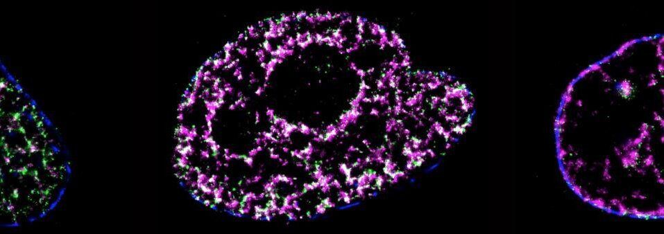
DNA replication is a process of critical importance to the cell, and must be coordinated precisely to ensure that genomic information is duplicated once and only once during each cell cycle. Using super-resolution technology a University of Technology Sydney led team has directly visualized the process of DNA replication in single human cells.

This is the first quantitative characterization to date of the spatio-temporal organization, morphology, and in situ epigenetic signatures of individual replication foci (RFi) in single human at the nanoscale.

The results of the study, published in PNAS (Proceedings of the National Academy of Sciences) give new insight into a poorly understood area of DNA replication namely how replication origin sites are chosen from thousands of possible sites.

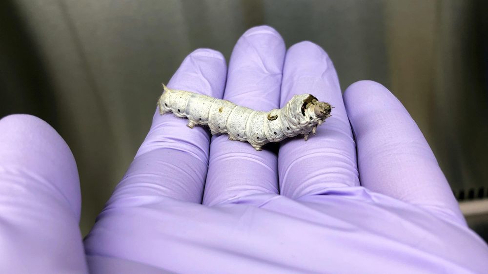
Scientists identify an important protein that increases “bacterial virulence,” when mutated, changing harmless bacteria to harmful ones.

As far as humans are concerned, bacteria can be classified as either harmful, pathogenic bacteria and harmless or beneficial non-pathogenic bacteria. To develop better treatments for diseases caused by pathogenic bacteria, we need to have a good grasp on the mechanisms that cause some bacteria to be virulent. Scientists have identified genes that cause virulence, or capability to cause disease, but they do not fully know how bacteria evolve to become pathogenic.

To find out, Professor Chikara Kaito and his team of scientists from Okayama University, Japan, used a process called experimental evolution to identify molecular mechanisms that cells develop to gain useful traits, and published their findings in PLoS Pathogens. “We’re excited by this research because no one has ever looked at virulence evolution of bacteria in an animal; studies before us looked at the evolution in cells,” said Prof Kaito.

Xiang-Dong Fu, PhD, has never been more excited about something in his entire career. He has long studied the basic biology of RNA, a genetic cousin of DNA, and the proteins that bind it. But a single discovery has launched Fu into a completely new field: neuroscience.

For decades, Fu and his team at University of California San Diego School of Medicine studied a protein called PTB, which is well known for binding RNA and influencing which genes are turned “on” or “off” in a cell. To study the role of a protein like PTB, scientists often manipulate cells to reduce the amount of that protein, and then watch to see what happens.


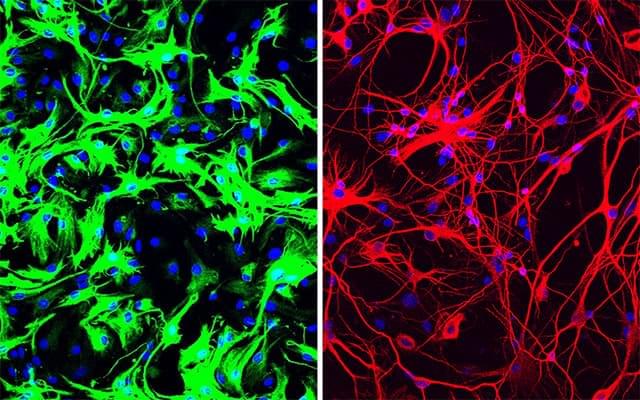
But then he noticed something odd after a couple of weeks — there were very few fibroblasts left. Almost the whole dish was instead filled with neurons.

In this serendipitous way, the team discovered that inhibiting or deleting just a single gene, the gene that encodes PTB, transforms several types of mouse cells directly into neurons.

More recently, Fu and Hao Qian, PhD, another postdoctoral researcher in his lab, took the finding a big step forward, applying it in what could one day be a new therapeutic approach for Parkinson’s disease and other neurodegenerative diseases. Just a single treatment to inhibit PTB in mice converted native astrocytes, star-shaped support cells of the brain, into neurons that produce the neurotransmitter dopamine. As a result, the mice’s Parkinson’s disease symptoms disappeared.

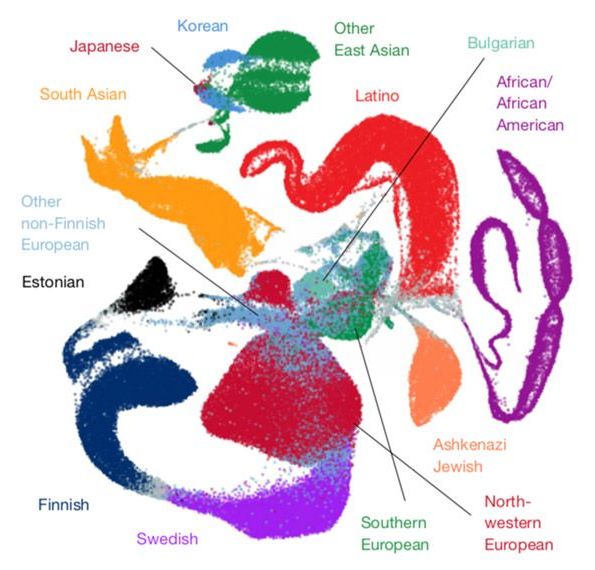
A trove of DNA sequences from 141,456 people — and counting — offers researchers an unparalleled look at genetic variation across the general population1,2. The resource has been helping researchers to identify variants that contribute to autism since it was released online about four years ago3,4.

The genomes of autistic people harbor hundreds of potentially harmful mutations. But to firmly connect a specific variant to the condition, researchers need to see if it is common among typical people — a sign that that variant may actually be benign.

In 2014, researchers debuted one of the first tools to probe the prevalence of a mutation in the general population. Known as the Exome Aggregation Consortium (ExAC), it contained 60,000 sequences of exomes — the protein-coding regions of the genome5.

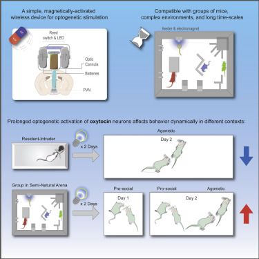
Complex behavioral phenotyping techniques are becoming more prevalent in the field of behavioral neuroscience, and thus methods for manipulating neuronal activity must be adapted to fit into such paradigms. Here, we present a head-mounted, magnetically activated device for wireless optogenetic manipulation that is compact, simple to construct, and suitable for use in group-living mice in an enriched semi-natural arena over several days. Using this device, we demonstrate that repeated activation of oxytocin neurons in male mice can have different effects on pro-social and agonistic behaviors, depending on the social context. Our findings support the social salience hypothesis of oxytocin and emphasize the importance of the environment in the study of social neuromodulators. Our wireless optogenetic device can be easily adapted for use in a variety of behavioral paradigms, which are normally hindered by tethered light delivery or a limited environment.

The world’s largest genetic study into chronic fatigue syndrome is to be launched in the UK after receiving £3.2m of funding from the Medical Research Council and National Institute for Health Research.

The research aims to shine a light on the debilitating long-term condition, about which little is known, by collecting DNA samples from 20,000 people who have CFS, also known as myalgic encephalomyelitis (ME).

CFS is believed to affect about 250,000 people in the UK and has been estimated to cost the economy billions of pounds each year. Individuals experience exhaustion that is not helped by rest, with one in four so severely affected they are unable to leave the house and, frequently, unable to leave their bed. Other symptoms include, pain, mental fogginess, light and noise sensitivities, as well as trouble with memory and sleep. No effective treatment exists.

New research untangles the complex code the brain uses to distinguish between a vast array of smells, offering a scientific explanation for how it separates baby powder from bleach, lemon from orange, or freshly cut grass from freshly brewed coffee.

A single scent can trigger a complex chain of events in what’s known as the olfactory bulb, the brain’s control center for smell. To unravel the intricacies of that process, researchers in the U.S. and Italy turned to a technique known as optogenetics, which uses light to control neurons in the brain. In research on mice, they used light to trick the brain into thinking it smelled a particular scent, then studied brain activity to understand the role different neurons play in a mouse’s ability to recognize that scent. Their findings were published Thursday in Science.

When we encounter a certain smell, it stimulates a specific pattern of activity among tiny spheres known as glomeruli, which are found in the olfactory bulb. The odor plays across these glomeruli like a melody across piano keys: Just as a tune is made distinct by which keys are pressed and at what point in the melody, a scent is made distinct by which glomeruli are activated and in what order.