Toggle light / dark theme

HOUSTON, May 1, 2020 — Rice University researchers plan to reconfigure their wastewater-treatment technology to capture and deactivate the virus that causes COVID-19. Their chemical-free nanotechnology, introduced earlier this year as a way to kill bacterial “superbugs” and degrade their antibiotic resistance genes in wastewater, will use graphitic carbon nitride to selectively adsorb viruses and then disable them by activating nearby catalysts with light. The team believes that this photocatalytic approach to disinfection — what it calls the “trap-and-zap” treatment approach — could be used to recognize coronaviruses that cause not only COVID-19 but also MERS and SARS.

German researchers have identified a strain of bacterium that not only breaks down toxic plastic, but also uses it as food to fuel the process, according to The Guardian.

The scientists discovered the strain of bacteria, known as pseudomonas bacteria, at a dump site loaded with plastic waste, where they noticed that it was attacking polyurethane. Polyurethane’s are ubiquitous in plastic products because they are pliable and durable. However, when they reach the end of their usefulness and end up in landfills, they decompose slowly and slowly release toxic chemicals into the soil as they degrade. They are also notoriously difficult to recycle, according to Courthouse News.

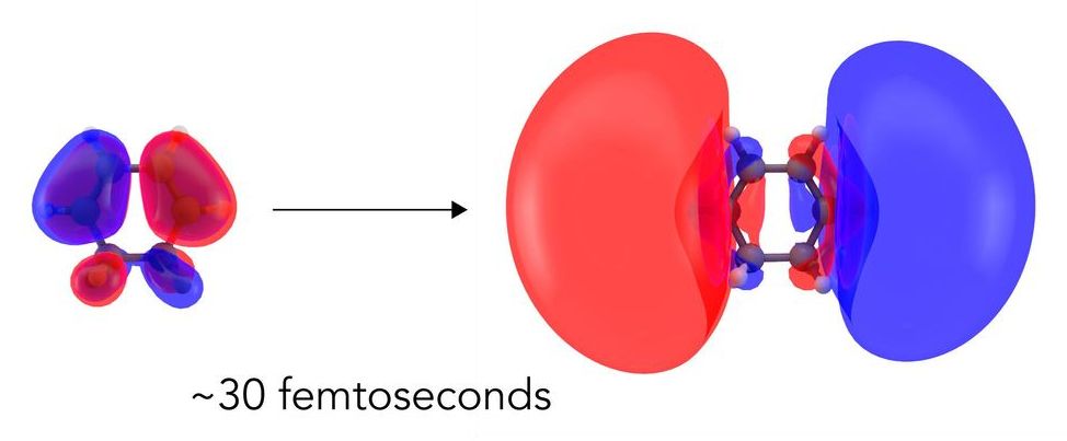
The first step in many light-driven chemical reactions, like the ones that power photosynthesis and human vision, is a shift in the arrangement of a molecule’s electrons as they absorb the light’s energy. This subtle rearrangement paves the way for everything that follows and determines how the reaction proceeds.

Now scientists have seen this first step directly for the first time, observing how the molecule’s electron cloud balloons out before any of the in the molecule respond.

While this response has been predicted theoretically and detected indirectly, this is the first time it’s been directly imaged with X-rays in a process known as molecular movie-making, whose ultimate goal is to observe how both electrons and nuclei act in real time when chemical bonds form or break.

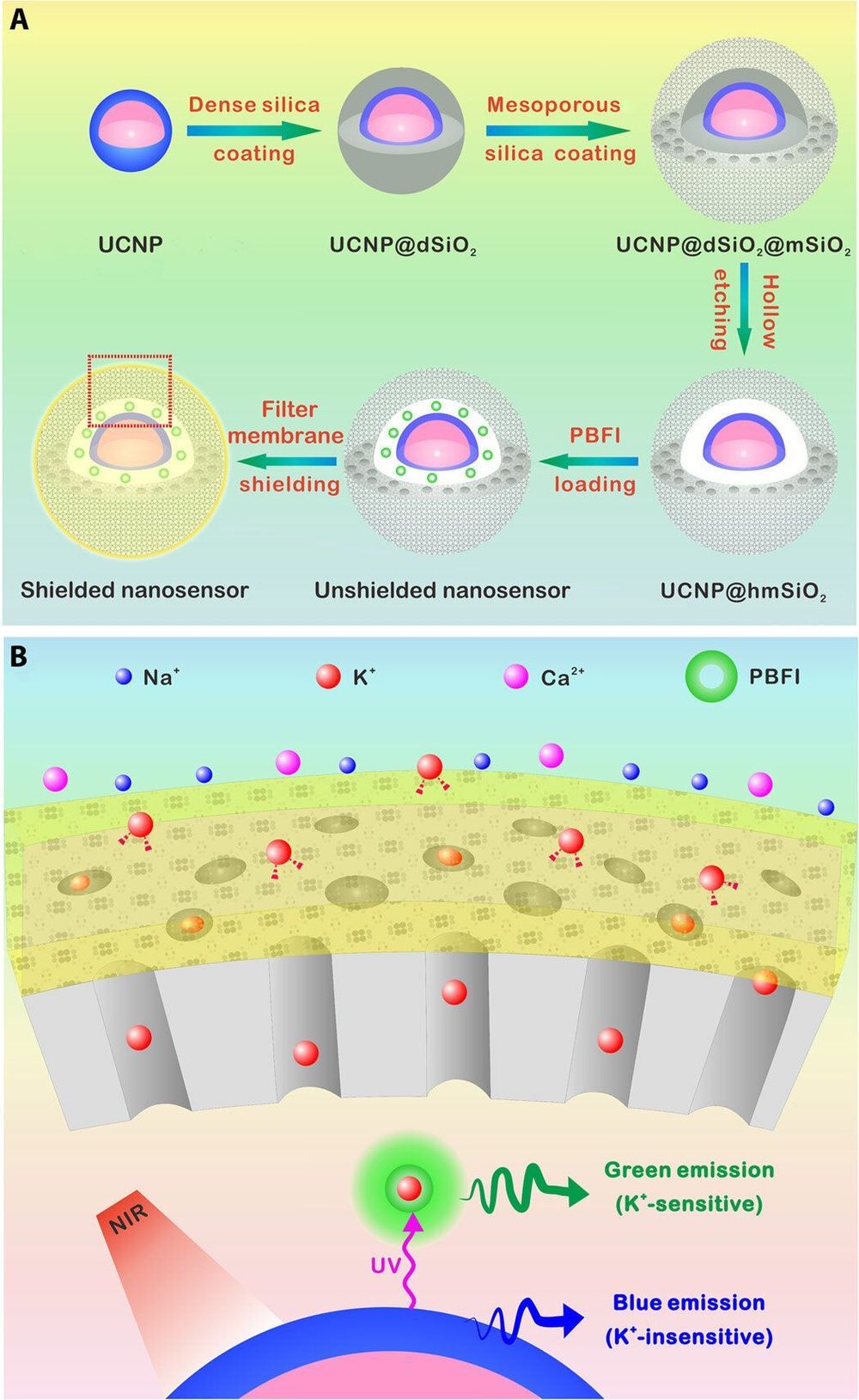
Researchers have developed a number of potassium ion (K+) probes to detect fluctuating K+ concentrations during a variety of biological processes. However, such probes are not sensitive enough to detect physiological fluctuations in living animals and it is not easy to monitor deep tissues with short-wavelength excitations that are in use so far. In a new report, Jianan Liu and a team of researchers in neuroscience, chemistry, and molecular engineering in China, describe a highly sensitive and selective nanosensor for near infrared (NIR) K+ ion imaging in living cells and animals. The team constructed the nanosensor by encapsulating upconversion nanoparticles (UCNPs) and a commercial potassium ion indicator in the hollow cavity of mesoporous silica nanoparticles and coated them with a K+ selective filter membrane. The membrane adsorbed K+ from the medium and filtered away any interfering cations. In its mechanism of action, UCNPs converted NIR to ultraviolet (UV) light to excite the potassium ion indicator and detect fluctuating potassium ion concentrations in cultured cells and in animal models of disease including mice and zebrafish larvae. The results are now published on Science Advances.

The most abundant intracellular cation potassium (K+) is extremely crucial in a variety of biological processes including neural transmission, heartbeat, muscle contraction and kidney function. Variations in the intracellular or extracellular K+ concentration (referred herein as [K+]) suggest abnormal physiological functions including heart dysfunction, cancer, and diabetes. As a result, researchers are keen to develop effective strategies to monitor the dynamics of [K+] fluctuations, specifically with direct optical imaging.

Most existing probes are not sensitive to K+ detection under physiological conditions and cannot differentiate fluctuations between [K+] and the accompanying sodium ion ([Na+]) during transmembrane transport in the Na+/K+ pumps. While fluorescence lifetime imaging can distinguish K+ and Na+ in water solution, the method requires specialized instruments. Most K+ sensors are also activated with short wavelength light including ultraviolet (UV) or visible light—leading to significant scattering and limited penetration depth when examining living tissues. In contrast, the proposed near-infrared (NIR) imaging technique will offer unique advantages during deep tissue imaging as a plausible alternative.

Using machine learning three groups, including researchers at IBM and DeepMind, have simulated atoms and small molecules more accurately than existing quantum chemistry methods. In separate papers on the arXiv preprint server the teams each use neural networks to represent wave functions of electrons that surround the molecules’ atoms. This wave function is the mathematical solution of the Schrödinger equation, which describes the probabilities of where electrons can be found around molecules. It offers the tantalising hope of ‘solving chemistry’ altogether, simulating reactions with complete accuracy. Normally that goal would require impractically large amounts of computing power. The new studies now offer a compromise of relatively high accuracy at a reasonable amount of processing power.

Each group only simulates simple systems, with ethene among the most complex, and they all emphasise that the approaches are at their very earliest stages. ‘If we’re able to understand how materials work at the most fundamental, atomic level, we could better design everything from photovoltaics to drug molecules,’ says James Spencer from DeepMind in London, UK. ‘While this work doesn’t achieve that quite yet, we think it’s a step in that direction.’

Two approaches appeared on arXiv just a few days apart in September 2019, both combining deep machine learning and Quantum Monte Carlo (QMC) methods. Researchers at DeepMind, part of the Alphabet group of companies that owns Google, and Imperial College London call theirs Fermi Net. They posted an updated preprint paper describing it in early March 2020.1 Frank Noé’s team at the Free University of Berlin, Germany, calls its approach, which directly incorporates physical knowledge about wave functions, PauliNet.2

The synthesis of plastic precursors, such as polymers, involves specialized catalysts. However, the traditional batch-based method of finding and screening the right ones for a given result consumes liters of solvent, generates large quantities of chemical waste, and is an expensive, time-consuming process involving multiple trials.

Ryan Hartman, professor of chemical and at the NYU Tandon School of Engineering, and his laboratory developed a lab-based “intelligent microsystem” employing , for modeling that shows promise for eliminating this costly process and minimizing environmental harm.

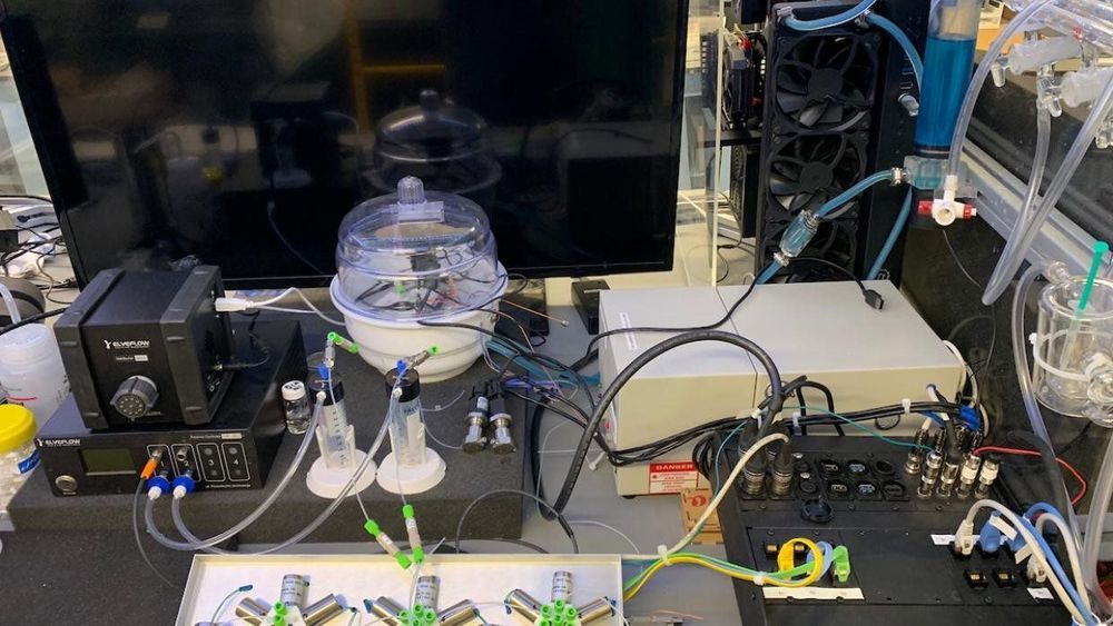
In their research, “Combining automated microfluidic experimentation with machine learning for efficient polymerization design,” published in Nature Machine Intelligence, the collaborators, including doctoral student Benjamin Rizkin, employed a custom-designed, rapidly prototyped microreactor in conjunction with automation and in situ infrared thermography to study exothermic (heat generating) polymerization—reactions that are notoriously difficult to control when limited experimental kinetic data are available. By pairing efficient microfluidic technology with machine learning algorithms to obtain high-fidelity datasets based on minimal iterations, they were able to reduce chemical waste by two orders of magnitude and catalytic discovery from weeks to hours.
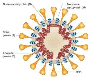
Scientists at the University of Alberta have shown that the drug remdesivir is highly effective in stopping the replication mechanism of the coronavirus that causes COVID-19, according to new research published today in the Journal of Biological Chemistry.

The paper follows closely on research published by the same lab in late February that demonstrated how the drug worked against the Middle East Respiratory Syndrome (MERS) virus, a related coronavirus.

“We were optimistic that we would see the same results against the SARS-CoV-2 virus,” said Matthias Götte, chair of medical microbiology and immunology at U of A.

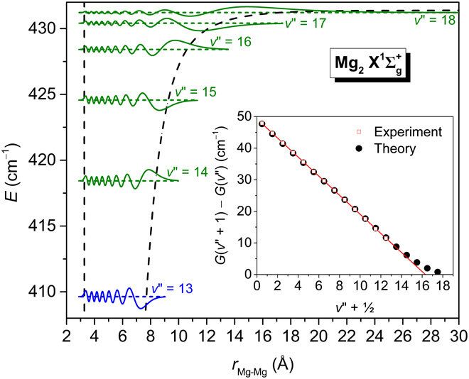
High vibrational states of the Magnesium dimer (Mg2) are an important system in studies of fundamental physics, although they have eluded experimental characterization for half a century. Experimental physicists have so far resolved the first 14 vibrational states of Mg2, despite reports that the ground-state may support five additional levels. In a new report, Stephen H. Yuwono and a research team in the departments of physics and chemistry at the Michigan State University, U.S., presented highly accurate initial potential energy curves for the ground and excited electron states of Mg2. They centered the experimental investigations on calculations of state-of-the-art coupled-cluster (CC) and full configuration interaction computations of the Mg2 dimer. The ground-state potential confirmed the existence of 19 vibrational states with minimal deviation between previously calculated rovibrational values and experimentally derived data. The computations are now published on Science Advances and provide guidance to experimentally detect previously unresolved vibrational levels.

Background

Weakly bound alkaline-earth (AE2) dimers can function as probes of fundamental physics phenomena, such as ultracold collisions, doped helium nanodroplets, binary reactions and even optical lattice clocks and quantum gravity. The magnesium dimer is important for such applications since it has several desirable characteristics including nontoxicity and an absence of hyperfine structure in the most abundant 24 Mg isotope that typically facilitates the analysis of binary collisions and other quantum phenomena. However, the status of Mg2 as a prototype heavier AE2 species is complicated since scientists have not been able to experimentally characterize its high vibrational levels and ground-state potential energy curve (PEC) for so long.

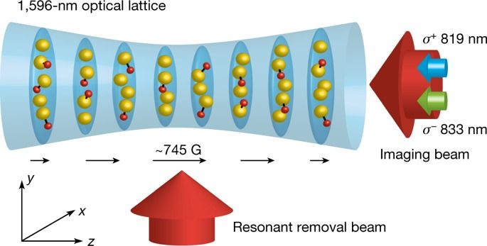
Since the original work on Bose–Einstein condensation1,2, the use of quantum degenerate gases of atoms has enabled the quantum emulation of important systems in condensed matter and nuclear physics, as well as the study of many-body states that have no analogue in other fields of physics3. Ultracold molecules in the micro- and nanokelvin regimes are expected to bring powerful capabilities to quantum emulation4 and quantum computing5, owing to their rich internal degrees of freedom compared to atoms, and to facilitate precision measurement and the study of quantum chemistry6. Quantum gases of ultracold atoms can be created using collision-based cooling schemes such as evaporative cooling, but thermalization and collisional cooling have not yet been realized for ultracold molecules. Other techniques, such as the use of supersonic jets and cryogenic buffer gases, have reached temperatures limited to above 10 millikelvin7,8. Here we show cooling of NaLi molecules to micro- and nanokelvin temperatures through collisions with ultracold Na atoms, with both molecules and atoms prepared in their stretched hyperfine spin states. We find a lower bound on the ratio of elastic to inelastic molecule–atom collisions that is greater than 50—large enough to support sustained collisional cooling. By employing two stages of evaporation, we increase the phase-space density of the molecules by a factor of 20, achieving temperatures as low as 220 nanokelvin. The favourable collisional properties of the Na–NaLi system could enable the creation of deeply quantum degenerate dipolar molecules and raises the possibility of using stretched spin states in the cooling of other molecules.

In an effort to make highly sensitive sensors to measure sugar and other vital signs of human health, Iowa State University’s Sonal Padalkar figured out how to deposit nanomaterials on cloth and paper.

Feedback from a peer-reviewed paper published by ACS Sustainable Chemistry and Engineering describing her new fabrication technology mentioned the metal-oxide nanomaterials the assistant professor of mechanical engineering was working with—including , cerium oxide and copper oxide, all at scales down to billionths of a meter—also have .

“I might as well see if I can do something else with this technology,” Padalkar said. “And that’s how I started studying antimicrobial uses.”