Toggle light / dark theme

New concept delivers continuous electricity with an approach that reduces cost and risk

San Diego, March 29, 2021 – Fusion energy is heating up. In the past few months, both the U.S. Department of Energy’s (DOE) Fusion Energy Sciences Advisory Committee (FESAC) and the National Academies of Sciences, Engineering, and Medicine (NASEM) released reports calling for aggressive development of fusion energy in the U.S.

Now, scientists at the DIII-D National Fusion Facility have released a new design for a compact fusion reactor that can generate electricity and help define the technology necessary for commercial fusion power. The approach is based on the “Advanced Tokamak” concept pioneered by the DIII-D program, which enables a higher-performance, self-sustaining configuration that holds energy more efficiently than in typical pulsed configurations, allowing it to be built at a reduced scale and cost.

UC scientists and physicians hope to permanently cure patients of sickle cell disease by using CRISPR-Cas9 to replace a defective gene with the normal version.


In 2014, two years after her Nobel Prize-winning invention of CRISPR-Cas9 genome editing, Jennifer Doudna thought the technology was mature enough to tackle a cure for a devastating hereditary disorder, sickle cell disease, that afflicts millions of people around the world, most of them of African descent. Some 100000 Black people in the U.S. are afflicted with the disease.

Mobilizing colleagues in the then-new Innovative Genome Institute (IGI) — a joint research collaboration between the University of California, Berkeley, and UC San Francisco — they sought to repair the single mutation that makes red blood cells warp and clog arteries, causing excruciating pain and often death. Available treatments today typically involve regular transfusions, though bone marrow transplants can cure those who can find a matched donor.

After six years of work, that experimental treatment has now been approved for clinical trials by the U.S. Food and Drug Administration, enabling the first tests in humans of a CRISPR-based therapy to directly correct the mutation in the beta-globin gene responsible for sickle cell disease. Beta-globin is one of the proteins in the hemoglobin complex responsible for carrying oxygen throughout the body.

Univ. of Toronto Researcher: “I did not realize quite how bad [the lack of reproducibility and poor quality in research papers] was.”


Many areas of science have been facing a reproducibility crisis over the past two years, and machine learning and AI are no exception. That has been highlighted by recent efforts to identify papers with results that are reproducible and those that are not.

Two new analyses put the spotlight on machine learning in health research, where lack of reproducibility and poor quality is especially alarming. “If a doctor is using machine learning or an artificial intelligence tool to aid in patient care, and that tool does not perform up to the standards reported during the research process, then that could risk harm to the patient, and it could generally lower the quality of care,” says Marzyeh Ghassemi of the University of Toronto.

In a paper describing her team’s analysis of 511 other papers, Ghassemi’s team reported that machine learning papers in healthcare were reproducible far less often than in other machine learning subfields. The group’s findings were published this week in the journal Science Translational Medicine. And in a systematic review published in Nature Machine Intelligence, 85 percent of studies using machine learning to detect COVID-19 in chest scans failed a reproducibility and quality check, and none of the models was near ready for use in clinics, the authors say.

Didn’t watch the video.


Prof David R. Liu, Professor at Harvard University, the Broad Institute, and HHMI was interviewed by the Sheeky Science Show. In the interview, they discussed how to make precise genome editing safe & efficient using the latest CRISPR tech advances in base editing and prime editing and taking it to the clinic (e.g Beam Therapeutics). They talked about the next frontier, epigenome editing.

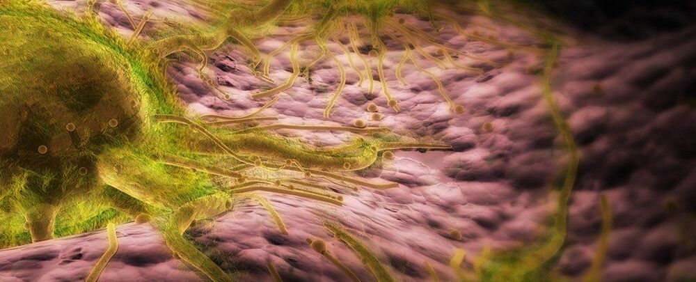
**Five years ago, scientists created a single-celled synthetic organism that, with only 473 genes, was the simplest living cell ever known.** However, this bacteria-like organism behaved strangely when growing and dividing, producing cells with wildly different shapes and sizes.

Now, scientists have identified seven genes that can be added to tame the cells’ unruly nature, causing them to neatly divide into uniform orbs. This achievement, a collaboration between the J. Craig Venter Institute (JCVI), the National Institute of Standards and Technology (NIST) and the Massachusetts Institute of Technology (MIT) Center for Bits and Atoms, was described in the journal Cell.

Identifying these genes is an important step toward engineering synthetic cells that do useful things. Such cells could act as small factories that produce drugs, foods and fuels; detect disease and produce drugs to treat it while living inside the body; and function as tiny computers.

But to design and build a cell that does exactly what you want it to do, it helps to have a list of essential parts and know how they fit together.

“We want to understand the fundamental design rules of life,” said Elizabeth Strychalski, a co-author on the study and leader of NIST’s Cellular Engineering Group. “If this cell can help us to discover and understand those rules, then we’re off to the races.”

Scientists at JCVI constructed the first cell with a synthetic genome in 2010. They didn’t build that cell completely from scratch. Instead, they started with cells from a very simple type of bacteria called a mycoplasma. They destroyed the DNA in those cells and replaced it with DNA that was designed on a computer and synthesized in a lab. This was the first organism in the history of life on Earth to have an entirely synthetic genome. They called it JCVI-syn1.0.

***Continue reading in the article, fascinating — DL***.

Researchers in Australia have discovered a gene responsible for a particularly aggressive type of hormone-sensitive breast cancer which has tragically low survival rates.

“Hopefully this will dramatically improve the poor outcomes these patients currently suffer,” said Harry Perkins Institute of Medical Research epigeneticist Pilar Blancafort.

It’s hard to overstate just how different cancers can be from one another. Even under the umbrella of ‘breast cancer’ lie several types, such as hormone receptor sensitive, HER2 positive, or non-hormone sensitive breast cancer; within those groups, there are even more types that can respond to treatments differently from one another.

Sargramostim/GM-CSF is prescribed to boost white blood cells after cancer treatments or exposure to radiation. The protein stimulates the bone marrow to make more macrophages and granulocytes, specific types of white blood cells, and progenitor cells that repair blood vessels. These white blood cells circulate throughout the body and remove cells, bacteria and amyloid deposits and also repairing blood vessels.


The team carried out a randomized, double-blind, placebo-controlled Phase II trial (NCT01409915) to test the safety and efficacy of Sargramostim treatment in participants with mild-moderate Alzheimer’s disease.

Study participants were either administered Sargramostim at the standard FDA dose of 250 μg/m2/day by subcutaneous injection, or saline for five days a week for three weeks. The study included 20 participants in the test and placebo group. Most participants in the study were recruited and treated at CU Anschutz with a few from the University of South Florida. The CU Anschutz researchers then conducted and studied multiple neurological, neuropsychological, cell, cytokine, Alzheimer’s pathology biomarkers and neuroimaging assessments.

The investigators found that short-term Sargramostim treatment increased innate and other immune cells, modulated cytokine measures, and was safe and well-tolerated by participants. They also found cognition memory improved by almost two points in the 30 point Mini-Mental State Exam. Brain amyloid, tangles, neurodegeneration, and measures of blood biomarkers of Alzheimer’s disease, all improved toward normal.

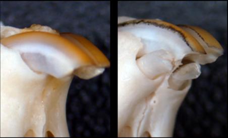
Experiments with this antibody revealed that BMP signaling is essential for determining the number of teeth in mice. Moreover, a single administration was enough to generate a whole tooth.


Japan — The tooth fairy is a welcome guest for any child who has lost a tooth. Not only will the fairy leave a small gift under the pillow, but the child can be assured of a new tooth in a few months. The same cannot be said of adults who have lost their teeth.

A new study by scientists at Kyoto University and the University of Fukui, however, may offer some hope. The team reports that an antibody for one gene — uterine sensitization associated gene-1 or USAG-1 — can stimulate tooth growth in mice suffering from tooth agenesis, a congenital condition. The paper was published in Science Advances.

Although the normal adult mouth has 32 teeth, about 1% of the population has more or fewer due to congenital conditions. Scientists have explored the genetic causes for cases having too many teeth as clues for regenerating teeth in adults.

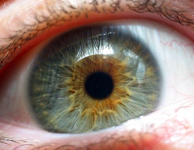
Age-related macular degeneration (AMD), which leads to a loss of central vision, is the most frequent cause of blindness in adults 50 years of age or older, affecting an estimated 196 million people worldwide. There is no cure, though treatment can slow the onset and preserve some vision.

Recently, however, researchers at the University of Rochester have made an important breakthrough in the quest for an AMD cure. Their first three-dimensional (3D) lab model mimics the part of the human retina affected in macular degeneration.

Their model combines stem cell-derived retinal tissue and vascular networks from human patients with bioengineered synthetic materials in a three-dimensional “matrix.” Notably, using patient-derived 3D retinal tissue allowed the researchers to investigate the underlying mechanisms involved in advanced neovascular macular degeneration, the wet form of macular degeneration, which is the more debilitating and blinding form of the disease.

More than 25 years ago, biologists in Arkansas began to report dozens of bald eagles paralyzed, convulsing, or dead. Their brains were pocked with lesions never seen before in eagles. The disease was soon found in other birds across the southeastern United States. Eventually, researchers linked the deaths to a new species of cyanobacteria growing on an invasive aquatic weed that is spreading across the country. The problem persists, with the disease detected regularly in a few birds, yet the culprit’s chemical weapon has remained unknown.

Today in Science, a team identifies a novel neurotoxin produced by the cyanobacteria and shows that it harms not just birds, but fish and invertebrates, too. “This research is a very, very impressive piece of scientific detective work,” says microbiologist Susanna Wood of the Cawthron Institute. An unusual feature of the toxic molecule is the presence of bromine, which is scarce in lakes and rarely found in cyanobacteria. One possible explanation: the cyanobacteria produce the toxin from a bromide-containing herbicide that lake managers use to control the weed.

The discovery highlights the threat of toxic cyanobacteria that grow in sediment and on plants, Wood says, where routine water quality monitoring might miss them. The finding also equips researchers to survey lakes, wildlife, and other cyanobacteria for the new toxin. “It will be very useful,” says Judy Westrick, a chemist who studies cyanobacterial toxins at Wayne State University and was not involved in the new research. “I started jumping because I got so excited.”