Toggle light / dark theme

The metazoan mitochondrial genome (mitogenome) is an ideal model system for comparative and evolutionary genomic research. The typical mitogenome of metazoans encodes a conserved set of 37 genes for 13 protein-coding genes (PCGs), two ribosomal RNA (rRNA) genes, and 22 transfer RNA (tRNA) genes1, with genome-level characters, such as genome size, gene content, and gene order, display high diversity in some lineages2,3. Gene rearrangements are observed frequently in some groups, while gene duplication and loss are distributed sporadically in limited lineages such as Bivalvia, Cephalopod, and Afrobatrachia4,5,6. These remaining duplicate genes and pseudogenes represent important data for exploring the evolutionary history and mechanisms of gene rearrangement and recruitment. For the arrangement of mitochondrial genes, the variation in relative positions of PCGs and rRNA genes are more limited compared with that of tRNA genes across organisms within a phylum7. The tRNA genes with characteristics of diverse changes in relative position, gene content, and secondary structure, are considered as an important tool in studying the evolution of mitogenome, in particular to the rearrangement mechanism8,9,10. Additionally, its variation is usually linked to evolutionary relationships in a wide range of lineages at different taxonomic levels suggesting these features of tRNA could be utilized as useful phylogenetic markers11.

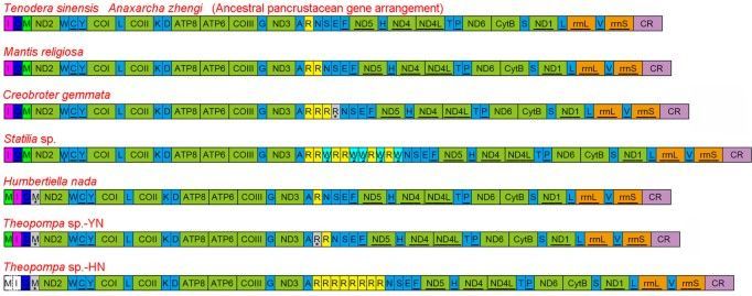
The extensive gene rearrangements (including PCGs and RNA) of insect mitogenomes have been detected in several lineages within the Diptera (Trichoceridae, Cecidomyiidae), Hemiptera (Enicocephalidae), Hymenoptera, Thysanoptera, Psocoptera and Phthiraptera12,13,14,15,16,17,18, while most of investigated mitogenomes share the same gene order with the hypothesized ancestral pancrustacean mitogenome arrangement19 or possess rare tRNA rearrangement. Previously reported dictyopteran mitogenomes consistently display the typical ancestral gene order and content, however only two species are praying mantises and the rest are cockroaches and termites. Members of the Mantodea, a separate lineage within the Dictyoptera, have evolved many unique morphological and behavioural features as the ambush and pursuit predators20,21,22. A better understanding of the diversity of mitogenome evolution in this enigmatic order underlines the need for exploring more taxa with the diverse praying mantis.

Herein, we report eight new mitogenomes from Mantodea and describe their general characteristics. Two new gene rearrangements and reassignment of tRNA genes are described, and evolutionary mechanisms for the gene rearrangements and duplication are discussed. Further, we examine the relationship between tRNA gene duplication and codon usage, and investigate whether these tRNA features vary with phylogeny.
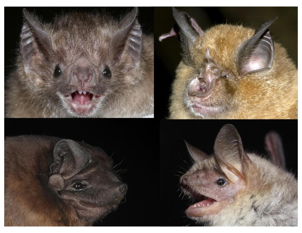
A new study led by University of Maryland and UCLA researchers found that DNA from tissue samples can be used to accurately predict the age of bats in the wild. The study also showed age-related changes to the DNA of long-lived species are different from those in short-lived species, especially in regions of the genome near genes associated with cancer and immunity. This work provides new insight into causes of age-related declines.

This is the first research paper to show that animals in the wild can be accurately aged using an epigenetic clock, which predicts age based on specific changes to DNA. This work provides a new tool for biologists studying animals in the wild. In addition, the results provide insight into possible mechanisms behind the exceptional longevity of many bat species. The study appears in the March 12, 2021, issue of the journal Nature Communications.

“We hoped that these epigenetic changes would be predictive of age,” said Gerald Wilkinson, a professor of biology at UMD and co-lead author of the paper. “But now we have the data to show that instead of having to follow animals over their lifetime to be sure of their age, you can just go out and take a tiny sample of an individual in the wild and be able to know its age, which allows us to ask all kinds of questions we couldn’t before.”

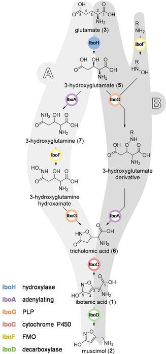
:Ibotenic Acid Biosynthesis in the Fly Agaric Is Initiated by Glutamate Hydroxylation.


The fly agaric, Amanita muscaria, is widely known for its content of the psychoactive metabolites ibotenic acid and muscimol. However, their biosynthetic pathway and the respective enzymes are entirely unknown. 50 years ago, the biosynthesis was hypothesized to start with 3‐hydroxyglutamate. Here, we build on this hypothesis by the identification and recombinant production of a glutamate hydroxylase from A. muscaria. The hydroxylase gene is surrounded by six further biosynthetic genes, which we link to the production of ibotenic acid and muscimol using recent genomic and transcriptomic data. Our results pinpoint the genetic basis for ibotenic acid formation and thus provide new insights into a decades‐old question concerning a centuries‐old drug.

Keywords: biosynthesis, enzyme catalysis, fly agaric, hydroxylation, ibotenic acid.

Summary: Researchers found an increased inflammatory signal in patients with the C90rf72 subtype of ALS. The increased inflammatory biomarkers could be found in peripheral serum tests.

Source: Thomas Jefferson University.

Amyotrophic lateral sclerosis (ALS), also known as Lou Gehrig’s disease, is a neurodegenerative disease that strikes nearly 5000 people in the U.S. every year.

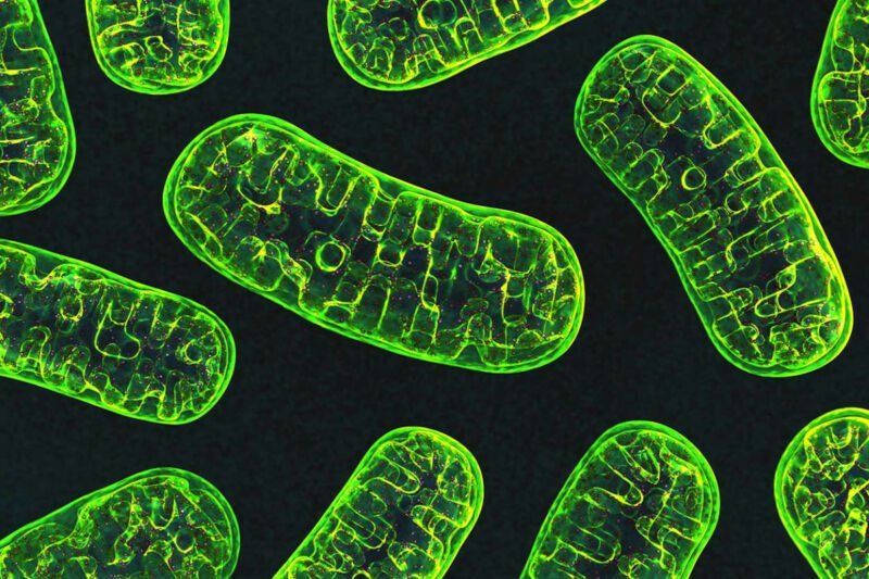
The inside of a mitochondria is made up of a folded membrane, which has evolved to produce the greatest surface area possible between two parts of the mitochondria known as the intermembrane space (the outer part) and the mitochondrial matrix (the inner part). To drastically oversimplify this entire process, the mitochondria uses glucose (and ethanol if it’s available) to pump hydrogen ions (with the occasional deuterium and tritium ion) across the membrane which separates these two compartments of the mitochondria (known as the cristae) into the intermembrane space. These hydrogen ions then flow back into the mitochondrial matrix through a very special protein called ATP synthase, which uses the electrostatic potential energy of the hydrogen ion to manufacture ATP.

Unfortunately, as we get older this inner membrane starts to decay and become smaller. As the cristae starts to shrink, there is less space for ATP synthase, which means there is less ATP produced, which ultimately means that our cells do not have enough energy to maintain all of our cellular functions. As you can imagine, this lack of energy is catastrophic for the health of the cell, and will eventually lead to either cell senescent (where the cell essentially becomes dormant), or complete cell death.

Numerous different suggestions have been put forward as to explain why exactly why mitochondria decay in this way, including mutations within the DNA of the mitochondria (they have their own chromosomes), as well as the build up of oxidative agents within the cell itself which cause direct damage to the mitochondria. However, a group of scientists lead by Dr Hazel Szeto have discovered that the decay of the mitochondrial cristae is linked to declining levels of a phospholipid (fat) called cardiolipin. It turns out that as we age, oxidative agents within our body destroy this phospholipid, which is essential for maintaining the folded inner membrane of the mitochondria.

Summary: A new imaging study reveals how the MFSD2A transporter protein provides a gateway for omega-3 fatty acids to enter the brain.

Source: Columbia University.

Spectacular images of a molecule that shuttles omega-3 fatty acids into the brain may open a doorway for delivering neurological therapeutics to the brain.

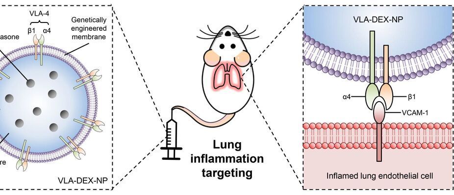
Nanoengineers at the University of California San Diego have developed immune cell-mimicking nanoparticles that target inflammation in the lungs and deliver drugs directly where they’re needed. As a proof of concept, the researchers filled the nanoparticles with the drug dexamethasone and administered them to mice with inflamed lung tissue. Inflammation was completely treated in mice given the nanoparticles, at a drug concentration where standard delivery methods did not have any efficacy.

The researchers reported their findings in Science Advances on June 16.

What’s special about these is that they are coated in a cell membrane that’s been genetically engineered to look for and bind to inflamed . They are the latest in the line of so-called cell membrane-coated nanoparticles that have been developed by the lab of UC San Diego nanoengineering professor Liangfang Zhang. His lab has previously used cell membrane-coated nanoparticles to absorb toxins produced by MRSA; treat sepsis; and train the immune system to fight cancer. But while these previous cell membranes were naturally derived from the body’s , the cell membranes used to coat this dexamethasone-filled nanoparticle were not.

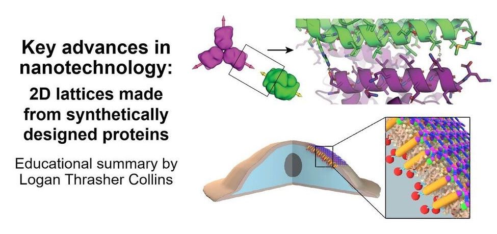
Check out my short video in which I explain some super exciting research in the area of nanotechnology: de novo protein lattices! I specifically discuss a journal article by Ben-Sasson et al. titled “Design of biologically active binary protein 2D materials”.


Here, I explain an exciting nanotechnology paper “Design of biologically active binary protein 2D materials” (https://doi.org/10.1038/s41586-020-03120-8).

Though I am not involved in this particular research myself, I have worked in adjacent areas such as de novo engineering of aggregating antimicrobial peptides, synthetic biology, nanotechnology-based tools for neuroscience, and gene therapy. I am endlessly fascinated by this kind of computationally driven de novo protein design and would love to incorporate it in my own research at some point in the future.

I am a PhD candidate at Washington University in St. Louis and the CTO of the startup company Conduit Computing. I am also a published science fiction writer and a futurist. To learn more about me, check out my website: https://logancollinsblog.com/.