Toggle light / dark theme

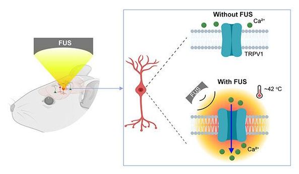
Using a mouse model, Chen and the team delivered a viral construct containing TRPV1 ion channels to genetically-selected neurons. Then, they delivered small burst of heat via low-intensity focused ultrasound to the select neurons in the brain via a wearable device. The heat, only a few degrees warmer than body temperature, activated the TRPV1 ion channel, which acted as a switch to turn the neurons on or off.


Neurological disorders such as Parkinson’s disease and epilepsy have had some treatment success with deep brain stimulation, but those require surgical device implantation. A multidisciplinary team at Washington University in St. Louis has developed a new brain stimulation technique using focused ultrasound that is able to turn specific types of neurons in the brain on and off and precisely control motor activity without surgical device implantation.

The team, led by Hong Chen, assistant professor of biomedical engineering in the McKelvey School of Engineering and of radiation oncology at the School of Medicine, is the first to provide direct evidence showing noninvasive, cell-type-specific activation of neurons in the brain of mammal by combining ultrasound-induced heating effect and genetics, which they have named sonothermogenetics. It is also the first work to show that the ultrasound-genetics combination can robustly control behavior by stimulating a specific target deep in the brain.

Results of the three years of research, which was funded in part by the National Institutes of Health’s BRAIN Initiative, were published online in Brain Stimulation May 11, 2021.
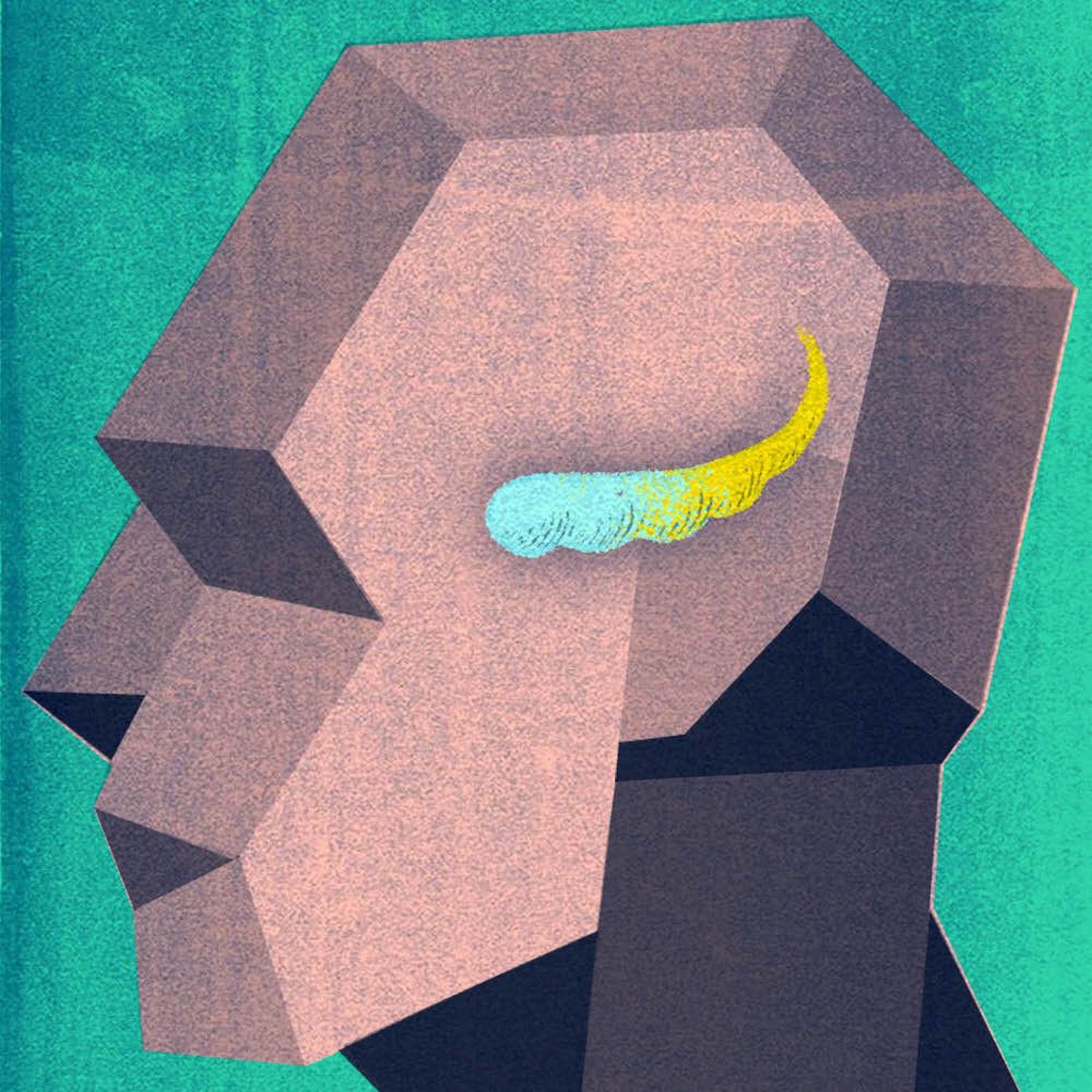
A study of gene activity in the brain’s hippocampus, led by UT Southwestern researchers, has identified marked differences between the region’s anterior and posterior portions. The findings, published today in Neuron, could shed light on a variety of brain disorders that involve the hippocampus and may eventually help lead to new, targeted treatments.

“These new data reveal molecular-level differences that allow us to view the anterior and posterior hippocampus in a whole new way,” says study leader Genevieve Konopka, Ph.D., associate professor of neuroscience at UTSW.

She and study co-leader Bradley C. Lega, M.D., associate professor of neurological surgery, neurology, and psychiatry, explain that the human hippocampus is typically considered a uniform structure with key roles in memory, spatial navigation, and regulation of emotions. However, some research has suggested that the two ends of the hippocampus—the anterior, which points downward toward the face, and the posterior, which points upward toward the back of the head—take on different jobs.

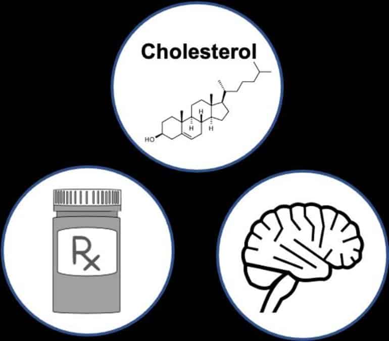
Summary: Disruptions in how the body converts cholesterol into bile acids may play a key role in the development of dementia.

Source: PLOS

The blood-brain barrier is impermeable to cholesterol, yet high blood cholesterol is associated with increased risk of Alzheimer’s disease and vascular dementia. However, the underlying mechanisms mediating this relationship are poorly understood.

There’s already a strong link between sleep and memory, and scientists have just found out more about how that relationship works: there are specific patterns of brain activity that open up windows on our past experiences, fixing them in our long-term memory.

These patterns involve the slow oscillations (SOs) of brain waves that normally accompany sleep, and the sharper sleep spindle bursts of activity that happen during dreamless slumber. It now seems that the precise way these two types of brain activity coordinate with each other makes a big difference in how well we remember something.

Our memories are essentially being reactivated during sleep via these two brain activity patterns, the researchers suggest, making us more likely to remember them. The stronger the reactivation, the more likely we are to be able to recall a memory later on.

More on thymus regeneration. Unless I understood wrong one patient’s epigenetic clock went from his mid 50’s to early 40’s.


Foresight Biotech & Health Extension Meeting sponsored by 100 Plus Capital.

2021 program & apply to join: https://foresight.org/biotech-health-extension-program/

Greg Fahy, Intervene Immune.
Thymus Rejuvenation Progress Update.

- Designed and led the TRIIM trial; Published the first report of thymus regeneration in a normal human; Granted patents on methods for and applications of human thymus regeneration.
- Fellow of the American Aging Association.
(since 2005), Former Director of the American Aging Association (16 years)
- Editor-in-Chief, The Future of Aging: Pathways to Human Life Extension.
- Awarded the Society for Cryobiology’s Luyet Medal in 2016.
- In 2009, showed indefinite survival of rabbit kidney transplanted after cooling to-130°Celsius; Led 21CM team as co-winner of Small Mammal Brain Preservation Prize, 2018 winner of Large Mammal Brain Preservation Prize.

Zoom Transcription: INSERT LINK

Join us:
► Twitter: https://twitter.com/foresightinst.
► Facebook: https://www.facebook.com/foresightinst.
► Instagram: https://www.instagram.com/existentialhope/
► LinkedIn: https://www.linkedin.com/company/foresight-institute.
► Support to join: https://foresight.org/donate/membership/

Foresight Institute advances technologies for the long-term future of life, focusing on molecular machine nanotechnology, biotechnology, and computer science.

Circa 2016


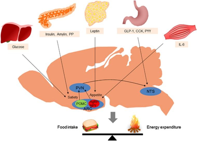
Accumulated evidence from genetic animal models suggests that the brain, particularly the hypothalamus, has a key role in the homeostatic regulation of energy and glucose metabolism. The brain integrates multiple metabolic inputs from the periphery through nutrients, gut-derived satiety signals and adiposity-related hormones. The brain modulates various aspects of metabolism, such as food intake, energy expenditure, insulin secretion, hepatic glucose production and glucose/fatty acid metabolism in adipose tissue and skeletal muscle. Highly coordinated interactions between the brain and peripheral metabolic organs are critical for the maintenance of energy and glucose homeostasis. Defective crosstalk between the brain and peripheral organs contributes to the development of obesity and type 2 diabetes. Here we comprehensively review the above topics, discussing the main findings related to the role of the brain in the homeostatic regulation of energy and glucose metabolism.

In normal individuals, food intake and energy expenditure are tightly regulated by homeostatic mechanisms to maintain energy balance. Substantial evidence indicates that the brain, particularly the hypothalamus, is primarily responsible for the regulation of energy homeostasis.1 The brain monitors changes in the body energy state by sensing alterations in the plasma levels of key metabolic hormones and nutrients. Specialized neuronal networks in the brain coordinate adaptive changes in food intake and energy expenditure in response to altered metabolic conditions ( Figure 1 ).2, 3.

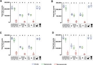
Various insect species serve as valuable model systems for investigating the cellular and molecular mechanisms by which a brain controls sophisticated behaviors. In particular, the nervous system of Drosophila melanogaster has been extensively studied, yet experiments aimed at determining the number of neurons in the Drosophila brain are surprisingly lacking. Using isotropic fractionator coupled with immunohistochemistry, we counted the total number of neuronal and non-neuronal cells in the whole brain, central brain, and optic lobe of Drosophila melanogaster. For comparison, we also counted neuronal populations in three divergent mosquito species: Aedes aegypti, Anopheles coluzzii and Culex quinquefasciatus. The average number of neurons in a whole adult brain was determined to be 199380 ±3400 cells in D. melanogaster, 217910 ±6180 cells in Ae. aegypti, 223020 ± 4650 cells in An. coluzzii and 225911±7220 cells in C. quinquefasciatus. The mean neuronal cell count in the central brain vs. optic lobes for D. melanogaster (101140 ±3650 vs. 107270 ± 2720), Ae. aegypti (109140 ± 3550 vs. 112000 ± 4280), An. coluzzii (105130 ± 3670 vs. 107140 ± 3090), and C. quinquefasciatus (108530 ±7990 vs. 110670 ± 3950) was also estimated. Each insect brain was comprised of 89% ± 2% neurons out of its total cell population. Isotropic fractionation analyses did not identify obvious sexual dimorphism in the neuronal and non-neuronal cell population of these insects. Our study provides experimental evidence for the total number of neurons in Drosophila and mosquito brains.

Citation: Raji JI, Potter CJ (2021) The number of neurons in Drosophila and mosquito brains. PLoS ONE 16: e0250381. https://doi.org/10.1371/journal.pone.

Editor: Matthieu Louis, University of California Santa Barbara, UNITED STATES.