Toggle light / dark theme

The researchers hypothesized that due to abnormal excitement of the Meynert basal ganglia, SB enters the brain and activates anticholinergic action to suppress abnormal acetylcholine secretion of acetylcholine-memory-related circuits centered on the Meynert basal ganglia, eliminating the flashbacks.


Fortunately, a group of Japanese researchers from the Sogo PTSD Institute, Medical Corporation Sogokai, Japan led by Dr. Masanobu Sogo appear to have made a breakthrough in PTSD treatment.

They have identified a drug called trihexyphenidyl, that can significantly reduce the flashbacks and nightmares experienced by patients with PTSD, according to a study published in Brain and Behavior.

Trihexyphenidyl is a central anticholinergic drug used to manage disorders like parkinsonism, and alleviate several side-effects induced by drugs acting on the central nervous system (CNS). It acts by blocking the activity of a neurotransmitter, acetylcholine, in the CNS. Interestingly, it has been available for therapeutic use for around 66 years.

A previous study led by Lee found losing just 16 minutes of sleep could impact job performance. Her previous findings also show that minor sleep loss can decrease daily mindfulness, which is a critical recourse for managing stress and maintaining healthy routines.


Summary: Three consecutive nights of sleep loss can have a negative impact on both mental and physical health. Sleep deprivation can lead to an increase in anger, frustration, and anxiety. Additionally, those who experienced sleep loss reported a change in physical wellbeing, including gastrointestinal and respiratory problems.

Source: USF

All it takes is three consecutive nights of sleep loss to cause your mental and physical well-being to greatly deteriorate.

A new study published in Annals of Behavioral Medicine looked at the consequences of sleeping fewer than six hours for eight consecutive nights – the minimum duration of sleep that experts say is necessary to support optimal health in average adults.

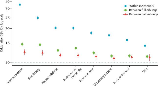
This mapping of aetiological sources of cross-disorder overlap can guide future research aiming to identify specific mechanisms contributing to risk of physical conditions in people with ADHD, which could ultimately inform preventive and lifestyle intervention efforts. Our findings highlight the importance of assessing the presence of physical conditions in patients with ADHD.

Swedish Research Council; Swedish Brain Foundation; Swedish Research Council for Health, Working Life, and Welfare; Stockholm County Council; StratNeuro; EU Horizon 2020 research and innovation programme; National Institute of Mental Health.

Possibilities.


Summary: Ketamine and exposure to 60-hertz flickering light show promise as a potentially new, non-invasive therapy to help rejuvenate the aging brain.

Source: IST Austria

Can you remember the smell of flowers in your grandmother’s garden or the tune your grandpa always used to whistle? Some childhood memories are seemingly ingrained into your brain. In fact, there are critical periods in which the brain learns and saves profound cognitive routines and memories. The structure responsible for saving them is called the perineuronal net.

This extracellular structure envelops certain neurons, thereby stabilizes existing connections – the synapses – between them and prevents new ones from forming. But what if we could remove the perineuronal net and restore the adaptability of a young brain? The neuroscientist Sandra Siegert and her research group at IST Austria now published two promising techniques to do so.

To create high-resolution, 3D images of tissues such as the brain, researchers often use two-photon microscopy, which involves aiming a high-intensity laser at the specimen to induce fluorescence excitation. However, scanning deep within the brain can be difficult because light scatters off of tissues as it goes deeper, making images blurry.

Two-photon imaging is also time-consuming, as it usually requires scanning individual pixels one at a time. A team of MIT and Harvard University researchers has now developed a modified version of two-photon imaging that can image deeper within tissue and perform the imaging much more quickly than what was previously possible.

This kind of imaging could allow scientists to more rapidly obtain high-resolution of structures such as vessels and individual neurons within the brain, the researchers say.

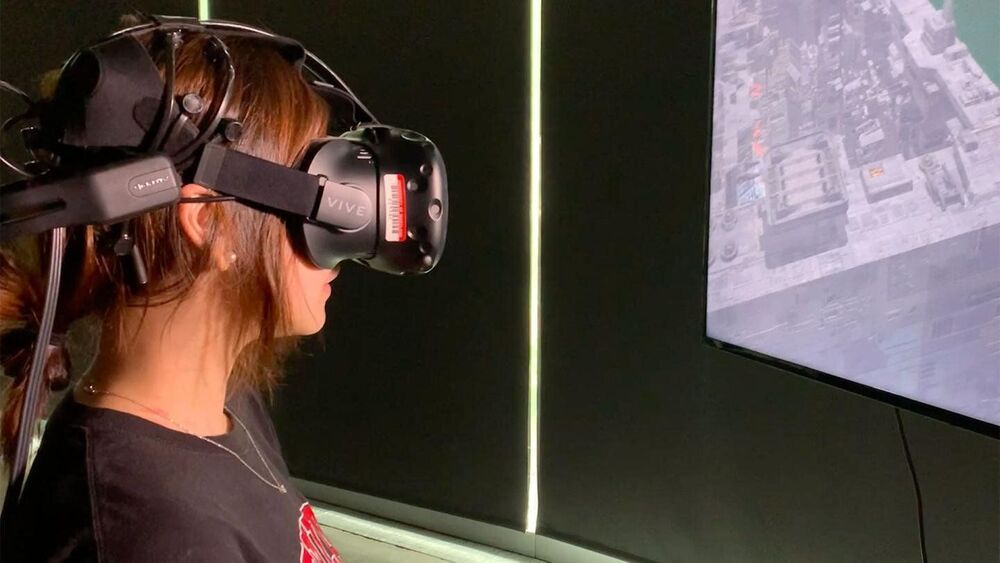
If a virtual world has ever left you feeling nauseous or disorientated, you’re familiar with cybersickness, and you’re hardly alone. The intensity of virtual reality (VR)—whether that’s standing on the edge of a waterfall in Yosemite or engaging in tank combat with your friends—creates a stomach-churning challenge for 30–80% of users.

In a first-of-its kind study, researchers at the University of Maryland recorded VR users’ using electroencephalography (EEG) to better understand and work toward solutions to prevent cybersickness. The research was conducted by Eric Krokos, who received his Ph.D. in computer science in 2018, and Amitabh Varshney, a professor of and dean of UMD’s College of Computer, Mathematical, and Natural Sciences.

Their study, “Quantifying VR cybersickness using EEG,” was recently published in the journal Virtual Reality.

Circa 2017


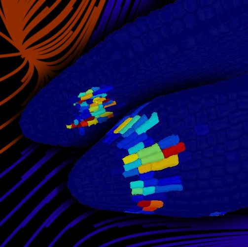
A new study by scientists at the University of Birmingham has revealed a group of cells that function as a ‘brain’ for plant embryos, capable of assessing environmental conditions and dictating when seeds will germinate.

A plant’s decision about when to germinate is one of the most important it will make during its life. Too soon, and the plant may be damaged by harsh winter conditions; too late, and it may be out-competed by other, more precocious plants.

In a study published today in Proceedings of the National Academy of Sciences (PNAS), scientists from the University of Birmingham have shown that this trade-off between speed and accuracy is controlled by a small group of within the that operate in similar way to the human brain.

How does the human brain keep track of the order of events in a sequence?

New research suggests that ‘time cells’ – neurons in the hippocampus thought to represent temporal information – could be the glue that sticks our memories together in the right sequence so that we can properly recall the correct order in which things happened.

Evidence for these kinds of sequence-tracking time cells was previously found in rats, where specific neuron assemblies are thought to support the recollection of events and the planning of action sequences – but less is known about how episodic memory is encoded in the human brain.

“Cannabis may contribute to increased risk for mental disorders, which has actually been shown in schizophrenia,” Penzes said. “Conversely, cannabis could be beneficial in some brain disorders, which prompted trials of medical marijuana in patients with autism.”


Summary: Findings reveal a role the endocannabinoid system plays in a range of psychiatric disorders, including schizophrenia, bipolar disorder, and ASD.

Source: Northwestern University

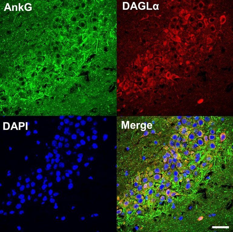
Northwestern Medicine scientists discovered an unexpected connection between a synapse protein that has been implicated in neuropsychiatric disorders and the endocannabinoid pathway, according to a study published in Biological Psychiatry.

These findings suggest a role for the endocannabinoid system in conditions including bipolar disorder, according to Peter Penzes, PhD, the Ruth and Evelyn Dunbar Professor of Psychiatry and Behavioral Sciences, professor of Physiology and Pharmacology, and senior author of the study.