Toggle light / dark theme

IBM Watson is known for its work in identifying cancer treatments and beating contestants on Jeopardy! But now the computing system has expertise in a new area of research: neuroscience.

Watson discovered five genes linked to ALS, sometimes called Lou Gehrig’s disease, IBM announced on Wednesday. The tech company worked with researchers at the Barrow Neurological Institute in Phoenix, Arizona. The discovery is Watson’s first in any type of neuroscience, and suggests that Watson could make discoveries in research of other neurological diseases.

SEE ALSO: This high-tech E.L.F. is guiding confused shoppers with the help of IBM’s Watson.

Read more

Nice.


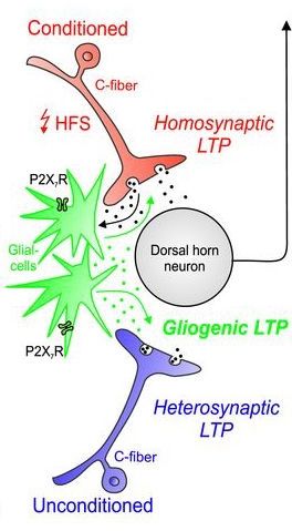
Chronic pain is thought to involve the long-lasting strengthening of synapses, akin to what happens during the formation of new memories. This phenomenon, known as long-term potentiation (LTP), is triggered when neurons on both sides of a synapse are active at the same time. But now, Jürgen Sandkühler, Medical University of Vienna, Austria, and colleagues provide evidence that LTP in nociceptive circuits arises in a different way.

By simultaneously activating two types of glial cells―astrocytes and microglia―the researchers were able to produce LTP at synapses that connect peripheral C-fibers and lamina I neurons in the dorsal horn spinal cord. They also showed that with high-frequency stimulation of C-fibers, glial cells strengthen active and inactive synapses through their release of the NMDA receptor co-agonist D-serine and the cytokine tumor necrosis factor (TNF). Moreover, these molecules traveled to distant synapses, perhaps explaining why pain hypersensitivity can develop in areas surrounding or far away from an injury.

“This paper is going to stimulate a lot of discussion that will lead to important advances for all of us in the pain field,” said Theodore Price, The University of Texas at Dallas, US, who was not involved in the study. “It raises questions for my lab in our day-to-day research that we can address immediately. That’s ultimately the true measure of a really good paper.”

Read more

More proof that Precision Medicine can predict and solve complex health issues.


Brain scans could help predict response to psychotherapy for anxiety and depression.

brain-scan-treatment-research

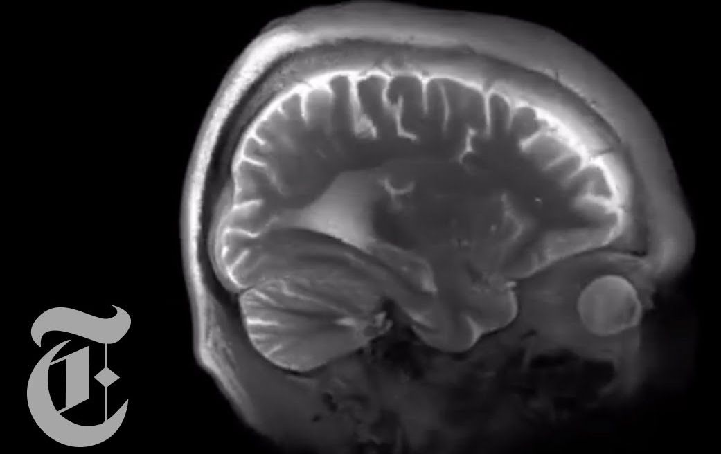
Nov. 10, 2016 – Brain imaging scans may one day provide useful information on the response to psychotherapy in patients with depression or anxiety, according to a review of current research in the November/December issue of the Harvard Review of Psychiatry, published by Wolters Kluwer.

Read more

Progress with Alzheimers and this time approaching it from the direction of Tau as a target rather than Beta Amyloid. This therapy has been tested in people and whilst it is only the first step hopefully this will lead to an effective treatment for this horrific diseases and and end to the suffering it brings.


Progress towards immunotherapies that can clear tau for Alzheimers here. Most therapies are focused on misfolded amyloid-β proteins but this particular approach targets Tau and the first in human test has proceeded!

“The authors of the study have developed a vaccine that stimulates the production of an antibody that specifically targets pathological tau, discovering its “Achilles’ heel”. It is able to do this because healthy tau undergoes a series of changes to its structure forming a new region that the antibody attacks. This new region (the “Achilles’ heel”), while not present in healthy tau, is present in diseased tau early on. Therefore, the antibody tackles all the different varieties of pathological tau. In addition to this important specificity, the antibody is coupled to a carrier molecule that generates a considerable immune response with the added benefit that it is not present in humans, thus avoiding the development of an immune reaction towards the body itself.”

#aging #crowdfundthecure

Read more

Excellent overview on BMI technology.


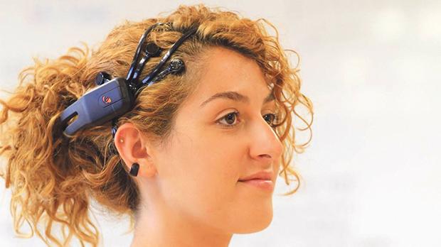
Less than a century ago, Hans Berger, a German psychiatrist, was placing silver foil electrodes on his patients’ heads and observing small ripples of continuous electrical voltage emerging from these. These were the first human brain waves to ever be recorded. Since Hans Berger’s first recordings, our knowledge on the brain structure and function has developed considerably. We now have a much clearer understanding of the neuronal sources that generate these electrical signals and the technology that is now available allows us to get a much denser and accurate picture of how these electrical signals change in time and across the human scalp.

The recording and analysis of brain signals has advanced to a level where people are now able to control and interact with devices around them with the use of their brain signals. The field of brain-computer interfaces has in fact garnered huge interest during the past two decades, and the development of low-cost hardware solutions together with the continuously evolving signal analysis techniques, have brought this technology closer to market than ever before.

Research in the field of brain-computer interfaces was primarily propelled by the need of finding novel communication channels for individuals suffering from severe mobility disorders as in the case of patients with locked-in syndrome. People suffering from the condition have a perfectly functioning brain but are trapped inside their body, which no longer responds to the signals being transmitted from their brain.

Read more

Wild.


The expression “once bitten, twice shy” is an illustration of how a bad experience can induce fear and caution. How to effectively reduce the memory of aversive events is a fundamental question in neuroscience. Scientists in China are reporting that by transplanting mouse embryonic interneurons into the brains of mice and combining that procedure with training to lessen fear, they can help to reduce the fear response. The study is being published December 8 in Neuron.

“Anxiety and fear-related disorders such as post-traumatic stress disorder [PTSD] cause great suffering and impose high costs to society,” says Yong-Chun Yu, a professor at the Institutes of Brain Science at Fudan University in Shanghai and the study’s senior author. “Pharmacological and behavioral treatments of PTSD can reduce symptoms, but many people tend to relapse. There’s a pressing need for new strategies to treat these refractory cases.”

In the study, the researchers used traditional conditioning to instill fear in the mice. They exposed them to a sound as a neutral stimulus, followed by a mild shock to the foot. To determine the level of fear, they measured the amount of time the mice exhibited freezing behavior–the natural sympathetic fear response in prey animals that is indicated by crouching. They then conducted fear extinction training, in which the mice were exposed to the sound but not the shock. After a few rounds, the freezing response times were significantly reduced.

Read more

A severely brain injured woman, who recovered the ability to communicate using her left eye, restored connections and function of the areas of her brain responsible for producing expressive language and responding to human speech, according to new research from Weill Cornell Medicine scientists.

The study, published Dec. 7 in Science Translational Medicine, began 21 months after Margaret Worthen suffered massive strokes, and her continuing recovery was tracked for nearly three years. The research signifies the first time that scientists have captured the restoration of communication of a minimally conscious patient by measuring aspects of brain structure and function before and after communication resumed. It also raises the question of whether other patients in chronic care facilities who appear to be minimally responsive or unresponsive may harbor organized, higher-level brain function.

“From the beginning of Margaret’s attempt to communicate, through the course of our study, we were able to show reorganization of the areas of her brain responsible for expressive language, as well as an exceptionally large change in the correlation across the brain areas in response to human speech,” said study lead author Daniel Thengone, the Fred Plum Fellow in Systems Neurology and Neuroscience in the Feil Family Brain and Mind Research Institute at Weill Cornell Medicine. Adds senior study author Dr. Nicholas D. Schiff, the Jerold B. Katz Professor of Neurology and Neuroscience in the Feil Family Brain and Mind Research Institute: “This is a unique demonstration of plastic change in the brain of an adult starting years after a severe brain injury. We showed a convergence of measurements over years and at multiple time points, revealing an evolving biological process of recovery.”

Read more