Ha! Take that Mark Zuckerberg! (the CEO who said anyone older than 29 years old is not sharp enough for FB)
A recent research revealed that some old people have brain functions fifty years younger than their physical age.

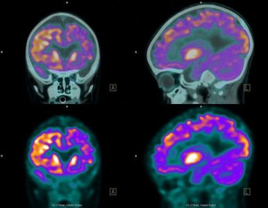
Alright my neuro research & deep-mind learning friends out their; you may wish to read this find; especially as we continue our mapping and mimicking brain functions in systems as well as look at brain enhancement technologies as this is good to know as we try to boost innovation via technologies.
The most creative individuals have more nerve connections between the right and left sides of their brains, reveal researchers in the United States who analyzed connections in 68 different brain regions.
Long believed to be key in fostering imagination and intuition, as well as artistic awareness, and visual and auditive approaches, the right hemisphere isn’t the only part of the brain with a role to play in determining creativity, according to new research from Duke University.
In reality, it is the ability of the brain—a veritable information superhighway comprising a complex network of wired connections—to increase connections between neurons on the two sides of the brain that could set highly creative people apart, the study concludes.

Are you over sleeping? If yes, be careful.
Sleeping for extended amounts of time may be an early indicator of cognitive decline in older people, especially among those with lower education levels, researchers report.
Elderly participants who consistently slept more than 9 hours a night had double the dementia risk over a decade of follow-up in an analysis of data from the Framingham Heart Study.
Longer sleepers also had smaller brain volumes in the study of close to 2,500 older men and women, now online in Neurology.
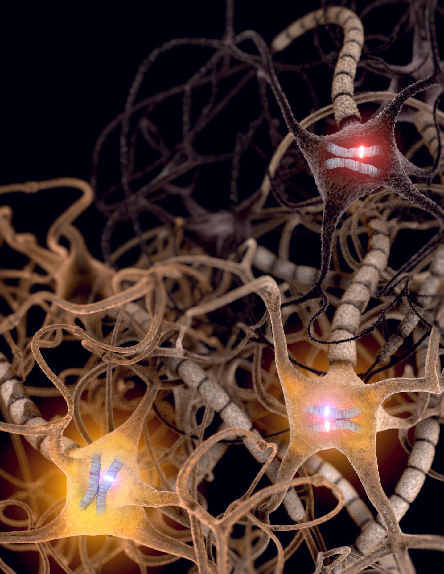
Well, in my immediate family; we get science, math, and futurists talents from my dad. And, there does seem to be a pattern in my immediate family with this; not sure about others. Would love to know though.
SALT LAKE CITY — Most kids say they love their mom and dad equally, but there are times when even the best prefers one parent over the other. The same can be said for how the body’s cells treat our DNA instructions. It has long been thought that each copy — one inherited from mom and one from dad — is treated the same. A new study from scientists at the University of Utah School of Medicine shows that it is not uncommon for cells in the brain to preferentially activate one copy over the other. The finding breaks basic tenants of classic genetics and suggests new ways in which genetic mutations might cause brain disorders.
In at least one region of the newborn mouse brain, the new research shows, inequality seems to be the norm. About 85 percent of genes in the dorsal raphe nucleus, known for secreting the mood-controlling chemical serotonin, differentially activate their maternal and paternal gene copies. Ten days later in the juvenile brain, the landscape shifts, with both copies being activated equally for all but 10 percent of genes.
More than an oddity of the brain, the disparity also takes place at other sites in the body, including liver and muscle. It also occurs in humans.
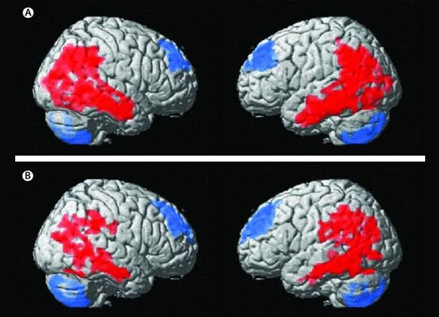
BMI is coming fast and will replace many devices we have today. Advances we making in deep brain development are huge markers that pushes the BMI needle forward for the day when IoT, Security, and big data analytics is a human brain’s and a secured Quantum Infrastructure and people (not servers sitting somewhere) owns and manages their most private of information. I love calling it the age of people empowerment as well as singularity.
Small study in 16 people suggests technique is safe and might help improve mood, anxiety and wellbeing, while increasing weight.
Deep brain stimulation might alter the brain circuits that drive anorexia nervosa symptoms and help improve patients’ mental and physical health, according to a small study published in The Lancet Psychiatry.
There is way more coming in BMI.
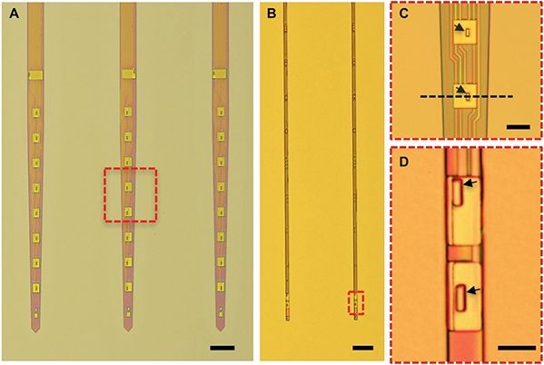
Researcher Dr. Luan and his interdisciplinary team from the University of Texas at Austin have developed an ultra flexible nanoelectronic thread (NET) that has the potential to offer a new type of the long-term neural implants. Neural probes are used to directly measure or even stimulate electrical activity in specific regions of the brain. However, despite the many advances in the field, issues with biocompatibility have limited the prospects and usefulness of the technology. Conventional probes induce scarring around the implant over time, which in turn increasingly impairs the devices’ ability to measure electrical impulses. This scarring is a result of conventional probes’ width, material, and lack of flexibility.
Due to the micrometer dimensions of the NET and its insulation, the implant doesn’t trigger the development of scar tissue and continues working for months at a time. Publishing in the journal Science Advances, the researchers describe the NET from 7 nm nanoprobes layered together and two to three layers of insulation. The layers together total to about 1 µm in thickness, which grants the probe flexibility that is almost 1000 times greater than conventional probes. This flexibility allows the device to remain in place without hindering the movement of or displacing surrounding tissue.
The probe, though significantly smaller, still records data using the same method as standard neural probes. The recording electrodes are evenly placed on the probe, and the collected electrical impulses are communicated to connected devices through microfabricated connections.
When Jan Scheuermann volunteered for an experimental brain implant, she had no idea she was making neuroscience history.
Scheuermann, 54 at the time of surgery, had been paralyzed for 14 years due to a neurological disease that severed the neural connections between her brain and muscles. She could still feel her body, but couldn’t move her limbs.
Unwilling to give up, Scheuermann had two button-sized electrical implants inserted into her motor cortex. The implants tethered her brain to a robotic arm through two bunches of cables that protruded out from her skull.
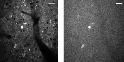
A three-photon microscopic video of neurons in a mouse brain. The imaging depth is approximately 1 millimeter below the surface of the brain. The firing of the neurons is captured by an indicator that is based on green fluorescent protein GFP, which glows brighter as the neuron sends a signal.
Nearly four years ago, then-President Obama launched the BRAIN (Brain Research through Advancing Innovative Neurotechnologies) Initiative, to “accelerate the development and application of new technologies that will enable researchers to produce dynamic pictures of the brain.”
Several of the program’s initial funding awards went to Cornell’s Chris Xu, the Mong Family Foundation Director of Cornell Neurotech – Engineering, and professor and director of undergraduate studies in applied and engineering physics. Xu’s projects aimed to develop new imaging techniques to achieve large scale, noninvasive imaging of neuronal activity.
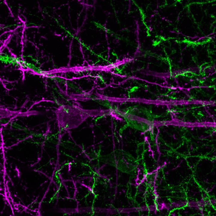
Green: NAc-projecting prefrontal cortex neurons become active during the presentation of a reward-predictive cue, and this activity drives reward-seeking behavior. Purple: PVT-projecting prefrontal cortex neurons inhibited during reward-predictive cue. Credit: The Stuber Lab (UNC School of Medicine)
The prefrontal cortex, a large and recently evolved structure that wraps the front of the brain, has powerful “executive” control over behavior, particularly in humans. The details of how it exerts that control have been elusive, but UNC School of Medicine scientists, publishing today in Nature, have now uncovered some of those details, using sophisticated techniques for recording and controlling the activity of neurons in live mice.

Got OCD; check your genes for a mutation.
A new Northwestern Medicine study found evidence suggesting how neural dysfunction in a certain region of the brain can lead to obsessive and repetitive behaviors much like obsessive-compulsive disorder (OCD).
Both in humans and in mice, there is a circuit in the brain called the corticostriatal connection that regulates habitual and repetitive actions. The study found certain synaptic receptors are important for the development of this brain circuit. If these receptors are eliminated in mice, they exhibit obsessive behavior, such as over-grooming.