Toggle light / dark theme

http://spie.org/bios.

Boyden’s award-winning research has led to tools that can activate or silence neurons with light, enabling the causal assessment of how specific neurons contribute to normal and pathological brain functions.

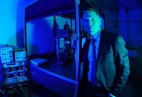
Ed Boyden is the founder and principal investigator of the Synthetic Neurobiology Group at Massachusetts Institute of Technology (MIT). The group develops tools for controlling and observing the dynamic circuits of the brain, and uses these neurotechnologies to understand how cognition and emotion arise from brain network operation, as well as to enable systematic repair of intractable brain disorders such as epilepsy, Parkinson’s disease, post-traumatic stress disorder, and chronic pain.

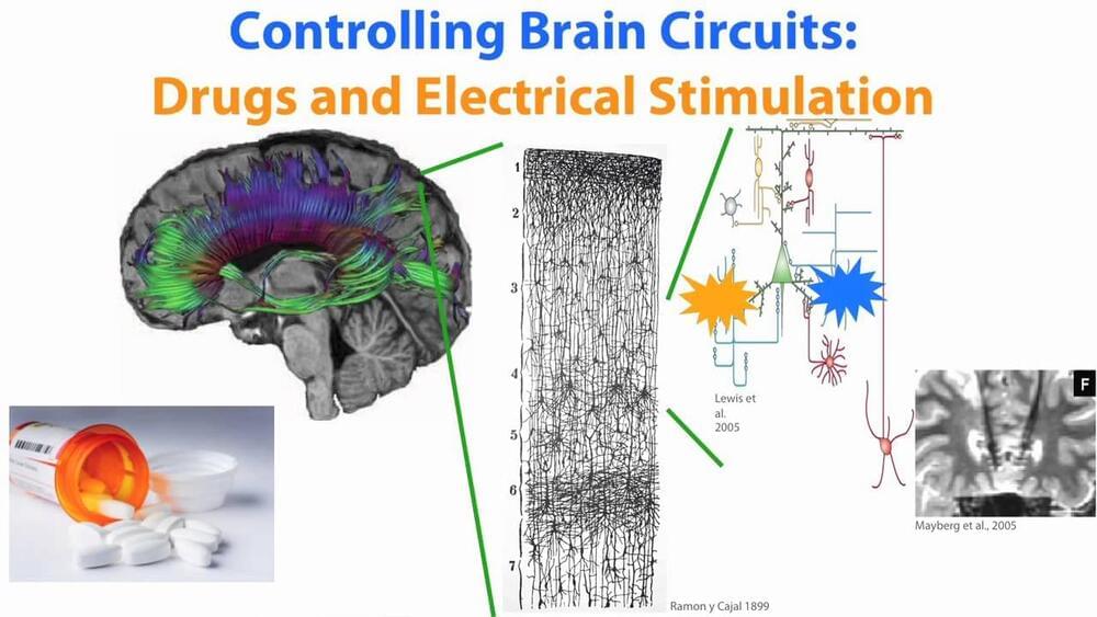
Many disorders of the brain currently are treated with drugs or electrical stimulation. Nearly a quarter of million people have implanted electrical probes in their brains for such stimulation. The problem with this approach is that it targets large areas of the brain instead of the discrete cells or location that cause the disorder. Boyden works on implementing light-stimulated processes in the brain to address these disorders at the cellular level. The method utilizes adeno-associated viruses (AAV) to create light-sensitive centers in the brain which can then be stimulated by light pulses. Very small optical waveguides (fibers) can then be introduced in the brain to stimulate these sites.

Boyden was named to the “Top 35 Innovators Under the Age of 35″ by Technology Review and to the “Top 20 Brains Under Age 40″ by Discover, and has received the NIH Director’s New Innovator Award, the Society for Neuroscience Research Award for Innovation in Neuroscience, and the Paul Allen Distinguished Investigator Award, as well as numerous other recognitions. In early 2011, he was an invited speaker at the renowned TED conference, sharing the bill with a high-powered lineup that included presenters as diverse as Bill Gates and choreographer Julie Taymor.

He has contributed numerous articles to SPIE Proceedings, and was an invited speaker at the Biomedical Optics Hot Topics Session at SPIE Photonics West 2011.

Nick talks to Stanford psychiatrist and neuroscientist Dr. Karl Deisseroth. They discuss a range of topics about the brain, including autism, depression, bipolar disorder, dissociation, and more. They also talk about optogenetics, a technique Karl co-developed which uses light to control specific neurons in the brain, allowing neuroscientists to turn circuits in the brain on and off to reveal how the brain generates perception, emotion, and behavior. They also talk about Karls’ new book, “Projections: A Story of Human Emotion.”

Buy “Projections” by Karl Deisseroth: https://www.amazon.com/gp/product/1984853694/ref=as_li_tl?ie=UTF8&camp=1789&creative=9325&creativeASIN=1984853694&linkCode=as2&tag=lifeboatfound-20&linkId=8e84ac9937aeb22c6158be40e9fc7537
Support the podcast: https://www.patreon.com/nickjikomes.
Organize your e-book highlights & notes w/ the Readwise app (two months free with signup through link): readwise.io/nickjikomes.
Start your own podcast ($20 Amazon gift card after signup): https://www.buzzsprout.com/?referrer_…
Learn more about this episode’s sponsor, Dosist: https://dosist.com/

Mind & Matter shirts: (15% off w/ code “NICKJIKOMES”): https://www.etsy.com/shop/OURMIND?ref…

Connect with Nick Jikomes on Twitter:

Download the podcast & follow Nick:
https://nickjikomes.com

About Nick Jikomes:

Nick is a neuroscientist and podcast host. He is currently Director of Science & Innovation at Leafly, the world’s largest cannabis information resource. He received a Ph.D. in Neuroscience from Harvard University and a B.S. in Genetics from the University of Wisconsin-Madison.

0:00:00 Episode Intro.
0:03:44 Karl Deisseroth Intro.
0:07:19 “Projections” Book Synopsis.
0:13:02 Language & Psychiatry.
0:18:19 Psychiatry vs. Neurology vs. Neuroscience.
0:20:57 What is Optogenetics?
0:29:46 Optogenetics Video Example.
0:38:25 Can Optogenetics Be Used in Humans?
0:44:09 Anxiety.
0:50:34 Brain Basis of Complex Behaviors.
0:58:50 Importance of Basic Research.
1:08:46 Dissociation.
1:20:02 Treatment Resistant Depression.
1:23:30 Ketamine, Psychedelics & Depression.
1:28:24 Autism.
1:36:55 Final Thoughts

Let me back up a moment. I recently concurred with megapundit Steven Pinker that over the last two centuries we have achieved material, moral and intellectual progress, which should give us hope that we can achieve still more. I expected, and have gotten, pushback. Pessimists argue that our progress will prove to be ephemeral; that we will inevitably succumb to our own nastiness and stupidity and destroy ourselves.

Maybe, maybe not. Just for the sake of argument, let’s say that within the next century or two we solve our biggest problems, including tyranny, injustice, poverty, pandemics, climate change and war. Let’s say we create a world in which we can do pretty much anything we choose. Many will pursue pleasure, finding ever more exciting ways to enjoy themselves. Others may seek spiritual enlightenment or devote themselves to artistic expression.

No matter what our descendants choose to do, some will surely keep investigating the universe and everything in it, including us. How long can the quest for knowledge continue? Not long, I argued 25 years ago this month in The End of Science, which contends that particle physics, cosmology, neuroscience and other fields are bumping into fundamental limits. I still think I’m right, but I could be wrong. Below I describe the views of three physicists—Freeman Dyson, Roger Penrose and David Deutsch—who hold that knowledge seeking can continue for a long, long time, and possibly forever, even in the face of the heat death of the universe.

Jon Kaas, Gertrude Conaway Vanderbilt Chair in Social and Natural Sciences, Distinguished Centennial Professor of Psychology and associate professor of cell and developmental biology, received the Ralph W. Gerard Prize in Neuroscience, the highest recognition from the Society for Neuroscience, for his pathbreaking work in illuminating the structure and function of the cerebral cortex and plasticity in the developing and adult brain.

Through mapping the cerebral cortex in 30 mammalian species over his career, Kaas has shown the functional and structural organization of the visual and somatosensory—that is, sensations that span the body, such as warmth—systems in detail. Through detailed pictorial construction and electrophysical mapping, Kaas reversed a scientific dogma that brain plasticity only occurs in early life. This has led to new approaches to rehabilitation from brain damage after stroke, from macular degeneration or from motor system disorders and injuries.

“I’m pleased to share this award with Bob Desimone who has done such wonderful research, and who we once tried to convince to move to Vanderbilt,” Kaas said. “From my first days at Vanderbilt, I have worked with outstanding graduate students, undergraduates and postdocs, who made everything possible. The support of members of my Department and other faculty at Vanderbilt has been especially important.”

Advancing our understanding of the human brain will require new insights into how neural circuitry works in mammals, including laboratory mice. These investigations require monitoring brain activity with a microscope that provides resolution high enough to see individual neurons and their neighbors.

Two-photon fluorescence microscopy has significantly enhanced researchers’ ability to do just that, and the lab of Spencer LaVere Smith, an associate professor in the Department of Electrical and Computer Engineering at UC Santa Barbara, is a hotbed of research for advancing the technology. As principal investigator on the five-year, $9 million NSF-funded Next Generation Multiphoton Neuroimaging Consortium (Nemonic) hub, which was born of President Obama’s BRAIN Initiative and is headquartered at UCSB, Smith is working to “push the frontiers of multi-photon microscopy for neuroscience research.”

In the Nov. 17 issue of Nature Communications, Smith and his co-authors report the development of a new microscope they describe as “Dual Independent Enhanced Scan Engines for Large Field-of-view Two-Photon imaging (Diesel2p).” Their two-photon microscope provides unprecedented brain-imaging ability. The device has the largest field of view (up to 25 square millimeters) of any such instrument, allowing it to provide subcellular resolution of multiple areas of the brain.

A silicon device that can change skin tissue into blood vessels and nerve cells has advanced from prototype to standardized fabrication, meaning it can now be made in a consistent, reproducible way. As reported in Nature Protocols, this work, developed by researchers at the Indiana University School of Medicine, takes the device one step closer to potential use as a treatment for people with a variety of health concerns.

The technology, called tissue nanotransfection, is a non-invasive nanochip device that can reprogram tissue function by applying a harmless electric spark to deliver specific genes in a fraction of a second. In laboratory studies, the device successfully converted skin tissue into blood vessels to repair a badly injured leg. The technology is currently being used to reprogram tissue for different kinds of therapies, such as repairing brain damage caused by stroke or preventing and reversing nerve damage caused by diabetes.

Scientists in California tried to study Alzheimer’s disease from a different perspective and the results may have led them to the cause of the disease.

Researchers at the University of California-Riverside (UCR) recently published results from a study that looked at a protein called tau. By studying the different forms tau proteins take, researchers discovered the difference between people who developed dementia and those who didn’t.

The tau protein was critical for researchers because they wanted to understand what the protein could reveal about the mechanism behind plaques and tangles, two critical indicators doctors look for when diagnosing people with Alzheimer’s.

Circa 2019


Researchers of Sechenov University and University of Pittsburgh described the most promising strategies in applying genetic engineering for studying and treating Parkinson’s disease. This method can help evaluate the role of various cellular processes in pathology progression, develop new drugs and therapies, and estimate their efficacy using animal disease models. The study was published in Free Radical Biology and Medicine.

Parkinson’s disease is a neurodegenerative disorder accompanied by a wide array of motor and cognitive impairments. It develops mostly among elderly people (after the age of 55–60). Parkinson’s symptoms usually begin gradually and get worse over time. As the disease progresses, people may have difficulty controlling their movements, walking and talking and, more importantly, taking care of themselves. Although there is no cure for Parkinson’s disease, medicines, surgical treatment, and other therapies can often relieve some symptoms.

The disease is characterized by significant (up to 50–70%) loss of dopaminergic neurons, i.e. nerve cells that synthesize neurotransmitter dopamine which enables communication between the neurons. Another hallmark is the presence of Lewy bodies — oligomeric deposits of a protein called alpha-synuclein inside the neurons.

New BCI improves mental functioning, cognitive control, and relieves anxiety!


Hey it’s Han from WrySci HX presenting you with 5 awesome brain computer interface developments over the past year. Truly amazing stuff by all the researchers and am excited for what’s in store for the future. More below ↓↓↓

Subscribe! =]

Please consider supporting 🙏

Patreon: https://www.patreon.com/wrysci_hx.

Follow me on twitter: https://twitter.com/han_xavier_

Or better yet, consider supporting any of the following =]

https://www.researchamerica.org/

Summary: Findings shed new light on how brain states are regulated and how the brain can switch between them.

Source: University of Oregon

Even when at rest, the brain is never truly quiet.

New research in mice sheds light on the seemingly random brain signals that hum in the background of brains. These signals might help the brain switch between states of inattention or disengagement and states of optimal performance, UO researchers reported Oct. 14 in the journal Neuron.