Toggle light / dark theme

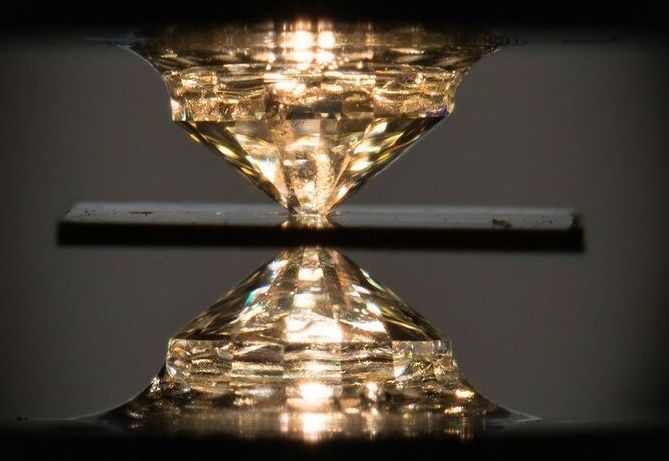
A novel metallic compound of hydrogen, carbon, and sulfur exhibited superconductivity at a balmy 59 degrees Fahrenheit—when pressurized between a pair of diamond anvils.

Via Quanta Magazine9 months ago.


Physicists finally achieved the long-sought goal, but there’s a catch: Their compound requires crushing pressures to keep from falling apart.

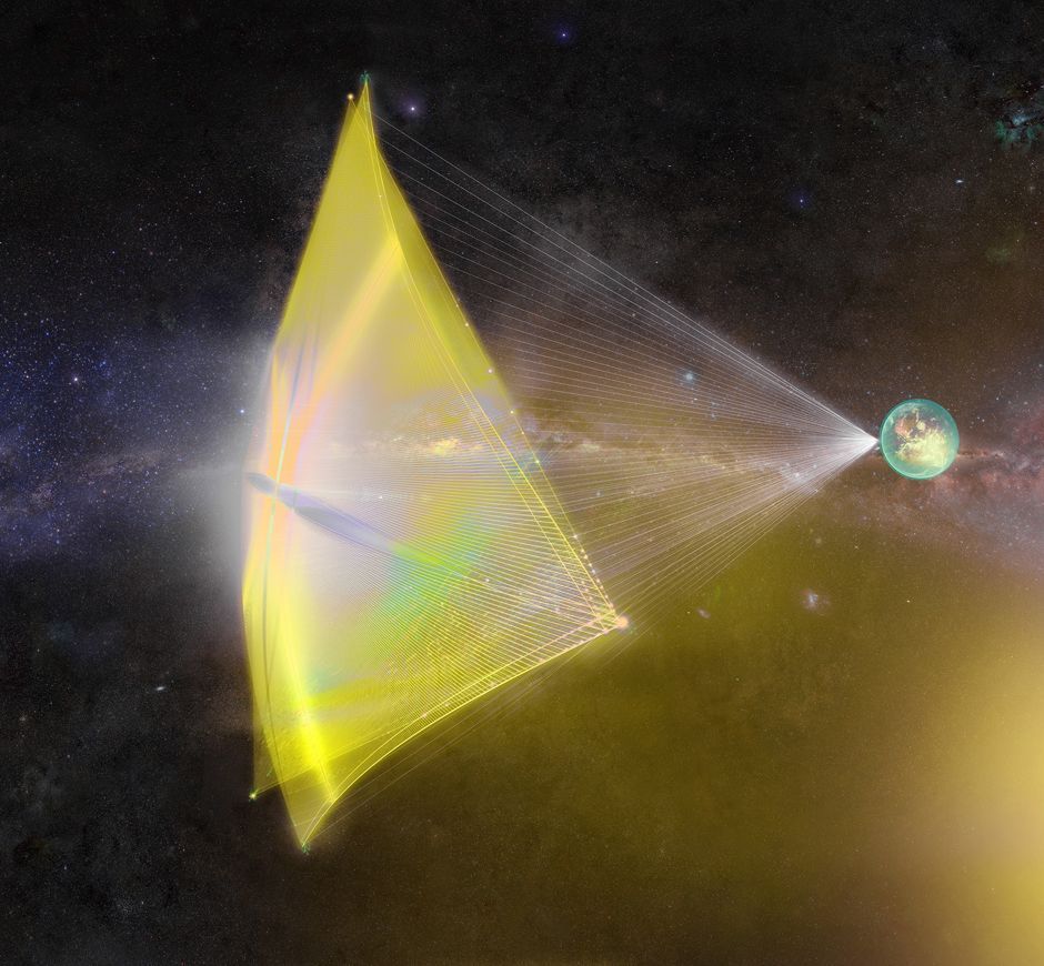
Solar sails have been receiving a lot of attention lately. In part that is due to a series of high profile missions that have successfully proven the concept. It’s also in part due to the high profile Breakthrough Starshot project, which is designing a solar sail powered mission to reach Alpha Centauri. But this versatile third propulsion system isn’t only useful for far flung adventures – it has advantages closer to home as well. A new paper by engineers at UCLA defines what those advantages are, and how we might be able to best utilize them.

The literal driving force behind some solar sail projects are lasers. These concentrated beams of light are perfect to provide a pushing force against a solar sail. However, they are also useful as weapons if scaled up too much, vaporizing anything in its path. As such, one of the main design constraints for solar sail systems is around materials that can withstand a high power laser blast, yet still be light enough to not burden the craft it is attached to with extra weight.

For the missions that graduate student Ho-Ting Tung and Dr. Artur Davoyan of UCLA’s Mechanical Engineering Department envision that weight is miniscule. They expect any sailing spacecraft to weigh less than 100 grams. That 100 grams would include a sail array that measures up to 10 cm square.

Late last year, a French company called Carmat received approval in Europe for its total artificial heart. It’s exactly what it sounds like: a heart made of synthetic and biological materials intended for implantation into people who need heart transplants. Now, just half a year later, the first US patient has received one of the hearts.

The transplant took place last week in a 39-year-old man at Duke University Hospital in North Carolina. The man didn’t go to the hospital expecting to have a heart transplant, but it ended up saving his life.

After experiencing unexpected heart failure, he was diagnosed with advanced coronary artery disease (when plaque builds up in the blood vessels that carry oxygen-rich blood to heart) and went in for bypass surgery (which implants a healthy blood vessel from another part of the body to redirect blood around a blocked artery).

Amazing.


While Venice may be home to the first 3D-printed concrete footbridge to be constructed entirely sans reinforcement or mortar, the similarly canal laced city of Amsterdam, not to be outdone, has unveiled the world’s first 3D-printed steel pedestrian bridge. The long-awaited project, first announced in 2015, was dedicated on the Oudezijds Achterburgwal canal in the city’s Red Light District on July 15. The ceremony was attended by Her Majesty the Queen of the Netherlands, Máxima, who was assisted by a ribbon-cutting robot during the festivities.

Spanning nearly 40 feet across the canal, the curving 6-ton stainless steel structure was constructed by Amsterdam-based 3D metal printing technology company MX3D using a wire arc additive manufacturing process that marries advanced robotics with welding. With the aid of four robots, the entire printing process took just six months. The completed bridge, designed by Joris Laarman Lab with Arup serving as lead engineer, was first unveiled in October 2018 during Dutch Design Week. Several load-testing rounds followed, the last of which was carried out in the fall of 2019 with plans to install the structure in early 2020. However, ongoing site prep work at the canal delayed the factory-produced bridge’s installation to just last week.

As reported by the Associated Press, the stainless steel structure, dubbed the MX3D Smart Bridge, will remain in place for at least two years while the footbridge that previously spanned the canal undergoes renovation.

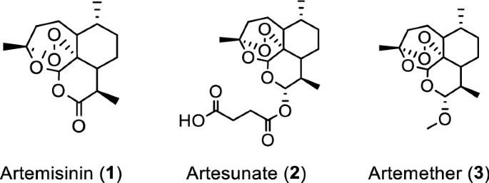
Artemisia annua plants grown from a cultivated seed line in Kentucky, USA, were extracted using either absolute ethanol or distilled water at 50 °C for 200 min and analyzed, as described in “Materials and methods” and Supplementary Information (Figures S1 and S2). Solids were removed by filtration and the solvents were evaporated. The extracted materials were dissolved in dimethylsulfoxide (DMSO) (ethanol extract) or a DMSO: water mixture (3:1 for aqueous extract) and filtered (see supporting information for details). Artemisinin (Fig. 1, (1)) was synthesized and purified following a published procedure or purchased17, while artesunate (Fig. 1, (2)) and artemether (Fig. 1, (3)) were only obtained from commercial sources.

Initial experiments were carried out at FU Berlin, Germany. To initially screen whether extracts and pure artemisinin were active against SARS-CoV-2, their antiviral activity was tested by pretreating VeroE6 cells at different time points during 120 min with selected concentrations of the extracts or compounds prior to infection with the first European SARS-CoV-2 isolated in München (SARS-CoV-2/human/Germany/BavPat 1/2020). The virus-drug mixture was then removed and cells were overlaid with medium containing 1.3% carboxymethylcellulose to prevent virus release into the medium. DMSO was used as a negative control. Plaque numbers were determined either by indirect immunofluorescence using a mixture of antibodies to SARS-CoV N protein18 or by staining with crystal violet19. The addition of either ethanolic or aqueous A. annua extracts prior to virus addition resulted in reduced plaque formation in a concentration dependent manner, while artemisinin exhibited little antiviral activity (Supplemental Tables S1 S8).

Concentration–response experiments were carried out at Copenhagen University Hospital-Hvidovre. In these experiments the Danish SARS-CoV-2 isolate SARS-CoV-2/human/Denmark/DK-AHH1/2020 was used employing a 96-well plate based concentration–response antiviral treatment assay, allowing for multiple replicates per concentration, as described in “Materials and methods” and Supplementary Information (Figures S3 and S4)20,21. Seven replicates were measured at each concentration and a range of concentrations was evaluated to increase data accuracy when compared to the plaque-reduction assay, which was carried out in duplicates. Extracts or compounds were added to VeroE6 cells either 1.5 h prior to (pretreatment (pt)) or 1 h post infection (treatment (t)), respectively, followed by a 2-day incubation of virus with extracts or compounds. Both protocols yielded similar results, with slightly lower median effective concentration (EC50) values observed for treatment assays.