Toggle light / dark theme

The new goal is to reverse aging, not only in animals, but in humans. And age reversal is essential, as significant age-related disruption has already occurred in most people due to changes in our gene expression profiles.

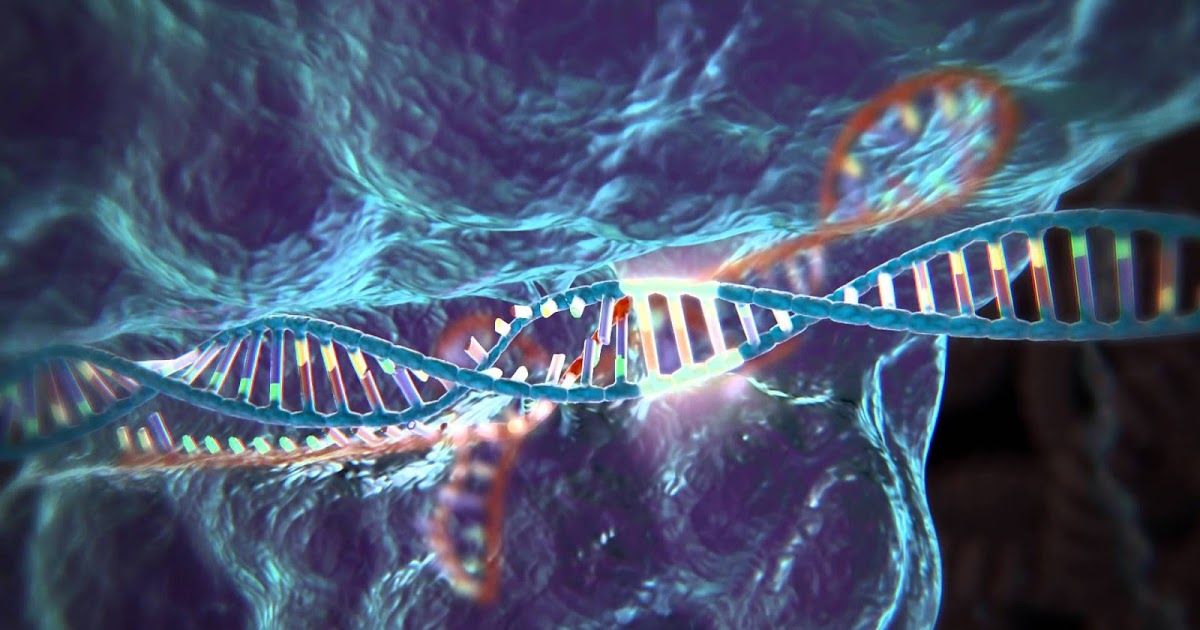
Gene expression patterns change with age. This influences the rate at which an individual ages, and also determines what senile disorders they are likely to contract. But innovative gene-editing methods based on a unique technology called CRISPR (clustered regularly interspaced short palindromic repeats) are now being successfully harnessed for use as an age-reversal therapy for humans.

In response to these breakthroughs, Life Extension® magazine sent biogerontologist Dr. Gregory M. Fahy to Harvard University to interview Dr. George Church, who is a leading developer of cutting-edge CRISPR techniques. Here, Dr. Church explains remarkable opportunities for transforming human aging that may begin to unfold sooner than most have imagined.
Read more

Very insightful for my deep mind/ neuro mapping friends.


If you have trouble learning a new language as an adult, maybe you can blame your genes and brain structure, a U.S. study suggested Monday.

The study by researchers at the University of Washington showed that genetic variations of the so-called COMT gene and a measure of the strength of the brain’s communications network — known as “white matter”— jointly accounted for 46 percent of the reason for why some college students performed better than others in the second language class.

“We are interested in understanding why individuals learn differently, including those who perform well and those who perform poorly,” said lead author Ping Mamiya, a research scientist at the UW’s Institute for Learning & Brain Sciences (I-LABS).

Read more

Diabetes 2 tie to Vitamin D issues.


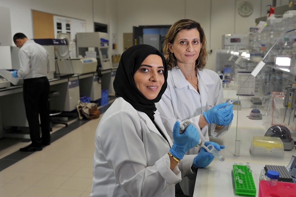
First study of Emiratis’ genes highlights the links between type 2 diabetes and a deficiency in vitamin D. Soon doctors may be able to prioritise care for those who are most at risk.

ABU DHABI // The first study to examine Emirati genes and the links between type 2 diabetes and vitamin D deficiency uncovered a genetic code that identifies those susceptible to the deficiency.

While studies internationally have found links between the two conditions, it is the first time the Emirati population has been looked at specifically.

Read more

Jumping genes — not jumping beans.


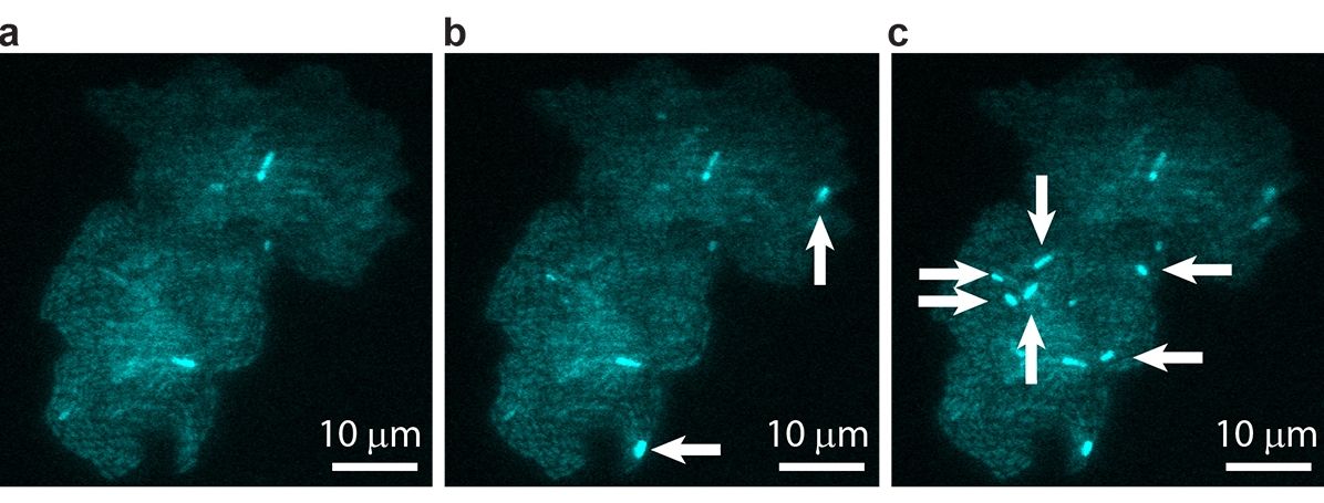
“Jumping genes” are ubiquitous. Every domain of life hosts these sequences of DNA that can “jump” from one position to another along a chromosome; in fact, nearly half the human genome is made up of jumping genes. Depending on their specific excision and insertion points, jumping genes can interrupt or trigger gene expression, driving genetic mutation and contributing to cell diversification. Since their discovery in the 1940s, researchers have been able to study the behavior of these jumping genes, generally known as transposons or transposable elements (TE), primarily through indirect methods that infer individual activity from bulk results. However, such techniques are not sensitive enough to determine precisely how or why the transposons jump, and what factors trigger their activity.

Reporting in the Proceedings of the National Academy of Sciences, scientists at the University of Illinois at Urbana-Champaign have observed jumping gene activity in real time within living . The study is the collaborative effort of physics professors Thomas Kuhlman and Nigel Goldenfeld, at the Center for the Physics of Living Cells, a National Science Foundation Physics Frontiers Center.

“In this study, we were able to see that there is actually more of this jumping gene action going on than might have been expected from previous studies,” said Kuhlman, whose team performed the in vivo experiments. “What’s more, we learned that the rates at which these genes jump depend sensitively on how the cells are growing—if there is food available for the cells to grow, for example. In other words, jumping gene activation isn’t entirely random, it’s dependent on environmental feedback.”

Read more

Using the CRISPR gene-editing tool, scientists from Harvard University have developed a technique that permanently records data into living cells. Incredibly, the information imprinted onto these microorganisms can be passed down to the next generation.

CRISPR/Cas9 is turning into an incredibly versatile tool. The cheap and easy-to-use molecular editing system that burst onto the biotech scene only a few years ago is being used for a host of applications, including genetic engineering, RNA editing, disease modeling, and fighting retroviruses like HIV. And now, as described in a new Science paper, it can also be used to turn lowly microorganisms into veritable hard drives.

http://io9.gizmodo.com/5935415/why-dna-is-the-future-of-data-storage

Read more

Very cool.


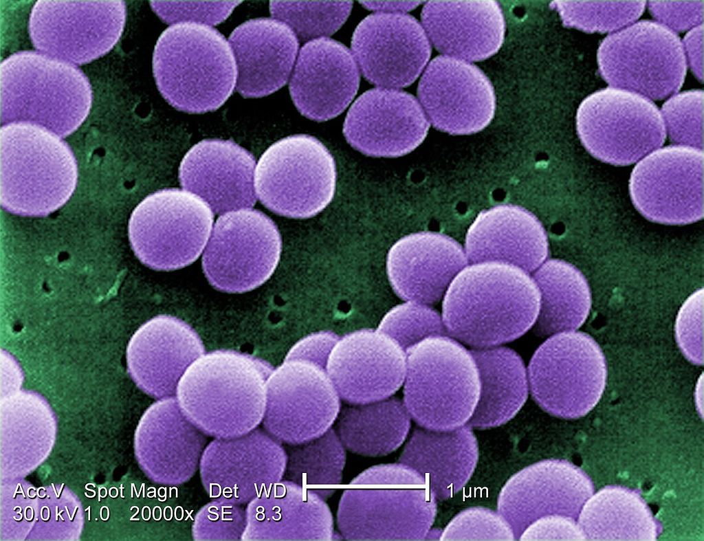
Pinpointing the type of bacteria that are at the root of an infection in clinical samples removed from living tissues, such as blood, urine or joint fluids, to quickly identify the best anti-microbial therapy still poses a formidable challenge. The standard method of culturing can take days to reveal pathogens, and they often fail to bring them to light altogether.

A team lead by Donald Ingber, M.D., Ph.D., at the Wyss Institute for Biologically Inspired Engineering at Harvard University now reports a method in PLoS, which enables the rapid isolation and concentration of infectious bacteria from complex clinical samples to help speed up bacterial identification, and it should be able to accelerate the determination of antibiotic susceptibilities as well.

“We leveraged FcMBL? the genetically engineered pathogen-binding protein we developed for our sepsis therapeutic device program? to develop a fast and simple technology to help overcome this diagnostic roadblock,” said Ingber, who is the Wyss Institute’s Founding Director, the Judah Folkman Professor of Vascular Biology at Harvard Medical School and the Vascular Biology Program at Boston Children’s Hospital, and Professor of Bioengineering at the Harvard John A. Paulson School of Engineering and Applied Sciences. “Using clinical samples of joint fluids, we were able to show that this method can be used to quickly and efficiently isolate bacterial pathogens for various kinds of subsequent analysis, including PCR, which is commonly used for molecular diagnostics in clinical laboratories.”

Read more

The ability to track molecular events inside the cells of living organisms offers a powerful window into fundamental biological processes, but methods for visualizing RNA in vivo without interfering with cell processes have been elusive. Now, researchers have developed a light-induced chemical reaction that accomplishes this feat in live zebrafish embryos (ACS Cent. Sci. 2016, DOI: 10.1021/acscentsci.6b00054). It is the first technique for detecting specific strings of nucleic acids in live vertebrates that doesn’t require genetically modifying the organism. What’s more, it’s sensitive enough to visualize the expression of microRNAs, small noncoding RNAs that act as puppetmasters of gene expression.

To do the reaction, chemical biologist Nicolas Winssinger, biochemist Marcos Gonzalez-Gaitan, and their colleagues at the University of Geneva designed two nucleic acid probes that each complement and bind to adjacent halves of a target microRNA sequence. The researchers conjugated one probe to a ruthenium complex that absorbs visible light and the other to a fluorogenic rhodamine that lights up when its azide bonds are cleaved. When the probes attach to the target sequence, the two reagents come close enough to react. Shining a light on the sample activates the ruthenium which then reduces the azide in the rhodamine conjugate, releasing its fluorescence. The dependence on external light allows researchers to control when the reporting reaction happens, Winssinger explains.

The team first developed the system three years ago (Chem. 2013, DOI: 10.1002/chem.201300060) for use in cultured cells; here, they adapted it for use in just-fertilized zebrafish embryos. “That’s really not trivial,” says Winssinger. The probes had to be nontoxic, stable for a day or more, and powerful enough to work even after being diluted through cell division.

Read more

There are various animals that can live for centuries or millenia.

Genetic engineering technology is rapidly improving and genome wide genetic engineering could become a reality within 10–20 years. It could be possible to replicate in humans the longevity genes and cancer immunity in certain animals.

The longest lived mammal is the bowhead whales. Some confirmed sources estimate bowhead whales to have lived at least to 211 years of age.

Read more

Interesting concept; my only concern is to individuals with nuero diseases or prone through genetics to have neuro diseases. For Dystonia patients/ victims who have copper compounds in their systems can potentially develop a form of secondary dystonia which can be terminal. Also, my years in the labs at ORNL taught us a lot about heavy metal exposures (including copper compounds); so I am a bit taken back by this article.


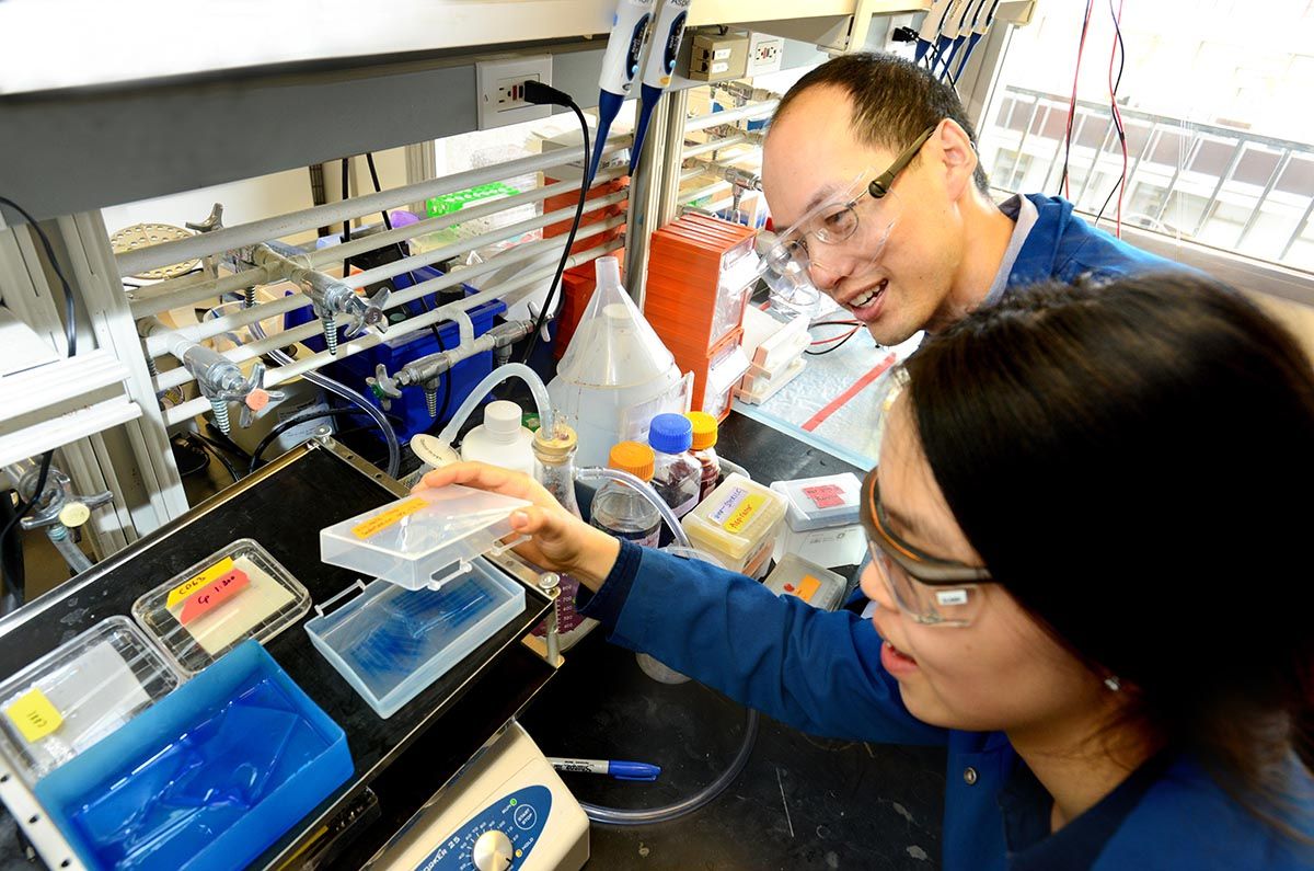
A new study is further burnishing copper’s reputation as an essential nutrient for human physiology. A research team led by a scientist at the Department of Energy’s Lawrence Berkeley National Laboratory (Berkeley Lab) and at the University of California, Berkeley, has found that copper plays a key role in metabolizing fat.

Long prized as a malleable, conductive metal used in cookware, electronics, jewelry and plumbing, has been gaining increasing attention over the past decade for its role in certain biological functions. It has been known that copper is needed to form red blood cells, absorb iron, develop connective tissue and support the immune system.

The new findings, to appear in the July print issue of Nature Chemical Biology but published online today, establishes for the first time copper’s role in .

Read more